Introduction
Pancreatic surgery has historically been associated with significant morbidity and mortality. However, advancements in robotic surgical technology have revolutionized the approach to complex pancreatic procedures. This case study highlights the successful implementation of a totally robotic spleen-sparing distal pancreatectomy for a patient with multiple pancreatic neuroendocrine tumors, demonstrating the precision and advantages of robotic-assisted surgery in preserving critical organs while effectively treating pathology.
Patient Presentation and History
A 58-year-old male with multiple comorbidities including type 2 diabetes, hypertension, and hyperlipidemia initially presented with back pain in 2019. Despite not exhibiting classic symptoms typically associated with pancreatic tumors (such as jaundice, weight loss, or digestive difficulties), diagnostic imaging revealed concerning findings that warranted further investigation.
The patient’s initial workup included:
- MRI and MRCP (Magnetic Resonance Cholangiopancreatography) scans that detected a non-functioning pancreatic tumor
- Identification of a large cystic lesion in the liver
- Institution of regular imaging surveillance due to the asymptomatic nature of the findings
Comprehensive Diagnostic Evaluation
In March 2024, following a period of watchful waiting with regular imaging follow-ups, the patient underwent a comprehensive diagnostic evaluation to better characterize the lesions and determine appropriate management.
Advanced Imaging and Tissue Sampling
- Endoscopic Ultrasound (EUS)
- Revealed a 3.4 cm cystic lesion in the pancreatic body and tail region
- Identified a concurrent liver lesion in the right lobe
- Facilitated fine needle aspiration (FNA) biopsies of both lesions
- Histopathological Analysis
- Pancreatic lesion: Confirmed a well-differentiated Grade 1 neuroendocrine tumor
- Liver lesion: Showed evidence of steatohepatitis without malignancy
- CT Scan of Abdomen and Pelvis
- Documented progression with multiple pancreatic lesions:
- 3.3 × 3.3 cm lesion in the mid-body
- 10 mm lesion in the tail
- 9 mm lesion in the proximal body
- Revealed a 4 × 3 cm lobulated cystic lesion in segment 6 of the liver
- Documented progression with multiple pancreatic lesions:
- DOTA PET Scan
- Demonstrated somatostatin receptor-expressing lesions in the pancreas
- Identified additional satellite lesions
- Provided functional characterization of the tumors
Multidisciplinary Approach to Treatment Planning
Despite negative tumor markers and normal serum Chromogranin-A levels (a biomarker typically elevated in neuroendocrine tumors), a multidisciplinary team (MDT) recommended surgical intervention based on the imaging findings and histological confirmation. The patient was specifically referred to the Department of Gastrointestinal Surgery for consideration of robotic surgery—an approach that would allow for precise tumor removal while potentially preserving the spleen.
Surgical Approach and Technical Considerations
Following thorough preoperative counseling and evaluation, the surgical team proceeded with a totally robotic spleen-sparing distal pancreatectomy. This approach offered several key advantages:
- Enhanced Visualization: The robotic system provided magnified, high-definition 3D visualization of the surgical field, essential for identifying and preserving the splenic vessels.
- Intraoperative Ultrasonography: Used to precisely locate the tumors and guide the extent of resection, ensuring complete removal of all lesions while preserving maximum healthy pancreatic tissue.
- Precision Dissection: The robotic instruments allowed for meticulous dissection around critical vascular structures, particularly important in separating the pancreas from the splenic vessels.
- Minimal Blood Loss: The procedure was completed with less than 100 mL of blood loss, significantly lower than traditional open approaches.
- Efficient Operative Time: Despite the complexity of the procedure, the operation was completed in 5 hours.
Surgical Technique: Preserving the Spleen While Removing Diseased Pancreas
The spleen-sparing technique employed in this case deserves special attention. In traditional distal pancreatectomy, the spleen is often removed along with the distal pancreas because of their shared blood supply. However, preserving the spleen offers significant immunological benefits, particularly important for long-term health and prevention of post-splenectomy infections.
The robotic approach facilitated:
- Warshaw Technique Modification: Preservation of the splenic vessels while completely mobilizing the pancreas from surrounding structures.
- Selective Vascular Control: Precise ligation of small pancreatic vessels without compromising the main splenic vasculature.
- Real-time Assessment: Continuous evaluation of splenic perfusion throughout the procedure.
Postoperative Course and Outcomes
The patient experienced an exceptionally smooth recovery:
- Minimal postoperative pain, effectively managed with standard analgesics
- Resumed oral intake on the first postoperative day
- Normalized bowel function by the second postoperative day
- Discharged home on the third postoperative day
- No evidence of pancreatic fistula or other complications
- Maintained excellent follow-up status with preserved glycemic control
Technical Challenges and Considerations
The case presented several technical challenges that highlight why robotic approaches offer advantages in complex pancreatic surgery:
- Patient Factors: The patient’s obesity represented a technical challenge that was mitigated by the enhanced dexterity and visualization of the robotic system.
- Multiple Pancreatic Neoplasms: The presence of several tumors throughout the distal pancreas required precise localization and comprehensive resection.
- Splenic Preservation: Maintaining adequate blood supply to the spleen while completely removing the distal pancreas was technically demanding, particularly due to the intricate vascular anatomy behind the pancreas.
Discussion: Advantages of Robotic Approach in Pancreatic Surgery
This case exemplifies the unique advantages of robotic-assisted surgery for complex pancreatic procedures:
Enhanced Technical Capabilities
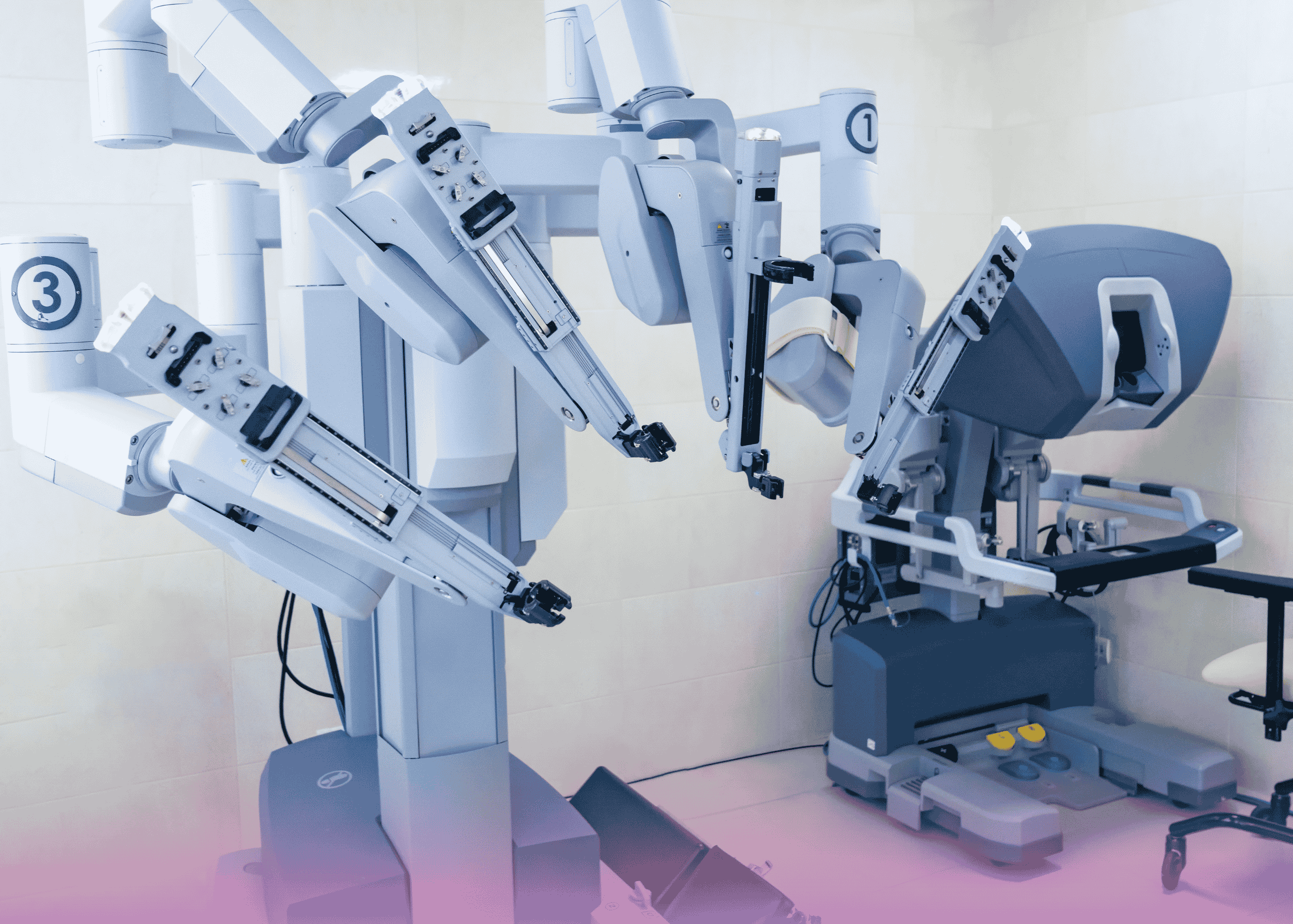
The da Vinci Robotic Surgical System offers:
- Superior Visualization: High-definition 3D imaging allows for better appreciation of fine tissue planes.
- Increased Dexterity: The robotic “wrists” enable 540-degree rotation and movement in tight spaces, critical when working around major vessels.
- Tremor Filtration: Elimination of natural hand tremor enhances precision during delicate dissection.
- Improved Ergonomics: The surgeon’s improved positioning reduces fatigue during lengthy procedures.
Clinical Benefits
Patients undergoing robotic pancreatic surgery often experience:
- Reduced blood loss compared to open procedures
- Lower conversion rates to open surgery compared to conventional laparoscopy
- Shorter hospital stays
- Faster return to normal activities
- Improved cosmetic outcomes due to smaller incisions
Organ Preservation Benefits
The spleen plays critical roles in:
- Immune function, particularly against encapsulated bacteria
- Blood filtration and recycling of red blood cells
- Serving as a blood reservoir
Preserving this organ provides long-term benefits to the patient, including reduced risk of overwhelming post-splenectomy infection (OPSI) and maintained hematological function.
Conclusion
This case demonstrates the successful application of robotic technology in performing a complex spleen-sparing distal pancreatectomy. The robotic approach facilitated precise tumor removal while preserving the immunologically important spleen, despite the technical challenges posed by the patient’s body habitus and the presence of multiple neoplasms.
The excellent postoperative outcome, characterized by minimal blood loss, short hospital stay, and rapid recovery, underscores the potential benefits of robotic surgery in selected patients with pancreatic pathology. As this technology continues to evolve and surgical teams gain more experience, robotic approaches may become increasingly important in the management of complex pancreatic diseases.
This case, managed by Dr. Ali Iyoob Valiyaveettil, Consultant & Head of Gastrointestinal Surgery, serves as an excellent example of how cutting-edge robotic technology can be leveraged to achieve optimal surgical outcomes while prioritizing organ preservation and minimizing surgical trauma.