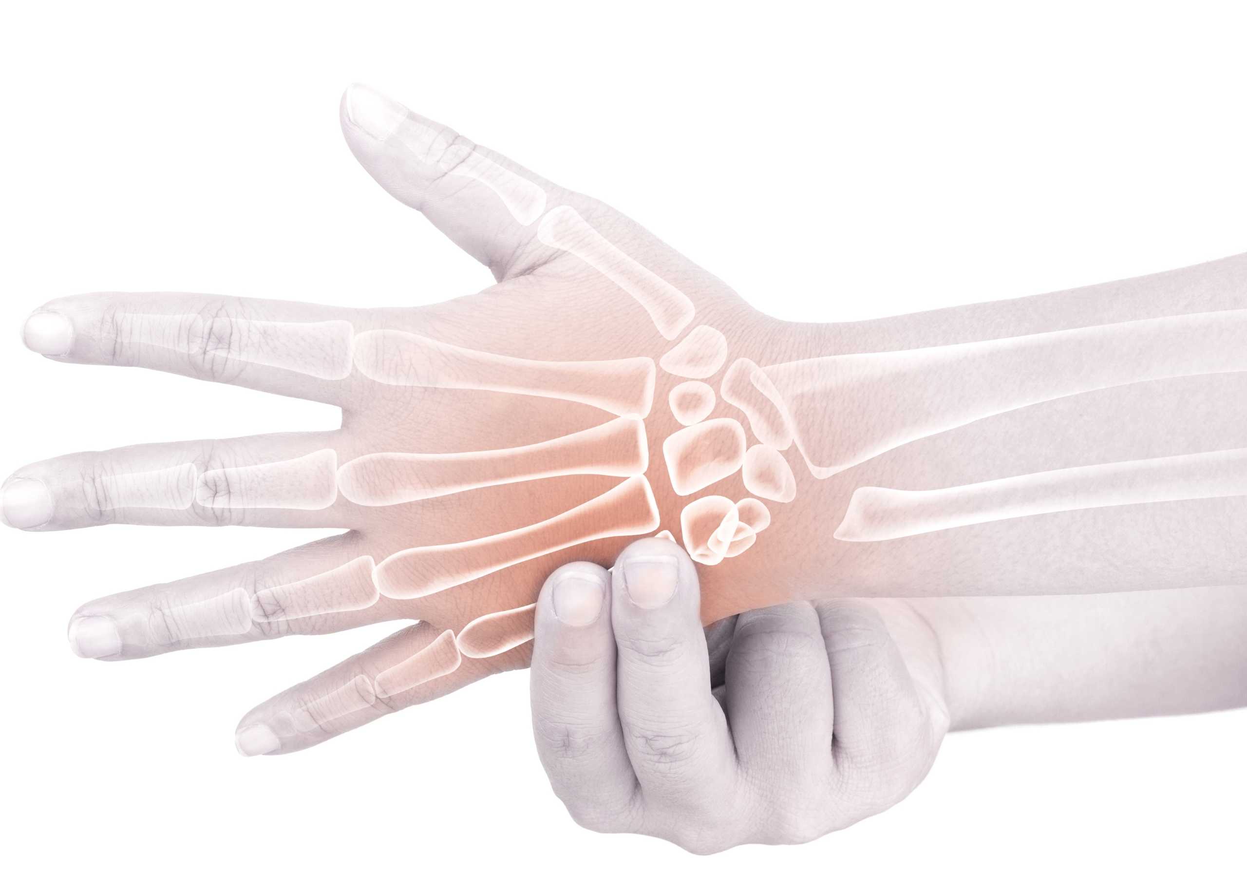
Most people experience pain at least once in their lifetime, whether in the form of back pain, knee pain or shoulder pain. Often, the disorders are fortunately benign and disappear after a brief period of rest, exercise and the observation of a few behavioral rules. If the pain does not disappear, the Pain Clinic team of specialists at the Burjeel Hospital for Advanced Surgery, Dubai, will accompany you with the help of proven treatment measures for the pain.
Pain is maintained by complex mechanisms and can have disastrous consequences on social, family and professional life. Patients referred to Pain Clinic should quickly receive a standardized assessment based on a holistic approach and a personalized self-management plan.
The Pain Clinic at the Burjeel Hospital for Advanced Surgery, Dubai, offers a global and comprehensive approach, from initial assessment to treatment of all types of orthopedic pain due to from sports injuries, varied conditions and all other day-to-day activities.
Here’s more you need to know about our Pain Clinic!
Our muscles, joints, ligaments and other parts are subjected to multiple stresses and can become the sites of ailments often due to sports injuries, monotonous load, age or other related factors. Our orthopedic surgeons team specializes in Sports Medicine and has expertise and experience in treating all kinds of Orthopedic pain through a modern diagnostic and therapeutic approach.
The Burjeel Hospital for Advanced Surgery’s Pain Clinic team directs patients to the most appropriate care as needed. The coaching encourages patients to take control of their pain care plan gradually. The goal is to develop a care plan tailored to each patient’s specific needs based on evidence-based recommendations.
The first step is a correct diagnosis through a clinical examination and appropriate radiological and neurological screening, after which a treatment adjusted as needed can be initiated. Pain treatment options range from conservative treatment (including invasive pain treatment) to conventional surgical techniques, including various microsurgical, percutaneous and minimally invasive interventions.
In addition to the classic stabilization technique (dorsal or ventral), we use dynamic implants. The objective of each treatment is as short a rehabilitation as possible, a rapid return to professional activity and the minimization of the damage.
Burjeel Hospital for Advanced Surgery has a team of certified Orthopedic Surgeons and pain specialists experienced in treating varied conditions and injuries and related structures. Our team has the expertise in treating all forms of sports injuries, ranging from shoulders, ankles, knees, elbows to spinal injuries. The different spine conditions include lumbar and cervical disc herniation, narrow lumbar canal, narrow cervical canal, etc.
Palette of Treatments
- Muscle tears
- Repetitive injuries
- Tendon or Ligament injury
- Achilles tendonitis
- Wrist fracture
- Scapula fracture
- Clavicle fracture
- Ankle fracture
- Runners’ knee
- Tennis or golfers Elbow
- Rotator Cuff Injury
- Revision of failed ACL Reconstruction Surgery
- RSLAP Repair
Types of Pain Due to Sports Injuries Treated at the Pain Clinic
Different spinal pain conditions are treated at the pain clinic. If you recognize yourself in any of the spinal pain examples below, contact us at to book an appointment to get a precise diagnosis and a therapy proposal.
Muscle Tears
Muscle tears or fiber breakage can be caused by a blow or contusion or by a sudden muscle contraction. It consists of the rupture of the fibers that make up the muscle. It generally affects muscles of the lower limbs such as the gastrocnemius, the hamstrings and the quadriceps. Its severity is given fundamentally by the number of muscle fibers torn in the injury. The main symptom is sudden, sharp, localized pain. It is one of the most frequent injuries in all types of sports.
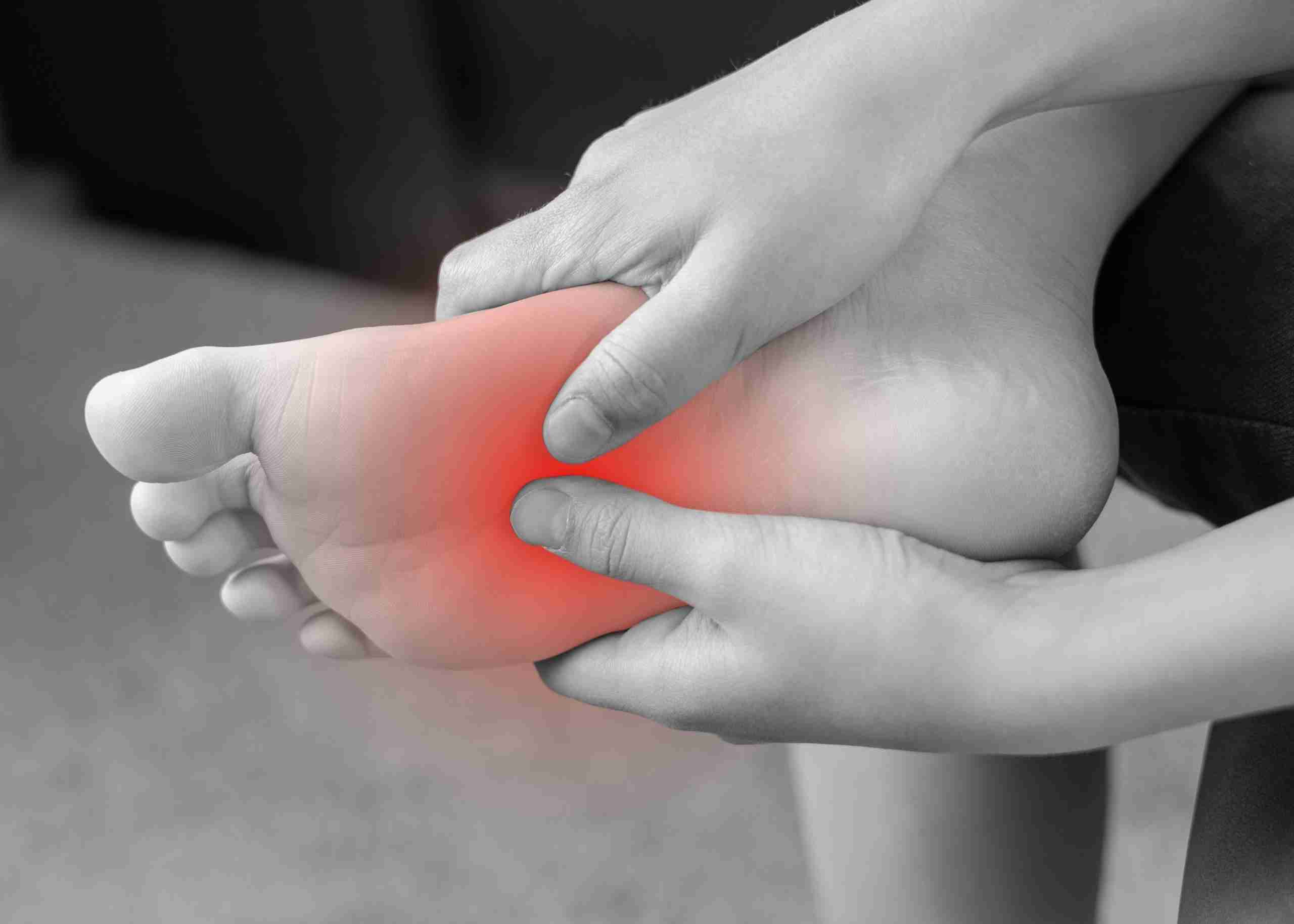
Ankle Sprain
This injury is very common in sports practice but also in daily activity. In most cases, it occurs due to an inward foot movement that causes tension in the ligaments. The sprain can be mild when there are micro-tears in the ligaments, moderate when there is a partial tear, and severe when the ligaments are completely torn. The most common symptoms are bruising and pain.
It occurs mainly in impact sports or in which the practice is carried out on unstable surfaces.
Tendinopathies
Popularly known as tendinitis, they consist of chronic inflammation of the tendon, that is, over a long period, giving rise to micro-tears in the tendon tissue. Tendonitis can lead to different sports injuries, such as:
- Achilles tendons
- lateral epicondylitis (tennis elbow)
- medial epicondylitis (golfer’s elbow)
- patellar tendonitis (jumper’s knee) or
- patellofemoral pain syndrome (runner’s knee)
Although none of these three injuries is exclusive to athletes, lateral epicondylitis is common in people who play racket sports. Medial epicondylitis is common in golfers or athletes who play racket sports or do weight training. Patellar tendinitis occurs in people who play sports that involve repeated jumping, such as basketball and volleyball. It happens when the tendon connecting your shinbone to your kneecap becomes inflamed. Runner’s knee occurs when your kneecap has veered off the patellar groove.
Fractures
Perhaps the most serious injury that can affect the bone. The most frequent causes of fractures are falls or excessive muscular activity that lasts over time. In addition to fractures, bone injuries include others, such as those caused by overload, which can inflame the periosteum (tissue that covers the bone) and lead to breakage.
In most cases, immobilization, rest and rehabilitation are required, as well as surgery in the most severe cases. In both cases, it requires a subsequent progressive recovery.
Knee Ligament Injuries
Ligaments are the fibrous tissue that joins bones together. Those that are most frequently injured are those of the knee, where we find the following ligaments:
- Collateral medial.
- Collateral lateral.
- Rear cross.
- Former crusader.
When the tear or rupture of one of the ligaments occurs, there is localized inflammation with severe pain and a sensation that the knee gives way when pressure is exerted on it.
Spinal Conditions
Back pain occurs in patients in various ways: from moderate discomfort to intense pain in the dorsal and/or lumbar region and sometimes in the legs, which can seriously hinder movement.
Cervical Pain occurs in the upper back area, such as the nape, shoulder area, and lateral neck area often progresses to the shoulder blades and can even present as a headache. It is a very common pain in young people often caused by maintaining incorrect postures or by sports practices. However, it can also occur due to a problem in the vertebrae, spinal discs, or a muscle complication, such as contractures.
When a patient presents back pain, it is recommended that other possible pathologies such as cardiac or pulmonary pathologies be ruled out. The causes that generate it are varied, including osteoarthritis, herniated disc, and narrow spinal canal, among others.
Pain Therapy at the Pain Clinic
The basis of each therapy is as precise diagnostic clarification as possible in cooperation with colleagues in the practices and the various specialties represented in the hospital.
Back pain can be felt in many ways due to different causes. The cause and therapy must, therefore, always be determined individually. We have a team of highly trained professionals with knowledge of pain techniques and procedures such as:
Pharmacological Treatments
Pharmacological treatments consist of the oral administration of specific drugs according to the pathology to be treated and the pathological history of the patient.
Pain medication provides daily relief without treating the condition. Inflammation medications are reserved for periods of crisis. They can be given systemically (tablets and suppositories are as effective as intramuscular injections) or locally, in the form of corticosteroid injections.
Joint and Spinal Injections
This intra-articular administration is applied to patients with osteoarthritis, tendinitis, bursitis or some patients with rheumatic diseases.
Targeted infiltration of painful structures, pain-carrying nerve fibers, facet joints, and nerve root envelopment under X-ray or CT guidance can often successfully eliminate severe pain. In addition to treatment, infiltrations are also used for diagnosis and determining the causes.
Radiofrequency
Radiofrequency is a procedure that uses high-frequency current through a needle to exert its therapeutic action on different joints or nerves.
Radiofrequency therapy allows the pain associated with irritated osteoarthritic facet joints to be lastingly and gently eliminated, the procedure can be carried out as part of outpatient treatment, and after a few hours, the patient can leave the hospital. Iliosacral joint and pelvic pain are often treated in the same way.
Rehabilitation
Rehabilitation, including physiotherapy, balneotherapy, tractions, occupational therapy, corset, neck brace, etc., is particularly important for pain. The means of rehabilitation differ according to the objective sought: to stop a congestive attack of osteoarthritis more quickly, strengthen the damaged bone, and reduce pain and daily discomfort. Physiotherapy, also known as physical therapy, is a science responsible for using all physical agents.
A surgical procedure is justified if pain is resistant to medical treatment for more than six months and affects the patient’s daily activities.
A Brief Overview of the Surgical Procedures Offered at the Pain Clinic
The Pain Clinic of Burjeel Hospital for Advanced Surgery has a team of Orthopedic surgeons specializing in treating osteoarticular disorders. The extensive expertise of our specialists in sports surgery has supported many athletes in sports practice.
Here’s a summary of the surgical procedures carried out at the Pain Clinic in the context of sports surgery.
Sports Surgery for Ankle Injuries
- Rupture of the Achilles tendon: The surgery involves reinserting the torn ends of the tendon.
- Ankle instability and ankle sprains: Sprains are the daily lot of athletes. They do not necessarily require surgical intervention, but in the case of repeated sprains leading to proven instability, surgery can be considered.
- Previous ankle conflict: Generally, surgery is unnecessary, but if non-surgical treatments fail to stop the pain, it can be considered.
Sports Surgery Related to Shoulder Injuries
- Instability of the shoulder: The shoulder can be dislocated. This type of trauma is benign and does not require surgery. However, as for the ankle, repeated dislocations can indicate instability, which can be the subject of an operation.
- Pathologies of the rotator cuff: These are the tendons that give the shoulder its exceptional mobility. Certain movements can seriously damage this system: tendonitis and tears can threaten the athlete.
Sports Surgery Related to Knee Injuries
- Sprains of the knee: This is a frequent trauma whose severity can be variable. A simple stretching of the ligament will not necessarily require surgery, but if it leads to serious issues, such as patellar instability, surgery is considered.
- Ligament Injuries: Depending on the nature of the affected ligament (interior or posterior), the management will be different. Consulting sports and knee surgery specialists is essential for a good diagnosis.
- Meniscal pathologies: Like the ligaments, the meniscus is a fundamental element of the knee that may be subjected to various traumas. Meniscal lesions are often tears that will usually undergo meniscectomy or meniscal suturing under arthroscopy.
Thanks to technological advancements and the use of minimally invasive techniques, there are shorter recovery periods than traditional surgeries.
Although results vary from person to person, many of the above procedures successfully relieve most or all of the pain and symptoms caused by the different sports injuries.
Burjeel Hospital for Advanced Surgery is a reference to pain care in Dubai, having a board-certified team of Orthopedic Surgeons with unparalleled expertise in treating a wide range of conditions and sports injuries. A highly qualified team is at your service with several qualified and complementary professionals who work in synergy to offer you the optimal care necessary to recover your health as quickly as possible.