A novel technique combining laparoscopic surgery with on-table flexible ureteroscopy has been successfully implemented at Lifecare Hospital, a Burjeel Holdings facility, offering new hope for patients with complex ureteric strictures.
Complex Case Presentation
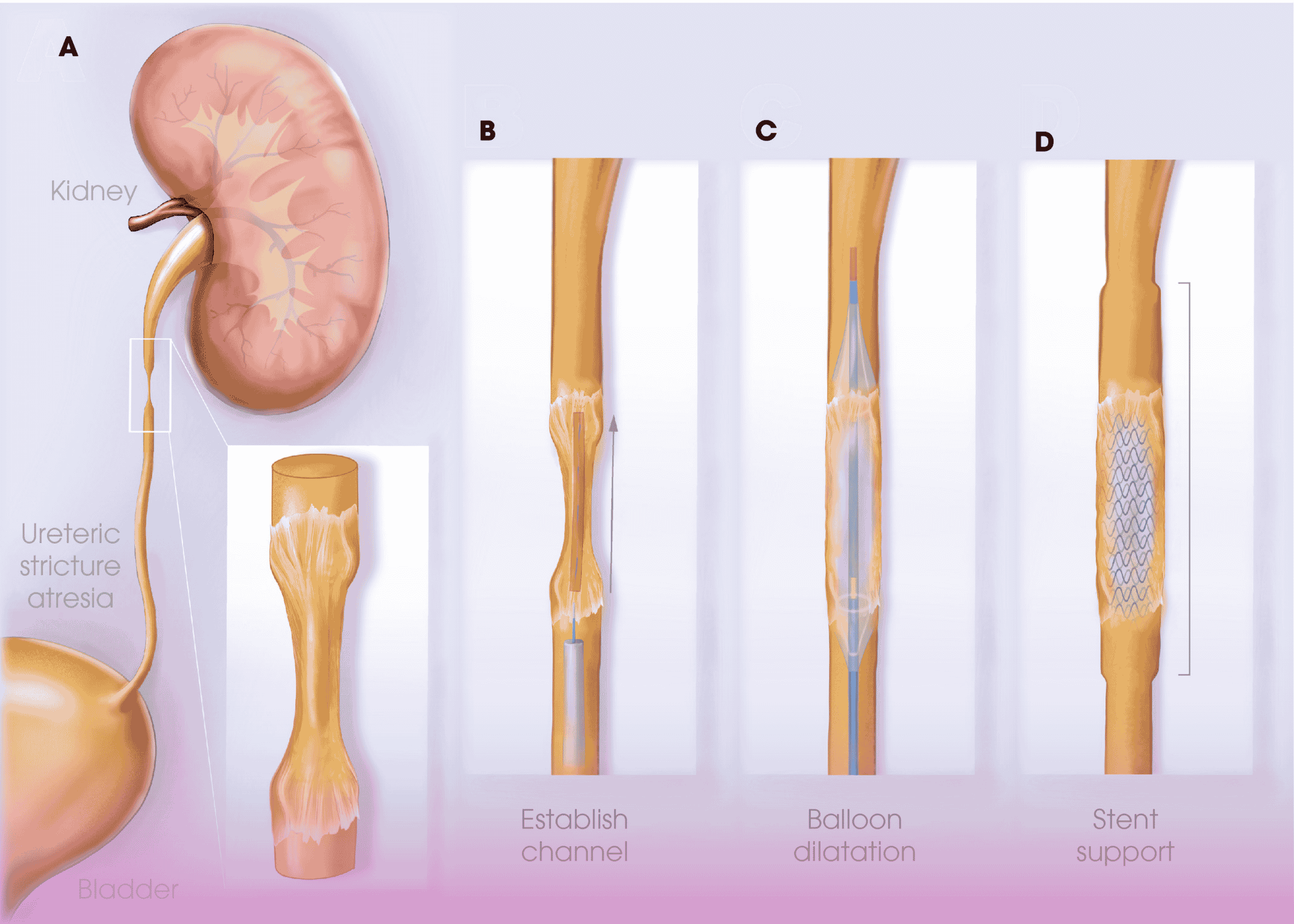
A 41-year-old male presented with persistent left flank pain and recurrent urinary tract infections over a one-year period. His medical history revealed a previous ureteroscopic intervention for a left mid-ureteric stricture that had developed secondary to an impacted ureteric stone. The initial management included balloon dilation and stenting, but despite these standard interventions, the patient experienced symptom recurrence indicative of stricture reformation.
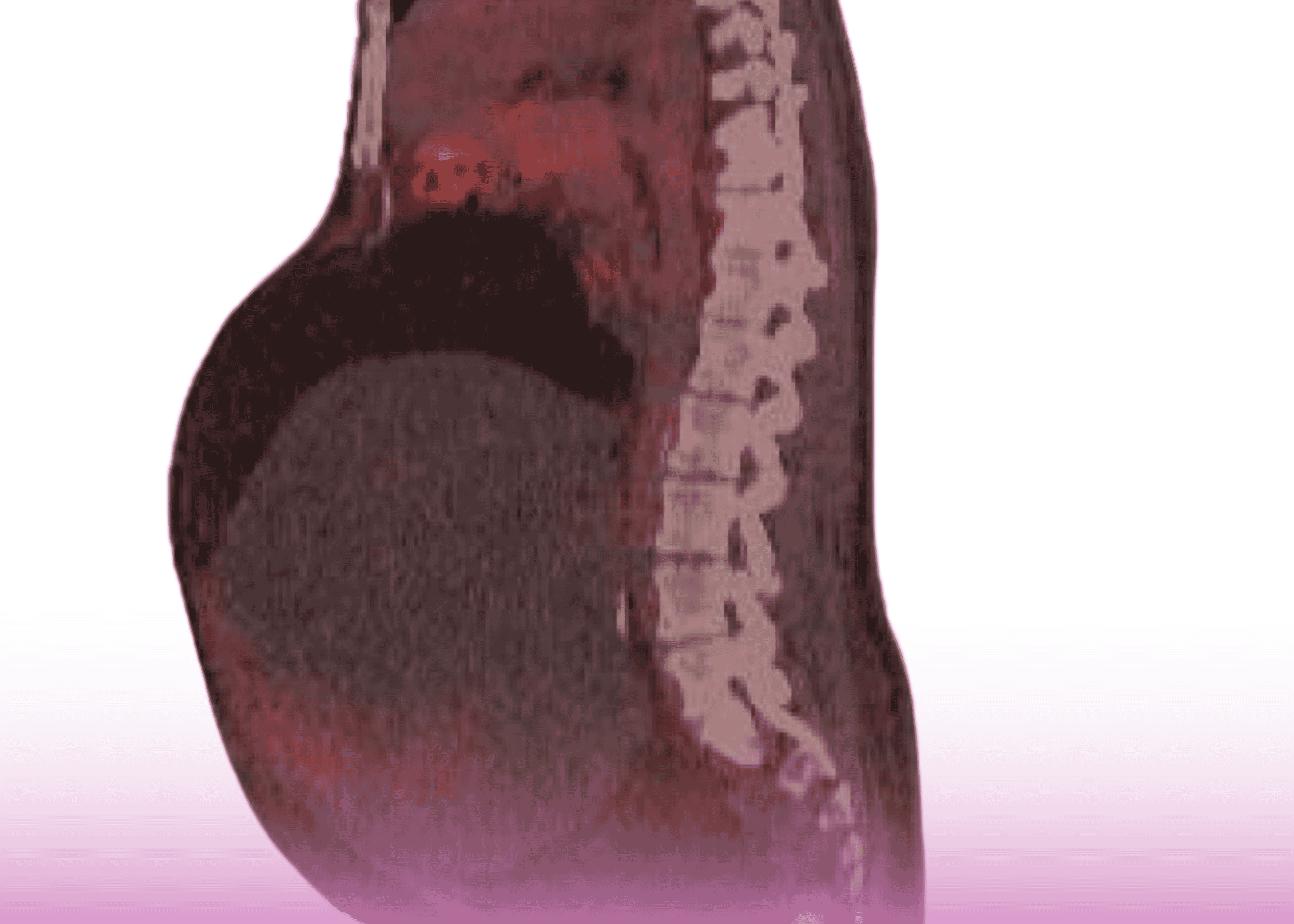
Diagnostic ultrasound demonstrated left-sided hydronephrosis, while previous intraoperative retrograde ureterogram had confirmed a mid-ureteric stricture approximately 2 cm in length with proximal ureteral dilation. Laboratory tests showed normal renal function with no active infection at the time of presentation.
Innovative Surgical Approach
The urology team at Lifecare Hospital, led by, Dr. Althaf Hussain and Urology Specialist, Dr. Anand Srivastava, implemented a novel combined approach that integrated both laparoscopic and endourological techniques in the same procedure.
Traditional management of recurrent ureteric strictures typically involves either:
- Endoscopic management (balloon dilation, endoureterotomy)
- Open surgical reconstruction (ureteroureterostomy, ureteral reimplantation)
- Standard laparoscopic repair
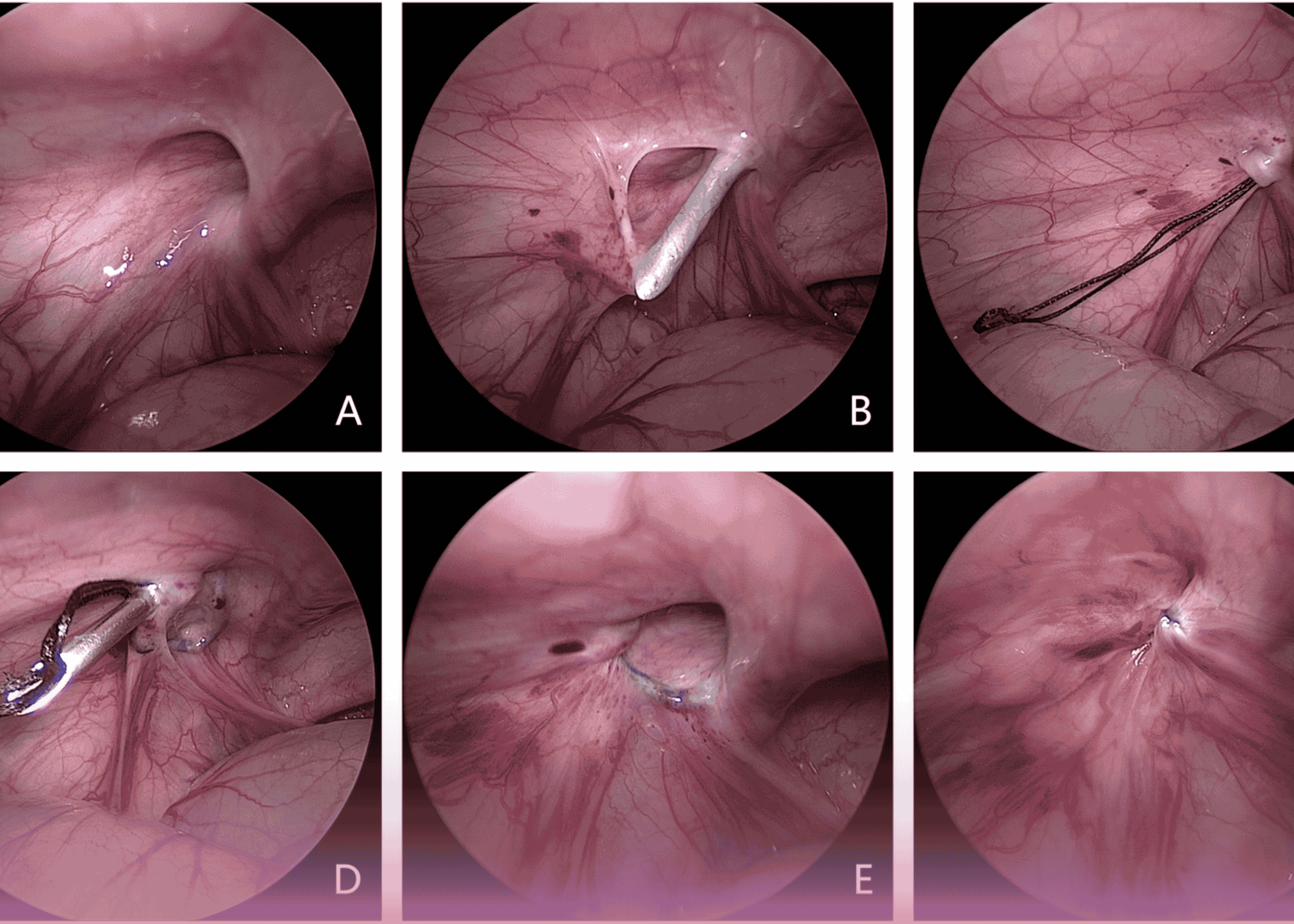
The innovative aspect of this case was the simultaneous use of flexible ureteroscopy during the laparoscopic procedure, providing enhanced visualization and precision that neither technique alone could achieve.
Technical Advantages
This combined approach offers several significant benefits over conventional methods:
Enhanced Visualization: The flexible ureteroscope provides real-time endoluminal imaging during laparoscopic dissection, allowing for precise identification of the stricture’s proximal and distal extent. This dual visualization prevents inadvertent injury to healthy segments of the ureter.
Minimally Invasive: The laparoscopic approach minimizes surgical trauma compared to open surgery, while the addition of flexible ureteroscopy reduces the need for extensive ureteral mobilization and dissection.
Improved Technical Precision: The combined technique enables more accurate placement of sutures during reconstruction and immediate confirmation of ureteral patency following repair.
Reduced Morbidity: By minimizing dissection and handling of the ureter, this approach potentially reduces the risk of devascularization, which can lead to recurrent stricture formation.
Advancing Urological Care
“Recurrent ureteric strictures, particularly in the mid-ureter, present significant therapeutic challenges,” notes Dr. Hussain. “This novel approach represents a promising advancement in the management of complex strictures that have failed conventional treatment.”
The technique demonstrated at Lifecare Hospital merits further investigation and could potentially become a standard approach for managing similar cases, offering patients the benefits of advanced laparoscopic surgery in urology performed by expert Urology Specialists with enhanced precision and potentially improved long-term outcomes.
This case exemplifies the commitment to surgical innovation within the Burjeel Holdings network, where specialists continuously explore new approaches to address challenging clinical scenarios and improve patient outcomes.