Type 2 Diabetes: Can the Risk Be Reduced?

In the UAE in 2021, about 1 million people had diabetes, and the predicted figure in 2040 is expected to be 2.2 million. There are two main types of diabetes mellitus, type1 and type2, type1 presents about 10% of diabetes mellitus, and type2 diabetes mellitus presents around 90%. Therefore, can we reduce the burden of type 2 diabetes mellitus on society and individuals? Do you believe we can reduce the estimated number of diabetes cases by 2040?

Does Type 2 Diabetes Occurs Solely as a Result of Genetics and Family History

Genetics and family history account for 15% of your risk of developing type 2 diabetes mellitus; however, lifestyle, diet, and exercise account for 85% of your risk. This indicates that 15% is considered a non-modifiable risk, while on the other hand, 85% is considered modifiable. So, let us look at how we can address the modifiable 85% risk:

First, for early detection, a blood checkup should be done every 6 months, including the diagnostic test HBA1C, which checks your mean blood glucose for the last 3 months and fasting and post-prandial blood sugar. Early detection will be achieved even in the pre-diabetes stage.

Second, exercise can work on increasing the sensation of insulin in your body, which helps improve the glucose absorption in your body cells, reduces the work burden on the pancreas, and can help you lose extra body weight if the person is obese.

Thirdly, weight loss is one of the most important key elements in reducing the risk of developing type 2 diabetes mellitus, as it helps improve insulin sensation at the cell level. The American Diabetes Association recommends that people with prediabetes lose at least 7% to 10% of their body weight to prevent disease progression. More weight loss will increase the health benefits even more.

Fourth, lifestyle and behavioral changes are the most difficult challenges in the journey. Therefore, a multidisciplinary team of a doctor, dietitian, psychologist, and diabetic educator is needed to empower you to achieve the ultimate goal.

Diet plays the most important role in managing blood glucose readings; having a realistic goal and meal plans will help you maintain the result of adequate blood glucose control.

In conclusion, prevention is better than cure. By working on all the mentioned elements, the burden of diabetes mellitus can be reduced in society and improve your quality of life.

Diabetes Expert at Burjeel Day Surgery Center in Abu Dhabi

Ms. May Abu Galala

Diabetes Educator


Years of Experience: 11

Nationality: Palestine

Languages Known: Arabic, English

Our Experts

See more

Cervical Cancer – Symptoms, Causes & Treatment

Cervical cancer is a disease in which malignant (cancer) cells form in the cervix, the lower part of the uterus. The cervix is the passageway that connects the vagina, uterus, and fallopian tubes. The uterus is where a baby grows before birth. Cervical cancer can lead to death if not treated, but it’s most often treatable when found early.

What Causes Cervical Cancer?

Cervical cancer is a disease that develops when cells become abnormal and multiply uncontrollably. These abnormal cells begin to crowd out healthy cells in the cervix responsible for protecting the uterus and fallopian tubes. Cervical cancer is most often caused by human papillomavirus (HPV), although other factors can also cause it.

What are the Symptoms of Cervical Cancer?

Cervical cancer symptoms include,

  • Bleeding after sexual intercourse or between menstrual periods
  • Difficulty urinating or pain in the lower abdomen or pelvis
  • Pain during sex, especially deep penetration
  • Prolonged menstrual bleeding or bleeding between periods
  • A lump or mass in the vagina

What are the Stages of Cervical Cancer?

There are three stages of cervical cancer: stage 0, stage 1, and stage 2.

Stage 1: In this stage, the cancer is confined to the cervix, the lower part of the uterus. Stage 1 is further divided into two sub-stages:

  • Stage 1A: The cancer is microscopic and can only be detected with a microscope.
  • Stage 1B: The cancer is visible to the naked eye or can be detected through imaging tests but remains confined to the cervix.

Stage 2: At this stage, the cancer begins to spread beyond the cervix but still remains within the pelvic area. Stage 2 is further divided into two sub-stages:

  • Stage 2A: The cancer has spread to nearby tissues, such as the upper part of the vagina.
  • Stage 2B: The cancer has reached the tissues next to the cervix, such as the parametrium (connective tissue surrounding the cervix) or the lower part of the vagina.

Stage 3: In this stage, the cancer extends further beyond the pelvis. Stage 3 is divided into three sub-stages:

  • Stage 3A: The cancer has reached the lower part of the vagina.
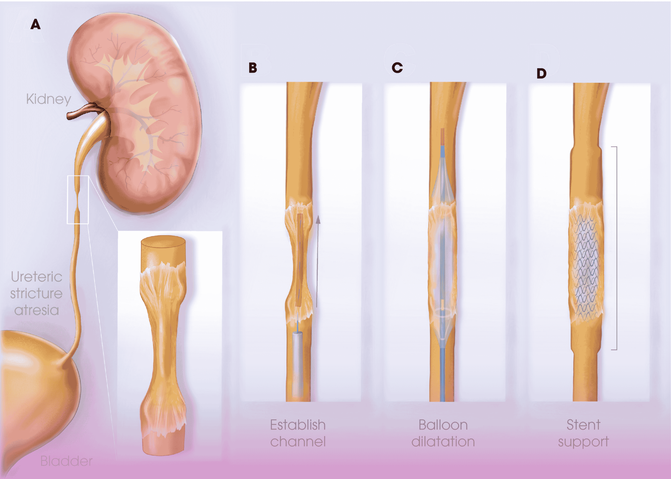
  • Stage 3B: The cancer has invaded the pelvic wall or caused hydronephrosis (buildup of urine in the kidney due to a blocked ureter).
  • Stage 3C: The cancer has spread to nearby lymph nodes.

Stage 4: This is the most advanced stage of cervical cancer, indicating that the cancer has spread to distant organs or tissues. Stage 4 is further divided into two sub-stages:

  • Stage 4A: The cancer has spread to nearby organs, such as the bladder or rectum.
  • Stage 4B: The cancer has metastasized to distant sites, such as the lungs, liver, or bones.

How is Cervical Cancer Diagnosed?

Cervical cancer is diagnosed through physical exams, tests, and procedures. The first step in the process is a pelvic exam. During this exam, your doctor will perform an internal examination to look for abnormalities in your cervix. If abnormalities occur, your doctor may order additional tests such as a pap smear or HPV test. If you have symptoms of cervical cancer, your doctor may recommend other tests such as an endocervical swab for cytology (a sample of cells) or imaging tests like an MRI or CT scan. It may also be diagnosed with colposcopy and biopsy. This procedure involves inserting a small magnifying instrument called a colposcope into the vagina to examine the cervical area. The doctor will then use a tiny tool to scrape cells from your cervix for testing.

What is Colposcopy?

A colposcopy is an examination of your cervix using a magnifying instrument called a colposcope. A biopsy removes tissue samples from your cervix to be examined under a microscope.

What are the First Steps in Treating Cervical Cancer?

You should first make an appointment with your primary care physician or gynecologist to discuss your diagnosis and treatment options.

Your doctor will likely order blood tests and a Pap smear to evaluate the stage of your cancer and determine if chemotherapy or radiation therapy is needed.

He or she may also recommend an MRI scan of your pelvis to check for any tumor spread into nearby lymph nodes. Once you have more information about the extent of your cancer, you may choose one of three treatment options: surgery, radiation therapy, or chemotherapy.

Surgery involves removing the tumor and some surrounding tissue; radiation therapy uses high-energy beams to destroy cancer cells; chemotherapy uses drugs that kill cancer cells by stopping them from dividing or growing.

Importance of Serial Cervical Smears for Early Detection of Cancer Cervix 

Cervical cancer is the second leading cause of death in women. Most women diagnosed with advanced stages of cancer have either not done screening or have not followed up on their abnormal smears. 

Here are some interesting facts about cancer cervix: 

  1. Unlike most other cancers, the cervix is slow-growing, and it has a pre-malignant/pre-invasive stage where it can be captured through smears and treated completely. The diagnoses of these pre-malignant lesions are made through serial pap smears in short paps smears.  
  2. Majority of these cancers, the etiological factor is found to be a virus called the Human Papillomavirus ( HPV). The high-risk HPV strains, predominantly 16 and 18, and a few other strains are responsible for causing the disease. One has to understand that the mere presence of HPV does not mean that the woman will develop cancer; subsequently, in the majority of cases, the virus dies a natural death and disappears when tested in a couple of years. Only in a small percentage do they persist and cause the disease.
  3. Vaccines are developed that protect against HPV 16, 18, and a few other high-risk HPV strains. Vaccination is ideally given before the girls get sexually active and young women up to 26 years of age.

 According to the UAE National Guidelines

  • All women should begin screening with cervical smears  at 21 years of age 
  • Women aged 21-30 should have pap smears every 3 years.  
  • After 30 years, the frequency of screening is again once in 3 years, but if HPV testing is also combined, the frequency could be once every 5 years. 

Since Abu Dhabi has a rapid population turnover, we, as general gynecologists, perform opportunistic testing; we take cervical smears when women present to us with unrelated complaints.  Few precautions, however, need to be exercised to get satisfactory reports with opportunistic testing. Avoid taking smears when women present with symptoms of vulvovaginitis. It is a good practice to schedule the women for smears after the infection is treated. Performed during an ongoing infection can result in the finding of abnormal cells, which will confuse the patient and the physician. It is important to understand that the presence of abnormal cells does not mean the presence of cancer but needs to be evaluated by an expert who has experience treating abnormal cervical smears and correctly interpreting them in the context in question.   

Our Experts

See more

Thyroid Disorders – Symptoms, Causes, and Treatment

Thyroid disorders are a group of conditions that affect the thyroid gland. Thyroid disorders can cause a wide range of symptoms and can affect children, adults, and seniors. They also may be temporary or permanent, depending on your disease type.

What Does the Thyroid Gland Do?

The thyroid gland is a butterfly-shaped organ in the neck that produces hormones that help regulate metabolism. It’s located just below Adam’s apple and above the collarbone. It consists of two lobes, each with its thyroid tissue. A part of the brain controls the thyroid called the pituitary gland, which sends out hormones that tell your thyroid to make more or less of certain protein-based hormones. The thyroid can be affected by infections, physical injury, or radiation exposure. If it becomes overactive (hyperthyroidism) or underactive (hypothyroidism), it can cause problems with metabolism and lead to other health issues.

What is the Function of Thyroid Hormone?

Thyroid hormones are essential for the body’s proper growth, development, and function. They influence nearly every cell in the body, affecting growth, metabolism, and other complex processes. A feedback mechanism controls the production of thyroid hormones. The pituitary gland secretes a hormone called thyrotropin (TSH).

TSH signals the thyroid gland to produce more thyroid hormone when levels fall too low or less when they are too high. The thyroid gland produces two types of thyroid hormone: triiodothyronine (T3) and thyroxine (T4). T3 is the active form of the hormone; it stimulates cellular activity in almost every organ system in the body. In contrast, T4 is inactive until it is converted into T3 in target tissues such as muscle, bone marrow, and fat cells.

What are the Symptoms of Thyroid Hormone?

Signs and symptoms of a thyroid problem can include weight gain, especially around the midsection.

  • Fatigue or lack of energy
  • Muscle weakness or cramps
  • Hair loss or thinning hair
  • Cold intolerance (feeling cold even when others are warm) and unexplained chills
  • Constipation or diarrhea, especially after eating, sometimes alternating between constipation and diarrhea.
  • Dry skin that may feel coarse to the touch

What are the Causes of Thyroid Disorders?

Thyroid disorders are caused by the thyroid gland not producing enough hormones or by an overactive thyroid gland. The most common causes include the following:

  • Iodine deficiency
  • Autoimmune thyroid diseases
  • Radiation exposure
  • Congenital hypothyroidism

What are the Types of Thyroid Disorders?

Graves’ Disease

Graves’ disease is a condition that causes hyperthyroidism or an overactive thyroid gland. The thyroid gland is in the front of your neck, beneath your Adam’s apple. When someone has Graves’ disease, the immune system mistakenly produces antibodies that attack the thyroid gland and cause it to produce too much thyroid hormone. This can lead to several symptoms, including:

  • Feeling overly energetic
  • Weight loss despite increased appetite
  • Hoarse voice (due to swelling in the throat)
  • Increased sweating
  • Irregular heartbeat (palpitations)

Hyperthyroidism

Hyperthyroidism is when the thyroid gland produces too much of the hormone thyroxine. The main treatment for hyperthyroidism is a medication that suppresses the production of thyroxine by the thyroid gland. Surgery may also remove part or all of the thyroid gland if the medication does not work well enough to control symptoms. The symptoms may include:

  • Weight loss
  • Heat intolerance
  • Increased appetite

Hypothyroidism

Hypothyroidism is an underactive thyroid gland, which means the thyroid doesn’t produce enough hormones. It’s a condition that occurs when the body fails to secrete enough thyroid hormones. The symptoms of hypothyroidism include:

  • Fatigue
  • Weakness
  • Constipation
  • Dry skin and hair
  • Weight gain and depression

Thyroid Cancer

Thyroid cancer is when abnormal cells form in the thyroid gland. The cause of thyroid cancer is not known. However, risk factors include exposure to radiation and certain chemicals, a family history of thyroid cancer, and being female and over 60 years old. Symptoms of thyroid cancer may include:

  • A lump or swelling in your neck
  • A sore throat that doesn’t go away with antibiotics
  • Painless swelling in your neck or underarm
  • Difficulty swallowing or breathing
  • A cough or hoarseness that doesn’t go away

Thyroid Nodules

Thyroid nodules are small lumps that develop on the thyroid gland. Thyroid nodules are common and can develop due to an enlarged thyroid gland (goiter), inflammation, or irritation, or they may be caused by other conditions that affect your thyroid. If you have a thyroid nodule, it’s important to see your doctor for an evaluation to determine whether it is cancerous. The symptoms include:

  • Difficulty swallowing
  • Enlarged neck (goiter)
  • Hoarseness or voice changes
  • Difficulty breathing
  • Pain in your neck or throat that feels like pressure or pain when you swallow

Hashimoto Thyroiditis

Hashimoto thyroiditis is when the immune system attacks the thyroid gland, causing thyroid inflammation. Hashimoto thyroiditis occurs when antibodies attack the cells in the thyroid gland, causing it to become inflamed. The antibodies are produced by a type of white blood cell called a T-lymphocyte. The body normally produces these antibodies to protect itself against viruses and bacteria, but in people with Hashimoto’s disease, these antibodies mistakenly attack healthy tissue. Hashimoto’s disease often appears gradually over time, with symptoms including:

  • A hoarse voice
  • Weight gain or difficulty losing weight
  • Fatigue Sensitivity to cold temperatures

Silent Lymphocytic Thyroiditis (SLT)

Silent Lymphocytic Thyroiditis (SLT) is an autoimmune disease that causes a gradual deterioration of the thyroid gland. It’s known colloquially as “silent” because it doesn’t present with any symptoms, and it’s only discovered when a doctor orders a blood test for another reason. SLT is most common in women aged 15-35 with no other health problems. It can also affect men, but at a lower rate than women. Symptoms include:

  • Low-grade fever
  • Weight gain/loss
  • Fatigue
  • Tenderness or pain in the neck

Goiter

Goiter is a condition that causes the thyroid gland to enlarge beyond its normal size. Goiter usually occurs when there’s an iodine deficiency in the diet or water supply. Other causes include:

  • The thyroid gland not producing enough thyroid hormone
  • Excess iodine in the diet
  • Excessive consumption of foods high in goitrogens (e.g., soy, cabbage, broccoli)

The most common symptom of goiter is swelling in the neck. The patient may also experience Difficulty Swallowing, Shortness of breath, and heart palpitations.

How are Thyroid Disorders Diagnosed?

Physical Examination

The doctor will work with several tests and procedures to determine whether or not you have a thyroid disorder. They will also look at factors like your family history, symptoms, and age to make a diagnosis. A physical exam is often the first step in diagnosing a thyroid disorder. Your doctor will look at your neck for signs of nodules or swollen lymph nodes and examine your throat and mouth for any unusual lumps or bumps. They may also listen to your heart and lungs and check your temperature, blood pressure, and weight.

Blood Tests

Diagnosis can be made through a blood test measuring thyroid-stimulating hormone (TSH), which is released by the pituitary gland in response to low levels of thyroid hormones. If the TSH level is greater than 4 mIU/L but less than 10 mIU/L, you may have hypothyroidism, which means your body isn’t producing enough thyroid hormone. If your TSH level is greater than 10 mIU/L but less than 50 mIU/L, you may have hyperthyroidism or too much thyroid hormone. If your TSH level is greater than 50 mIU/L and you also have symptoms such as weight loss or heart palpitations, then you may have Grave’s disease (hyperthyroidism). Suppose the symptoms don’t match any of these conditions. In that case, your doctor may order lab tests for thyroid antibodies—TSH receptor antibody (TRAb), thyroglobulin antibody (TgAb), anti microsomal antibody (AMA), thyroperoxidase antibody (TPOAb), and thyroid antimycobacterial peptide antibody (MAB).

Imaging Studies

Imaging tests can look at the thyroid gland’s size and shape, while thyroid ultrasound uses sound waves to create an image of the gland. They may also order imaging studies such as magnetic resonance imaging (MRI), or computed tomography (CT) scans if they suspect there’s something wrong with your pituitary gland or hypothalamus—the areas of the brain that control hormone production.

How are Thyroid Disorders Treated?

The treatment for thyroid disorders depends on the type of disorder, its severity, and the patient’s age. In most cases, treatment for thyroid disorders will involve taking a combination of medications and lifestyle changes to help manage symptoms. Medications used to treat thyroid disorders include

  • Levothyroxine – a synthetic T4 hormone that replaces what you don’t have enough of in your body; it’s often used to treat hypothyroidism.
  • Liothyronine – synthetic T3 hormone that replaces what you don’t have enough in your body; it’s often used to treat hypothyroidism.
  • Thyroid hormone replacement therapy (THRT) replaces T3 and T4 hormones; this is often used when someone has had their thyroid removed surgically or accidentally damaged by radiation treatment.
  • Antithyroid drugs – block the production or actions of thyroid hormones; they are prescribed as a last resort when hyperthyroidism is severe or uncontrolled by other treatments.

Thyroid Surgery

A thyroidectomy is a surgical procedure to remove the thyroid gland. Thyroidectomy is one of the most common surgeries performed on adults, and it’s often used to treat benign (non-cancerous) tumors and nodules in the thyroid gland. During a thyroidectomy, your doctor will remove all or part of your thyroid gland. Your surgeon may also remove lymph nodes from around your neck and any enlarged lymph nodes found near your vocal cords during surgery. This procedure can be done through a traditional “open” approach or a minimally invasive technique called robotic-assisted surgery (RAS), which uses computer-controlled surgical tools instead of traditional metal instruments. After surgery, you’ll stay overnight at the hospital for observation and then go home when you’re ready to resume normal activities. Most people can return to work within two weeks after having this surgery—but it depends on how many lymph nodes were removed during surgery and how quickly your body heals afterward.

At Burjeel Hospital in the United Arab Emirates, our endocrinologists are experts in diagnosing and treating all thyroid diseases, including hyperthyroidism (including Graves disease), adrenal disorders, hypothyroidism, hypopituitarism, and thyroid cancer. Our team of experts uses state-of-the-art techniques in their diagnosis, including ultrasound imaging and blood tests. We also provide comprehensive management for the treatment and surgical procedures for various thyroid conditions.

Our Experts

See more

Vitamin D Deficiency – Symptoms, Causes, Treatment

Vitamin D is a fat-soluble vitamin naturally produced by the skin when it is exposed to sunlight. Vitamin D is essential for the body’s calcium absorption, which helps keep bones strong and healthy. It also helps your body absorb calcium and phosphorus. Unfortunately, most people don’t get enough sunlight to create enough vitamin D in their bodies leading to vitamin D deficiency.

How Does the Body Get Vitamin D?  

The sun is the primary source of vitamin D in our bodies. When exposed to sunlight, our skin naturally produces vitamin D3, which helps the body absorb calcium and maintain a healthy bone structure. In addition to sunlight, some foods contain vitamin D. Fruits like cantaloupe and orange juice are fortified with vitamin D during processing. Other foods that contain small amounts of natural vitamin D include: 

  • Fatty fish like salmon and mackerel
  • Eggs
  • Mushrooms are exposed to ultraviolet light (portobello mushrooms)

How Does the Body Use Vitamin D?  

The body uses vitamin D in a variety of ways, including the following:  

  • Vitamin D helps maintain normal blood glucose levels and aids in calcium absorption
  • It also aids in the development of bone, muscle, and nerve cells
  • Vitamin D is essential for immune function and helps reduce inflammation

What are Normal Vitamin D Levels?  

According to the Vitamin D Council, most people with normal vitamin D levels will have serum 25(OH)D levels between 40 ng/mL and 100 ng/mL. Between 30 ng/mL and 40 ng/mL is considered deficient, while levels below 30 ng/mL are severely deficient.  

How Do You Measure Vitamin D?  

Vitamin D is tested using a blood test. The test called 25-hydroxyvitamin D measures the amount of vitamin D in your blood and helps to determine whether you have too little or too much.  

What is a Vitamin D Deficiency?  

It is a condition in which the body does not have enough vitamin D to meet its needs. A lack of vitamin D causes by diet or sunlight or a problem absorbing vitamin D from food or supplements.  

How Can I Know if My Vitamin D Level is Low?  

The symptoms of vitamin D deficiency can vary depending on the severity of the deficiency. Some of the symptoms include,  

  • Aches and pains  
  • Feeling tired all the time  
  • Sore muscles and joints  
  • Constipation  
  • Fatigue or drowsiness  

If you’re experiencing any of these symptoms, talk with your doctor about getting tested.  

Who is at Risk?  

Vitamin D deficiency affects people of all ages, but some groups are more likely to experience it than others. For example,  

  • Children and adolescents who do not receive enough sun exposure are at an increased risk for developing the condition.  
  • Older adults may also be at risk because the skin’s ability to produce vitamin D decreases with age.  
  • In addition, those who do not consume enough food sources (such as milk or fish) that contain vitamin D are also at risk for developing this condition.  

What are the Risk Factors?  

It has been linked to osteoporosis, cancer, diabetes, arthritis, heart disease, depression, autoimmune diseases such as asthma and multiple sclerosis, Alzheimer’s disease, osteoarthritis, and rheumatoid arthritis.  

How is it Treated?  

If you are deficient in vitamin D, you should talk to your doctor about taking a supplement. The doctor might prescribe a daily vitamin D supplement, which would be best for preventing future health problems related to the deficiency. The recommended dose differs for everyone, so consult an expert physician about what’s best for you.  

Vitamin D deficiency can lead to several health issues, including chronic pain, depression, fatigue, and more. It is estimated that 1 billion people worldwide are deficient in vitamin D. So if you suspect that you have low levels of vitamin D, you should look into it and consider consulting an endocrinologist to ensure optimal health.  

Our Experts

See more

Hand Arthritis: Symptoms, Causes, & Treatment

What is Arthritis of the Hand? 

Arthritis of the hand is an inflammatory condition that causes pain, swelling, and stiffness in the joints of your hands. It most commonly affects people over 50 but can occur at any age. 

The joints of your hands are made up of bones covered by a layer of cartilage. Cartilage acts as a cushion between the bones to help them move smoothly. When you have arthritis, this protective cartilage breaks down and wears away from use over time. Eventually, this can cause bone spurs or bony growth around the affected joint. 

Arthritis of the hand may affect one or more joints in your fingers or thumb. This can result in pain and stiffness, making it difficult for you to move your fingers or thumb normally. 

How Does Arthritis Affect the Hand? 

Arthritis of the hand can devastate a person’s quality of life. The joints in hand are small and unstable, which makes them vulnerable to injury and degeneration. When arthritis becomes advanced, it can cause severe pain and deformity.  

What are the Causes of Arthritis of the Hand? 

The exact cause of arthritis is not known, but several factors may contribute to its onset: 

Age: As you age, your body will naturally break down and become less efficient at fighting off an infection or repairing itself. This can lead to inflammation in joints where there is no longer enough tissue to repair them. 

Genetics: If you have family members who suffer from arthritis, you’re more likely to develop it. This is because genetics affect how your body responds to injury or infection. 

Injury: Inflammation can occur after an injury such as falling on an outstretched hand or wrist injury from sports like tennis or golfing, which puts significant stress on your joints. 

Obesity: Being overweight puts extra pressure on your joints because they are not designed to carry this additional weight. 

What is Rheumatoid Arthritis of the Hand? 

Rheumatoid arthritis of the hand is a painful condition that can cause significant disability. It’s usually caused by an autoimmune disorder, in which your body attacks itself for no apparent reason. 

Rheumatoid arthritis causes swelling, pain, and stiffness in the joints. It can also cause other symptoms, such as fatigue, fever, and weight loss. The symptoms may vary from mild to severe. There are several types of rheumatoid arthritis, including symmetric polyarthritis (SP), asymmetric polyarthritis (AP), systemic onset juvenile idiopathic arthritis (JIA), vasculitis-related polyarthritis (VRA), seronegative symmetric polyarthritis (SSP) and seronegative asymmetric polyarthritis (SAP). 

What is Psoriatic Arthritis of the Hand? 

Psoriatic arthritis of the hand is a form of psoriatic arthritis, an autoimmune disease that causes inflammation in joints and other tissues. It can affect any joint but most often affects your hands, wrists, knees, and feet. It can cause pain and swelling in your fingers and hands, worsening with time. If you have psoriatic arthritis of the hand, you may find it hard to use your hands for daily activities such as writing or opening jars. 

The exact cause of psoriatic arthritis is unknown. However, it’s thought that genes play an important role because people who are related may be more likely to develop this condition. Other factors like smoking or drinking alcohol may also contribute to developing this condition. 

What is Osteoarthritis of the Hand? 

Osteoarthritis of the hand is a medical condition that causes pain, stiffness, and swelling in your joints. It can be caused by previous injuries or overuse. 

The condition typically affects people over age 50. You may experience pain in the front and outside of your hand near your knuckles. The joint that’s affected most is usually the one you use most often, such as a finger or thumb joint. 

Osteoarthritis of the hand can also cause stiffness in your fingers and wrist and swelling around your joints. If you have osteoarthritis of a finger joint, it may feel like a lump under your skin when you touch it. 

What Parts of the Hand are Most Affected by Arthritis? 

Arthritis can affect any part of the hand, but it most commonly affects the joints. The joint most affected by arthritis is the wrist, followed by the knuckles, joints in the fingers and thumb, and the small joint at the base of each finger (called a carpometacarpal joint). 

What are the Symptoms of Arthritis in the Hands? 

Arthritis of the hands can cause pain, swelling, and stiffness in the joints. The symptoms get worse with age and can affect how you use your hands.  

The following are some common symptoms of arthritis in the hands:  

  • Pain – Arthritis is typically felt in the joints but can also be felt outside.  
  • Swelling occurs when fluid collects around your joint or between two bones. 
  • Stiffness can occur if you have arthritis in one or more joints because muscles around stiff joints tighten to support them.  
  • Tingling sensations – You may feel tingling sensations that radiate from your fingertips to other parts of your hand and coldness in one or more fingers or both hands at once. 

How is Arthritis of the Hand Diagnosed? 

Arthritis of the hand is diagnosed by a physical exam and sometimes X-rays if there are signs of inflammation or other problems. 

The doctor will usually check for tenderness, swelling, or deformity in the joints. X-rays can show how badly arthritic changes have affected bone structure.  

If you’re experiencing pain or stiffness in your hands and wrists, see your doctor for an evaluation. 

How is Arthritis of the Hand Treated? 

Arthritis of the hand can be treated with a variety of methods. The Goals of treatment are to: 

  • Decrease joint pain and stiffness. 
  • Improve mobility and function. 
  • Increase your quality of life. 
  • In the case of rheumatoid or psoriatic arthritis, to slow the progression of the disease. 

One way to treat this condition is through medication. The medications help reduce inflammation and pain in the joints and slow down damage to the cartilage. 

Another treatment option for arthritis of the hand is physical therapy. Physical therapy can help improve flexibility and strength in your hands, which helps decrease pain and stiffness from arthritis.  

A third treatment option for arthritis of the hand is surgery. This may be necessary if other treatments fail or if there are complications from your disease, such as loss of function in one or more fingers due to joint damage caused by arthritis in those fingers. The two most common types of hand arthritis surgery are: 

  • Fusion/Arthrodesis: This involves removing parts of the finger bones and replacing them with metal or plastic implants. This prevents movement between the bones, so there’s no pain. 
  • Arthroplasty: This is a more complex procedure where the end of each finger bone is removed and replaced with a metal rod or plate. The rod or plate helps stabilize the joint, relieves pressure on the nerves, and restores movement to your fingers. 

When the joints in your fingers and thumbs are affected by arthritis, it can be painful and difficult to use your hands, making everyday tasks more challenging. Our hand surgeons at Burjeel Hospital for Advanced Surgery in Dubai, UAE have years of experience treating arthritis and other conditions affecting your hands. They will work closely with you to determine the best treatment plan for your needs. If you suffer from arthritis of the hands or have questions about our services, please contact us today! 

Our Experts

See more

Wrist Arthritis – Symptoms, Causes & Treatment

What is Wrist Arthritis? 

Wrist arthritis is a degenerative disease that causes pain, swelling, and loss of motion in the wrist. It is most common in people over 50 and can occur due to injury or normal wear and tear on the joint. 

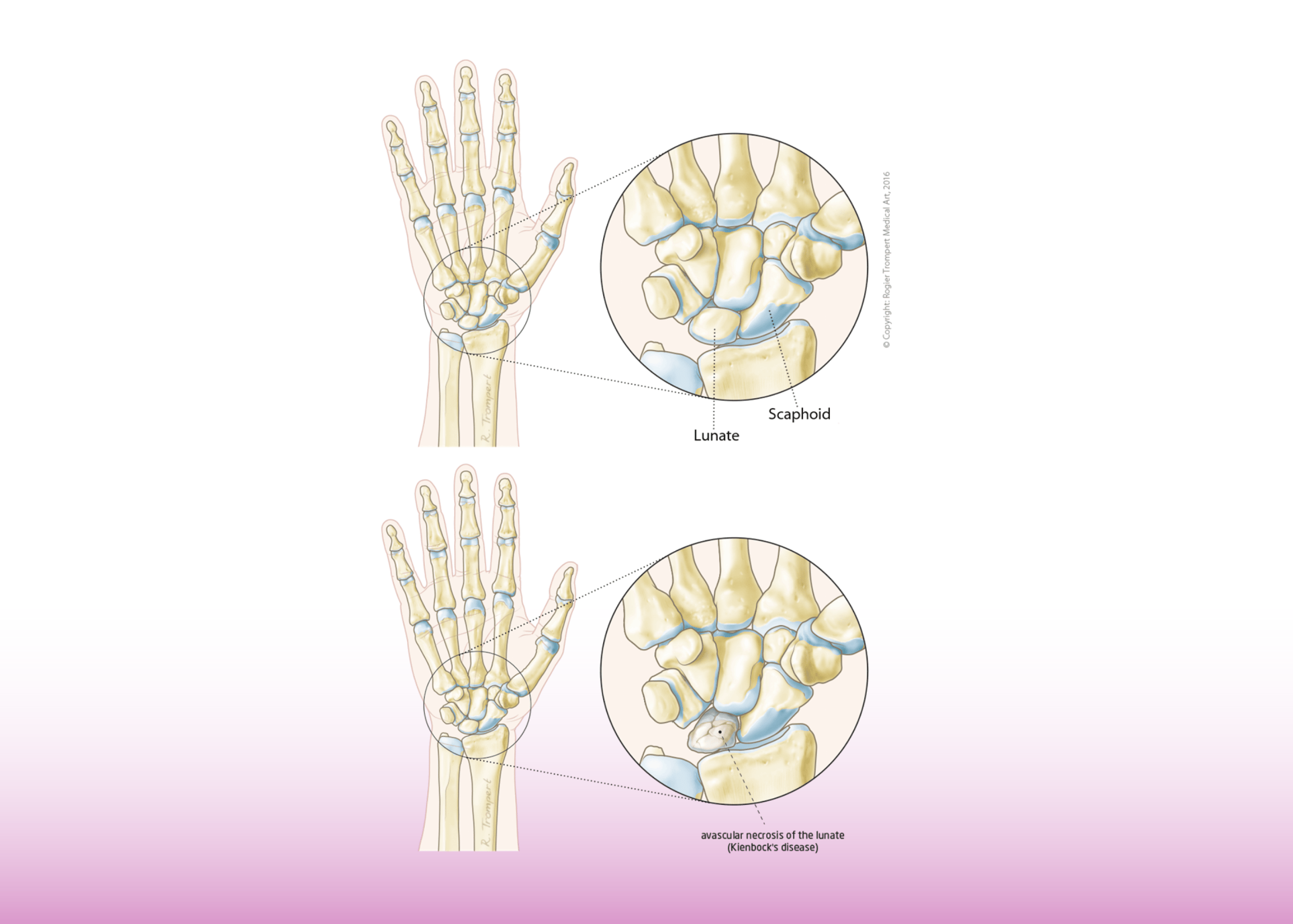
Wrists are made up of eight bones that connect at three different points: the base of the thumb (the carpus), the two halves of the forearm (the radius and ulna), and the hand itself (the metacarpals). The wrist’s range of motion allows us to write, type, play sports, and grip objects tightly. 

Because these joints are so complex, they are vulnerable to injury—especially when you consider all their moving parts: bones, ligaments, tendons, nerves and blood, and vessels.  

What are the Types of Wrist Arthritis? 

The three most common types of wrist arthritis are distal radioulnar, midcarpal, and radiocarpal. 

Distal radioulnar arthritis occurs when a ligament or tendon becomes inflamed and stiff. It’s usually caused by damage to the ulnar collateral ligament (UCL) on the outside of your elbow. 

Midcarpal arthritis involves damage to the bones in the middle of your wrist. This can be caused by overuse or injury. 

Radiocarpal arthritis is an injury that affects both sides of your wrist simultaneously. This is often caused by osteoarthritis (OA), a degenerative joint disease (DJD). 

What are the Causes of Wrist Arthritis? 

The causes of wrist arthritis are not completely understood, but many factors can contribute to the development of this condition. Some factors that may contribute include: 

Age – The older you get, the more likely your joints will begin to wear down over time. This is especially true if you have been involved in activities that stress your wrists over time (such as sports). 

Gender – Women tend to develop wrist arthritis earlier than men. 

Genetics – If your parents had wrist arthritis or other types of joint problems when they were younger, there’s a good chance you might develop it as well (even if they never told anyone). 

Obesity – Those who are overweight tend to have more joint problems because they carry extra weight around their body, putting more strain on their joints over time.” 

What are the Symptoms of Arthritis of the Wrist? 

The symptoms of arthritis of the wrist range from mild to severe and depend on the type of arthritis. The most common symptom is pain, which occurs early in the disease and can worsen with time. Other symptoms include: 

  • Swelling 
  • Loss of range of motion 
  • Stiffness 
  • Numbness or tingling in your fingers or hand 

How is Arthritis of the Wrist Diagnosed? 

To determine whether or not you have arthritis of the wrist, your doctor will perform a physical examination that includes checking for swelling, tenderness, and pain in the area around your wrist. 

In addition to performing a thorough examination, your doctor may recommend other diagnostic tests. These tests may include the following: 

X-rays – An x-ray can help determine if you have arthritis in your wrist by allowing your doctor to view the bones in your wrist and determine whether they are inflamed or damaged. 

MRI Scan – An MRI scan is an imaging technique that uses magnetic fields and radio waves to create detailed images of soft tissues in the body without using radiation as X-rays do. This test can help determine if there is inflammation or damage in the soft tissues around your wrist, as well as joint damage caused by osteoarthritis or rheumatoid arthritis. 

Blood Tests – Blood tests allow doctors to check levels of certain proteins (like C-reactive protein) that indicate inflammation has occurred somewhere in your body, including around joints like those found at the ends of bones where tendons attach them (ligaments). 

What are the Available Non-Surgical Treatment Options?

The non-surgical treatment options for wrist arthritis include: 

  • Pain Relievers: These medications reduce pain and inflammation in your wrists. Commonly prescribed pain relievers include ibuprofen, aspirin, naproxen sodium, and acetaminophen. 
  • Rest: Resting your wrist can reduce inflammation, but it’s important to ensure you’re using proper posture when resting your wrist so that you don’t cause further injury. 
  • Exercises: Exercises can be used to strengthen the muscles surrounding the joints in your wrist, which can help to improve mobility and decrease pain. Your doctor may also suggest exercises for stretching or strengthening other parts of your body that are affected by arthritis, such as your shoulders or neck (if they’ve been affected). 
  • Splints: Splints are devices that wrap around the joint to provide support while it heals; they’re often used after surgery on a joint because they help prevent future damage while allowing patients time off work during recovery periods (e.g., from surgery). 

What are the Available Surgical Treatment Options?

To effectively treat the condition, it’s essential to understand the different surgical treatment options available. 

Proximal row corpectomy – This procedure involves removing part of the proximal row of carpal bones. It is typically performed when there is little or no joint space between the bones in your wrist. 

Denervation surgery – This treatment option involves removing a nerve from your wrist joint. This surgery works well for people with rheumatoid arthritis who have a limited range of motion in their wrists. 

Fusion surgery – Fusion surgery is used to treat wrist arthritis that has progressed so far that there is no more joint space left between your bones in your wrist. The surgeon will remove the cartilage from your wrist joint and fuse the bones using metal hardware. 

Joint replacement – If arthroplasty has not been successful at treating your wrist arthritis symptoms, you may need to undergo joint replacement surgery to restore full function to your hand. 

Arthritis of the wrist is a common condition that affects many people. The hand and wrist surgeons at Burjeel Hospital for Advanced Surgery, Dubai, provide treatment for arthritis of the wrist, including wrist pain, carpal tunnel syndrome, fractures and dislocations, and tendonitis. 

Our Experts

See more

7 Common Myths and Misconceptions about Asthma in Kids

Here are seven common myths and misunderstandings about asthma in kids that you should know and understand.

Mythbuster 1: Asthma Cannot be Diagnosed in Children 

False: Although asthma can be difficult to diagnose, with supportive history from parents , clinical assessment and right tests, including breathing tests (Lung Functions) in most children asthma can be differentiated from other conditions.  

Mythbuster 2: Inhalers are Not for Children

False: We have good evidence from research that if inhalers are used appropriately with guidance from specialized teams, they will work to control asthma. Inhaled medicine is delivered directly into the lungs, so there are minimal side effects to other body systems. 

Mythbuster 3: Nebulizers are Better than Inhalers

True but False: Asthma can be well controlled on use of the preventive treatment including inhalers. Nebulizers have a role when child is acutely unwell and needs oxygen along with other treatments and best given in the hospital. Overuse of nebulized medicines at home can give false reassurance to parents and carers about child’s condition.

Mythbuster 4: Children with Asthma Should Avoid Exercise  

False: Exercise is an important element of children’s well-being. Children with asthma must have good control of their symptoms. Once this is achieved, they can participate in most physical activities to live a good quality of life. 

Mythbuster 5: Asthma Treatment is Only Needed When Children are Unwell  

False: There are two elements of asthma management in children, reliever and preventive treatment.  Preventive treatment is done when children don’t have asthma symptoms daily but to prevent future and recurrent asthma attacks. Most children need some preventive treatment to maintain good control of asthma.

Mythbuster 6: Asthma is a Psychological Problem   

False: Asthma is due to inflammation and tightening of the airway due to triggers that vary in every child. This can be due to animals, dust, pollen, or other contact or environmental triggers. 

Mythbuster 7: Inhalers and Other Asthma Treatments are Addictive 

False: Inhalers are not addictive. As asthma is a long-term condition, many children need ongoing treatment, but this does not mean these are addictive.

Asthma is a common condition that can be very scary for parents. You want to do everything you can to help your child, and it can be difficult to know what’s true and what’s not when it comes to asthma in children. We’ve compiled this list of seven common childhood myths and misconceptions about asthma. We hope this article has helped clear up some common myths and misconceptions about asthma in children and that you can now feel more confident about approaching the condition.

Please remember that this is not medical advice; if you think your child might have asthma or is having asthma, we recommend talking to our expert pediatric pulmonologist, who can help assess their condition.

Our Pediatric Pulmonologist at Burjeel Medical City, Abu Dhabi provides comprehensive care for kids with breathing problems and respiratory conditions, including asthma, chronic lung disease, cystic fibrosis, and sleep apnea. Our expert will work with you to create a treatment plan to help your child breathe more easily. 

Our Pediatric Pulmonology Expert


Dr. Ghulam Mujtaba

Consultant Pediatric Pulmonology


Years of Experience: 20

Nationality: Ireland

Languages Known: English, Urdu, Punjabi, Hindi

Thumb Arthritis – Symptoms, Causes & Treatment

What is Thumb Arthritis?

Thumb arthritis is a condition that causes the thumb joint to become inflamed and painful. It is usually caused by repetitive motion of the thumb, such as when you play an instrument or type on a keyboard. 

The symptoms of thumb arthritis include pain, swelling, and tenderness in the base of your thumb (the attachment area where it meets your wrist). You may also notice that your thumb becomes stiffer than normal when you try to move it. 

What Causes Thumb Arthritis? 

Thumb arthritis is caused by a breakdown of the joint cartilage and surrounding ligaments, which leads to excessive wear and tear on the joint. This causes pain and limited motion in the joint. 

The most common cause of thumb arthritis is osteoarthritis (OA) or degenerative joint disease (DJD). With OA, the protective cartilage layer that cushions your joints deteriorates over time due to age, injury, or overuse. This can result in bone rubbing against bone and causing pain.  

What are the Signs and Symptoms of Thumb Arthritis? 

The signs and symptoms of thumb arthritis include: 

  • Pain in your thumb joint when you move it or press on it 
  • Swelling in your thumb joint 
  • Stiffness in your thumb joint 
  • Decreased range of motion 
  • The enlarged or bony appearance of the joint at the base of your thumb 

How is Thumb Arthritis Diagnosed? 

Thumb arthritis is diagnosed through a physical exam, X-rays, and imaging tests. 

To diagnose thumb arthritis, a doctor will first perform a physical exam. They’ll look at the range of motion in your thumb and test the strength of your grip by asking you to pick up small objects with your thumb. This will help them determine if there are any changes in how your thumb moves or feels compared to before you were experiencing symptoms. 

Next, they’ll take an X-ray of your hand and wrist to assess whether changes in the bones or joints could be causing the pain. Suppose there are no obvious changes on the X-ray. In that case, they may recommend getting an MRI (magnetic resonance imaging) or CT (computed tomography) scan of your hand and wrist to provide more details about what might be causing your pain. 

How is it Treated? 

Thumb arthritis is treated with medication, physical therapy, and splinting. Based on the severity of the condition, surgery might also be required.  

Medications for thumb arthritis include corticosteroids, nonsteroidal anti-inflammatory drugs (NSAIDs), pain medications, and sometimes even steroids. These are used to reduce inflammation and pain. 

Physical therapy can help with range of motion exercises and strengthening exercises to keep your thumbs moving smoothly. 

A splint is better handmade that keeps your thumb in proper alignment while it heals. 

Several types of surgery can be performed on the thumb, depending on the nature of the injury. 

Arthrodesis: This procedure involves removing as much bone as possible from a joint before setting it into its new position, ensuring it maintains its alignment even after healing. Arthrodesis can treat arthritis and other conditions affecting your thumb joint. This old procedure is still useful for heavy workers. 

Trapeziectomy is another common type of surgery that involves removing only a portion of the bone rather than all of it. This type of procedure can be used to relieve pressure on nerves or tendons. 

Osteotomy involves breaking the bone (usually at the joint) and then realigning it to improve alignment and reduce pain. In some cases, the joint is fused after surgery.  

Arthroplasty: In this innovative surgery, the bone is replaced by an artificial implant that helps support your thumb during movement and prevents further damage from occurring because of osteoarthritis or other conditions affecting your joints. It preserves the length of the thumb and its strength. 

How Can I Prevent it from Getting Worse? 

To prevent the condition from worsening, you should start by taking care of your thumb joint. This means avoiding activities that may cause further damage to your joints. If you have a job that involves repetitive motion of your hands, try to reduce the amount of time spent performing those motions. You may also want to try using a splint at night to stabilize the joint while you sleep. 

You can also help prevent thumb arthritis from worsening by adopting an exercise routine that strengthens your hand and wrist muscles. Exercises help increase blood flow to your fingers and joints and improve flexibility, which will help reduce stress on the injured joint. 

Finally, suppose symptoms persist after these steps have been taken. In that case, it’s important to see a doctor specializing in hand conditions (such as a physical therapist or orthopedic surgeon) for further evaluation and treatment options. 

If you’re suffering from arthritis of your thumb, our hand surgeons at Burjeel SOS Hand Center in Dubai will work closely with you to determine which procedure suits your needs. We offer minimally invasive surgical procedures that allow us to treat patients without opening up their wounds or making long incisions during surgery. This means less pain, less scarring, and better results! 

Press & Media

UAE becomes the first Gulf country to offer artificial joint replacement of the thumb

Our Experts

See more

Mammogram – What You Need To Know

What Is a Mammogram?

A mammogram is a radiographic examination of the breast that can help detect breast cancer in its early stages. It is recommended for women at average risk of developing breast cancer. 

When Should I Get a Mammogram?

The American Cancer Society recommends mammograms yearly for women aged 45 to 54. Women 55 and older should switch to mammograms every two years or can continue yearly screening.

How Do I Prepare for a Mammogram?

Burjeel Hospitals are outfitted with cutting-edge diagnostic equipment, such as the HOLOGIC 3DimensionsTM mammography system. You can visit one of the closest facilities to have breast cancer screening.

Bring a list of the locations and dates of past mammograms, biopsies, or other breast procedures, as well as your previous medical records, if you visit one of our facilities for the first time.

Schedule your mammogram around a time when your breasts are unlikely to be sore or swollen to minimize discomfort and obtain high-quality images. Avoid the week leading up to your period.

Do not apply deodorant, antiperspirant, powders, lotions, creams, or perfumes beneath your arms or on or under your breasts on the exam day. Some of these contain substances that can appear as white spots on x-rays.

Before a mammogram, discuss any recent changes or issues with your breasts with the doctor. (If you have symptoms, diagnostic mammography may be necessary to obtain images of the area of concern.)

Make sure the doctor is aware of any aspect of your medical history that could increase your breast cancer risks, such as surgery, hormone use, a family history of breast cancer, or a previous diagnosis.

What Will Happen During a Mammogram?

The breast tissue is stretched out during mammography by compressing the breasts between two firm surfaces. Then, an X-ray is used to acquire 3D images, which are shown on a computer screen and analyzed for cancer indicators.

Will a Mammogram Hurt?

A mammogram is a painless procedure. You won’t feel anything during your mammogram. The only thing you might notice is the pressure of the breast compression. Women experience discomfort during a mammogram due to pressure on the breast tissue, which can be uncomfortable for some women. The risk of pain depends on the sensitivity of your breasts. Most women do not notice discomfort during their annual mammogram unless they have sensitive breasts or have previously undergone breast surgery. If you are concerned about pain during your mammogram, let your doctor know before your appointment so they can apply additional gel to reduce friction between the breast and the film plate or compression paddle that is used during the procedure.

What Happens After the Test?

After the mammogram screening test, you’ll be asked to wait for your results. You will receive them either immediately or within a week. Still, it’s important to know that they can take up to two weeks if the radiologist needs more information to make a diagnosis.

Suppose there are concerns about your mammogram result. In that case, you may be asked to come back for another appointment or for further tests such as ultrasound or biopsy (which involves taking cells from the tumor and examining them under a microscope).

Where Can I Get a Mammogram?

Our facilities are equipped with the latest mammography systems. The HOLOGIC 3DimensionsTM mammography system provides the highest image quality for breast imaging. It can deliver high-resolution images with greater detail than other systems, as well as provide more accurate detection and diagnosis of breast cancer.

A HOLOGIC 3Dimensions™ mammography system is a revolutionary technology that uses special software to create three-dimensional images of your breast tissue. The 3D images allow radiologists to visualize breast tissue more accurately than traditional film-based mammography, leading to earlier detection of abnormalities.

The HOLOGIC 3Dimensions™ system also features IQM™ software that provides real-time analysis and enhancement of all breast images taken during your exam, ensuring that only the clearest, most diagnostic images are reviewed by your radiologist.

Early Detection of Breast Cancer Can Improve Outcomes

Early detection of breast cancer can improve outcomes by helping to prevent the disease from spreading throughout the body. There are many options for early detection, including screening mammograms, breast self-exams, and clinical breast exams.

If you notice any changes in your breasts, such as lumps or swelling, you should make an appointment with your doctor as soon as possible.

If You are a Woman Above 40, it is Time to Get Your Annual Breast Cancer Screening.


    Our Experts

    See more

    Breast Cancer – What You Need To Know

    What is Breast Cancer?

    Breast cancer is the most common form of cancer in women. It occurs when cells in the breast become abnormal and start to grow out of control. The abnormal cells can be either benign (not cancer) or malignant (cancer). Breast cancer can be found in one breast or spread from one part of the body to another.

    What are the Types of Breast Cancer?

    There are several types of breast cancer, including:

    Angiosarcoma: A type of sarcoma (cancer of the soft tissue) that develops in the lining of blood vessels.

    Ductal Carcinoma In-Situ (DCIS): A noninvasive breast cancer that begins in the milk ducts and has not spread to other body parts.

    Inflammatory Breast Cancer: A rare form of breast cancer that develops quickly and grows more rapidly than other types.

    Invasive Lobular Carcinoma: A breast cancer that starts in the milk-producing glands’ lobules (tiny sacs).

    Lobular Carcinoma In-Situ (LCIS): A condition where abnormal cells appear in the lobules but do not grow into tumors or spread to other parts of the body.

    Male Breast Cancer: A rare form of cancer that affects men with breasts, most often those with Klinefelter syndrome or testicular feminization syndrome.

    Paget’s Disease of the Breast is a rare form of breast cancer in which the breast tissue becomes inflamed or infected, leading to a buildup of calcium deposits. It can also lead to changes in the skin, including thickening and discoloration.

    Recurrent Breast Cancer. Cancer cells return after treatment and surgery to remove them, causing more lumps or pain in the breasts.

    What are the Symptoms of Breast Cancer?

    It’s normal to be concerned about the symptoms of breast cancer. The most common symptom is a lump or mass in the breast, but there are others. Some of the other symptoms include:

    • lumps in lymph nodes (glands)
    • nipple discharge that is not milk
    • dimpling or puckering of the skin around the nipple
    • redness or flaking of the nipple
    • pain in any area of the breast, including the nipple
    • rash on your chest or under your arm
    • change in size or shape of your breast

    What are the Stages of Breast Cancer?

    Breast cancer is categorized into four stages based on the extent of the disease and the likely outcome.

    Stage I: The tumor is less than 2 centimeters across and has not spread to lymph nodes.

    Stage II: The tumor is between 2 and 5 centimeters across and has not spread to lymph nodes.

    Stage III: The tumor is greater than 5 centimeters across or has spread to one or more lymph nodes but not to distant organs.

    Stage IV: The tumor has spread to other body parts beyond the breast.

    What are the Risk Factors for Breast Cancer?

    Some of the risk factors for breast cancer include:

    Age—it’s most common in women over age 50, but it can occur at any age.

    Family History of Breast Cancer—if your mother or sister has had breast cancer, you are at higher risk of developing the disease yourself.

    Genetics—Women with a first-degree relative (mother, sister, daughter) who have had breast cancer are at greater risk of developing breast cancer. Women with BRCA1 or BRCA2 gene mutations are at higher risk of developing breast and ovarian cancers.

    Diet—certain foods may increase your risk of developing breast cancer; these include processed meats and red meats.

    Obesity—obese women are at higher risk for developing breast cancer than normal-weight women.

    How is it Diagnosed?

    It is often found through routine screenings such as mammograms and X-rays taken of your breasts. Mammograms can see early signs of breast cancer before you may feel any symptoms. 

    The American Cancer Society recommends that women get a mammogram every year starting at age 40 or in their 20s for those at high risk for developing this type of cancer A doctor will also recommend regular self-exams if you’re over 30 years old.

    It’s important to know what to expect during your diagnosis and treatment to feel prepared and supported throughout the process.

    Breast Exams

    A breast exam is an essential part of your breast health routine. You should schedule a visit with your doctor immediately if you notice any changes in your breasts, such as swelling or lumpiness. 

    Mammograms

    A mammogram is an X-ray of your breasts taken from different angles to check for abnormalities. A mammogram may lead doctors to perform additional testing, such as biopsies or ultrasounds, if they find something unusual during the procedure. These tests can help determine whether an abnormality is cancerous or benign (non-cancerous).

    Biopsy

    A biopsy removes tissue samples to examine them under a microscope for signs of cancerous cells.

    Breast Ultrasound

    An ultrasound uses sound waves to create an image of the breast tissue. Ultrasounds are usually recommended for women with dense breasts or who are pregnant because these conditions can make mammograms difficult to read accurately. 

    Breast MRI

    An MRI scan can also evaluate a woman’s breasts for signs of cancer or other conditions, such as a lump or tumor. MRI machines use strong magnetic fields and radio waves to create detailed images of soft tissues such as your breasts.

    How is it Treated?

    There are several treatment options that your doctor may recommend. These include surgery, radiation therapy, chemotherapy, hormone therapy, and targeted therapy.

    Surgery is the most common form of treatment. This procedure removes all or part of the tumor and any surrounding tissue. Your doctor may also take out lymph nodes if they think it is necessary.

    Radiation therapy uses high-energy beams to kill cancer cells to prevent them from growing back. It can be done alone or with other chemotherapy or hormone therapies.

    Chemotherapy is another treatment that uses drugs to kill cancer cells in the body and slow down their growth so they don’t spread as quickly. This type of treatment is often used with surgery or radiation therapy along with other treatments such as a hormone or targeted therapies, depending on the breast cancer you have been diagnosed with.

    Targeted therapies help destroy specific types of cells within your body by targeting certain proteins that are present on those cells only; this allows them to be destroyed without harming other healthy tissues nearby, which would happen if using traditional chemotherapy methods instead (such as most conventional chemo drugs).

    At Burjeel, we believe that cancer care is about treating the whole person, including their physical and emotional well-being. That’s why we offer a comprehensive range of services from leading oncology and palliative care experts.

    We are an ESMO Designated Centre of Oncology and Palliative Care. Our multi-disciplinary team of American and UK board-certified doctors includes medical oncologists, radiation oncologists, surgical oncologists, plastic surgeons, and palliative care specialists who work together to provide patients with the best possible care based on their needs.

    Early Detection Saves Lives. Get Screened Today!


      Our Experts

      See more