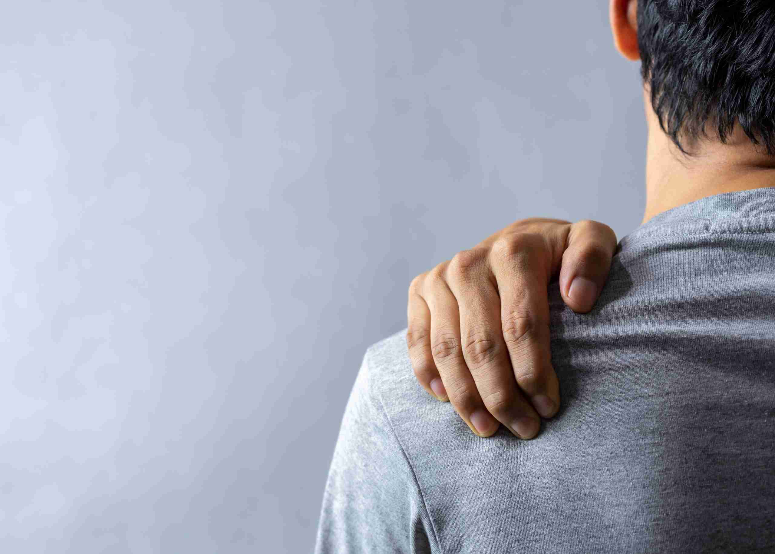
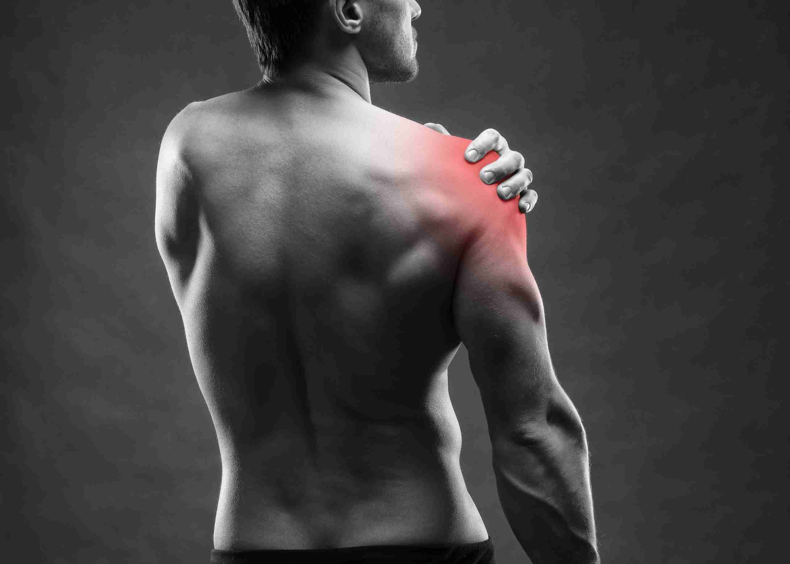
Tips for Healthy Eating Habits

There are several tips and tricks to eating healthy. Many of these ideas can be challenging to accomplish in today’s busy world. Still, they can give you the edge you’ve been looking for in a healthy lifestyle.

How Does a Healthy Plate Look Like?

  • ¼ of the plate with grains, choose at least 50% whole grains (whole wheat bread, Brown Rice, etc.).
  • ¼ of the plate with protein choices vegetarian or non-vegetarian, choose eggs, fish, lean meat, chicken, beans, cheese, and nuts.
  • ½ of the vessel to be loaded with colorful fruits and veggies.
  • Hydrate adequate. Drink water- 30ml/kg body weight is the requirement. Find yours and start to sip.

Fruit vs Fruit Juice

Fruit

  • 62 Kcal
  • More Fiber
  • Less concentrated fructose
  • Fewer calories
  • Lower glycemic index

Juice

  • 112 Kcal
  • Less fiber
  • More concentrated fructose
  • More calories
  • Higher glycemic index

How to Include Fruits in the Diet

  • Snack on fruits
  • Try fruit as dessert
  • Delight your sweet tooth by eating healthy with fresh fruit yogurt, parfaits, and dried fruits
  • Including fruit with breakfast or as dinner snacks is eating healthy

What is One Serving of Fruit?

  • 1 medium-size fruit
  • ½ cup fruit salad
  • ¼ cup Dried fruits
  • ½ cup fresh juice
  • Include at least two servings of fruit a day

Are You Drinking Enough Water?

How Much Water Should You Drink Per Day?

30 ml X Actual body weight (Kg) Note: Restrict fluids; if any medical conditions, or if recommended by the Physician

  • Thirst is the first signal of Dehydration
  • Check the urine color
  • The dark color indicates dehydration
  • Lighter the color, better hydration
  • Dry skin
  • Dry lips
  • Less urination
  • Feeling tired, dizzy, and headaches

How to increase water intake?

  • Keep a water bottle next to you or a reachable place.
  • Use a mobile app to remind and measure your intake.
  • Fancy bottles attract and improve the intake.
  • Infused water (Lemon, Ginger, and Mint) to enhance the taste.

What is the Healthy Thing to Snack on?

Most times the snacks are the unhealthiest stuff chosen. Portion-sized healthy snacks are a great way to follow small frequent meal patterns.

A healthy snack

  • Being between meal times is a good option to decrease hunger and prevent overeating at meal times.
  • boosts the metabolism, sustains the energy levels, and can overcome cravings.
  • to be chosen wisely, because unhealthy snacks lead to weight gain.
  • should be low in sugar, salt, and fat.
  • can be fruit or veggie salad or whole-grain snacks or low-fat dairy instead of junk.
  • can be a combination of Protein + Carbohydrate choices that can keep the stomach full for a longer time. Eg: Yogurt and fruits, Hummus with Vegetable sticks or cheese with fruit slices, etc.

Some sensible snack replacements:

  • Mixed nuts( 1 ounce)
  • Low-fat Yogurt/ laban (1 cup)
  • Fruits (1 serving)
  • Dark Chocolate (30 gm)
  • Air Popped Popcorn( 3 cups)

Why Is Fiber Good for You?

Benefits:

  • Helps maintain bowel health and Prevents constipation.
  • Helps in lowering blood cholesterol levels.
  • Helps in better control of blood sugar levels.
  • Keeps the stomach full.
  • Aids achieving healthy body weight.

Recommendation:

  • 14g of fiber for every 1000Kcal
  • Females: 25g/day Males: 38g /day

Tips to increase fiber in your diet

  • Include lean veg proteins-Beans, peas, and lentils.
  • Snack on nuts and seeds.
  • Increase your fruit and vegetable intake.
  • Have the fruit instead of the juice.
  • Don’t peel the fruit or veg before consuming it.
  • Go for whole-grain products (at least 50% can be whole grains).

How Physically Active Are You?

Inactive: less than 5,000 steps per day

Average (somewhat active): ranges from 7,500 to 9,999 steps per day

Active: more than 10,000 steps

Very active: more than 12,500 steps per day

Tips to Include More Steps to Your Day

  • Park farther away
  • Walk while waiting
  • Take the stairs
  • Consider a walk with the family
  • Take your pet for a walk
  • Take the farthest way
  • Talk in person. Rather than instant-messaging or emailing with coworkers, get up and walk to their desks
  • Walk during your kids’ activities
  • Start increasing your goal gradually by adding 500-1000 extra steps every 3-4 days

Health Benefits of Being Physically Active:

It reduces your risk of:

  • Heart diseases
  • Obesity
  • Diabetes
  • Depression
  • Elevated blood pressure
  • Elevated cholesterol levels

You are What You Eat

Plan Balanced Diet

  • Eating healthy means planning ahead and trying to include all the possible 5 groups in each meal- fiber-rich grains, lean protein choices, fruits, vegetables, and low-fat dairy products.

Adopt Healthy Cooking

  • Try to avoid deep-fried foods. Choose healthy cooking methods like baking, broiling, steaming, grilling, or roasting.

Utilize Time Efficiently to Plan

  • Chop fresh vegetables and fruits and store ahead for quick preparation.
  • Prepare the grocery list based on the requirement.

Flavor the Food

  • Use herbs, spices, low salt seasonings, or lemon juice to cook vegetables and meat.
  • Cut down the use of salt as much as possible.

Curb the Sweet Tooth

  • Include berries, dried fruits, and dark chocolate, and prepare healthy and quick desserts like yogurt parfait, granola parfaits, smoothies, chia seed pudding, etc instead of sugar-loaded stuff.

Learn to Read Nutrition Labels and Choose Wisely

  • Check for salt, sugar, and fat content of the packed foods. Look for the serving sizes to understand the portion sizes.

How to Improve Your Hair Health?

Nutritional deficiency may impact both hair structure and hair growth. Effects on hair growth include the result of sudden weight loss or decreased protein intake, niacin deficiency, or any underlying medical condition.

  • Eating healthy, and balanced diet with a lot of antioxidants might help prevent nutritional deficiencies.
  • Some major nutrients that benefit and add to our hair health are Vitamin A, C, Vitamin D, Vitamin E, Biotin, Iron, Zinc, Selenium, Proteins, and Omega 3 fatty acids.
  • Consuming foods rich in the above nutrients is beneficial, Eg: Citrus fruits, Oily fish, nuts and seeds, eggs, sweet potato, avocado, pumpkin, chia seeds, yogurt, spinach, asparagus, chickpeas, etc.
  • Avoid excess consumption of processed food that leads to your body getting deprived of nutrients.
  • Managing stress and developing good sleep patterns are add-on benefits.
  • Don’t depend on supplements without consulting the physician. Overdose of these supplements might be toxic.

Mind Your Salt

Salt is an important seasoning, flavoring agent, and preservative. Sodium in salt is one of the essential minerals and performs vital functions in the body. A diet high in salt (or sodium) can raise blood pressure, which can increase your risk of heart disease and stroke.

Tips to limit your sodium/salt intake

  • Avoid processed foods and buy fresh foods.
  • Rinse the sodium-containing canned foods.
  • When dining out, ask restaurants to limit the salt.
  • Portion control while eating out.
  • Cook your food whenever possible to avoid instant noodles/ soups/frozen meals.
  • Substitute the salt with spices, lemon, garlic, and herbs to flavor your food. This practice can make your eating healthy.
  • Always read the food label.
  • Try to have salt free/ low-salt snacks.

Reading the labels on your food package

  • Salt/sodium-free – less than 5mg of sodium per serving.
  • Very low sodium – 35mg of sodium or less per serving.
  • Low sodium – 140mg of sodium or less per serving.
  • Reduced sodium – at least 25% less sodium than the regular product.
  • Lightly salted – at least 50% less sodium than the regular product.

Eat Fat to Lose Fat

A small amount of fat is an essential and important part of eating healthy diet. The dietary reference intake for fats in adults is 20% to 35% of total calories per day. Fat is the source of essential fatty acids that our body cannot produce. It is important to include healthy fats in the diet.

Make sure you get most of your fat intake from unsaturated fat options. Unsaturated fats are found in plant foods and oily fish, and they are usually liquid at room temperature. They’re found in:

  • Oils from vegetables, nuts, and seeds, such as sunflower, safflower, olive, walnut, and corn oil
  • Spreads based on these oils
  • Nuts and seeds
  • Avocado
  • Oily fish such as herring, sardines, mackerel, salmon, and trout

Benefits of unsaturated fats:

  • Lower the risk of heart disease and stroke.
  • Lower bad LDL cholesterol levels, while increasing good HDL.
  • Support anti-Inflammatory Process
  • Essential for the absorption of A, D, E, and K vitamins
  • Optimize nerve and brain function
  • Improves and softens your skin

Keep Your Skin Radiant & Healthy

  • Water is “the forgotten nutrient”. It gets rids the body of harmful toxins that can clog the pores. Drink at least eight glasses of water a day.
  • Vitamin C keeps the skin look plump and wrinkle-free. Include citrus fruits, red peppers, and dark green leafy greens like kale, strawberries, and kiwis.
  • An inflamed gut means inflamed skin. Include probiotics regularly in the diet to heal the gut. Try sauerkraut, anything pickled, yogurt with live active cultures, kefir, and miso.
  • Prebiotics in the diet help to feed the healthy flora and restore the gut microbiome to a much more healthy state. Include Garlic, leeks, and asparagus—those are great sources of prebiotic fiber in the diet
  • Antioxidants protect against free-radical damage—doing wonders for the skin! Include Berries (Blueberries, blackberries, cranberries and goji berries, kale, beets, and spinach).

Is Coffee Bad for You?

World Health Organization (WHO) has taken coffee off the possible carcinogen list. And there’s increasing evidence that coffee might actually be good for you. Studies have found that coffee drinkers may have a reduced risk of:

  • Cardiovascular disease (including heart attack, heart failure, and stroke)
  • Type 2 diabetes
  • Alzheimer’s disease and Parkinson’s disease
  • Colon, uterine, and liver cancer
  • Cirrhosis
  • Avoid coffee if you have any health problems that require you to cut on caffeine
  • Don’t have coffee on an empty stomach
  • If you are a coffee drinker, make sure not to overconsume it, have it in moderation no more than 3-5 cups per day or on average 400mg of caffeine per day
  • Make sure to avoid the extra calories, sugar, and saturated fat in coffee that is loaded with whipped cream or flavored syrups
  • It is not necessary to drink coffee if you usually don’t as there are many other dietary strategies to improve your health

Anxiety – Symptoms, Causes & Treatment

What is Anxiety? 

Anxiety is characterized by a sense of unease, intense, excessive, and persistent worry and fear about everyday situations. It is frequently accompanied by nervous behavior that an upcoming event or circumstance can trigger. It is a normal stress response, but it becomes a disorder when it interferes with daily life. Experiencing occasional anxiety is a normal part of life. Anxiety disorders are the most common mental illness affecting millions of adults and young people. 

What Causes Anxiety? 

Anxiety attack is a complex condition that many factors can cause. It’s not uncommon for it to be caused by an underlying condition, such as depression or post-traumatic stress disorder (PTSD). Some of the most common causes of concern include: 

  • A traumatic event from the past 
  • Mental illnesses such as depression or PTSD 
  • Having trouble concentrating 
  • Having trouble sleeping 
  • Stress 
  • Being around other people who are anxious or angry 

What are the Symptoms of Anxiety?

The symptoms can vary from person to person. For example, some people may feel pressure in a crowd or around unfamiliar people, while others might experience it when they are alone. In general, though, the symptoms are: 

  • Feeling nervous, restless (feeling jittery), and tense. 
  • Nausea, stomach upset, and other GI symptoms. 
  • Difficulty concentrating 
  • Sleep problems (difficulty falling asleep or staying asleep) 
  • Fatigue 
  • Difficulty controlling worry. 
  • Trembling hands and knees 
  • Avoidance for things that trigger anxiety 
  • Feeling impending danger, panic, or doom.

What are the Types of Anxiety Disorders? 

There are many types of anxiety disorders, and the symptoms may vary according to the condition. Some common types include: 

Generalized Anxiety Disorder (GAD) is a condition that is characterized by: 

  • Excessive, uncontrollable worry and anxiety. 
  • Fatigue 
  • Trouble falling asleep or staying asleep 
  • Difficulty concentrating 
  • Other symptoms like restlessness and irritability accompany these feelings. 

The disorder can occur at any age but usually begins in early adulthood. The exact cause of Generalized Anxiety Disorder is unknown, but it may be related to genetic and environmental factors.  

People with this condition tend to think about events negatively and have difficulty controlling their thoughts. The most effective treatments for Generalized Anxiety Disorder include cognitive-behavioral therapy (CBT) and medication.  

CBT helps people learn how to change their thoughts, so they don’t focus on the negative aspects of situations as much. Medications such as antidepressants help reduce symptoms such as depression, irritability, and panic attacks that often occur alongside Generalized Anxiety Disorder. 

Panic Disorder: Panic Disorder is a mental illness that causes sudden feelings of terror and fear. The feelings are so intense that they can lead to a panic attack, including: 

  • Chest pain 
  • shortness of breath 
  • A strong sense of losing control and impending doom. 

Panic attacks usually last around 10-15 minutes, but they can be extremely debilitating for many people who get them on a regular basis.  These panic attacks may cause you to be concerned about them happening again or to avoid circumstances where they have already happened. Medication and therapy are used to treat panic disorder. 

Phobia-Related Disorders:  

The four most common phobia-related disorders are: 

Specific Phobia is an intense fear of a specific object or situation, such as snakes or heights. The person knows their fear is irrational, but they still experience it as though it were real. 

Social Phobia: Also known as Social anxiety disorder, is an anxiety disorder characterized by:

  • Significant and persistent fear of being judged or criticized by others 
  • This leads people to avoid what they usually do, such as going out with friends or meeting new people. 
  • Feel very nervous in social situations and think other people judge them harshly. 
  • They may worry about being embarrassed or judged by other people. Sometimes they may even fear that they will do something embarrassing or humiliating. 

It can be treated with Antidepressant medications and Cognitive behavior therapy. 

Agoraphobia: This disorder involves both specific and social fears, but it also involves worries about going outside alone or being away from home without help from another person who is nearby at all times (e.g., family members).  Agoraphobics are often associated with panic attacks when they leave their homes because they’re worried about what might happen if they’re alone and unable to contact someone immediately for help if needed (e.g., getting lost). 

Separation Anxiety Disorder:  It’s a childhood disorder characterized by excessive anxiety for the child’s developmental level. It is related to separation from parents or others who have parental roles. 

Selective Mutism:  Children consistently fail to speak in certain situations, such as school, even when they can speak in other situations, such as at home with close family members. This can interfere with school, work, and social functioning. 

Substance-Induced Anxiety Disorder: It is characterized by symptoms of intense anxiety or panic that are a direct result of misusing drugs, taking medications, exposure to a toxic substance, or withdrawal from drugs. 

Other Specified Anxiety Disorders and Unspecified Anxiety Disorders: These anxieties or phobias don’t meet the exact criteria for any other anxiety disorders but are significant enough to be distressing and disruptive. 

What are the Risk Factors? 

There are many risk factors including: 

Genetic Factors. People with anxiety disorders often have a family history of anxiety. If one or both parents have an anxiety disorder, they are more likely to develop one. 

Environmental Factors. Traumatic experiences like abuse or neglect during childhood can increase the likelihood of someone developing it later in life. Environmental factors like poverty or financial stress can also contribute to it. 

Brain Chemistry. Researchers believe some people may have a genetic predisposition toward developing certain mental illnesses. These people are thought to have an “anxiety sensitivity,” making them more likely to develop anxiety disorders or experience panic attacks when exposed to triggers such as stress or loud noises (see below). 

Trauma: Experiencing trauma during childhood or adolescence increases the risk of developing an anxiety disorder later in life. Post-traumatic stress disorder (PTSD) is particularly strongly linked to it. 

Personality. People with certain personality types are more prone than others are. 

Negative Thinking Patterns: Many people who suffer from severe anxiety have negative thoughts about themselves and their situation. This thinking can contribute to stress and make it harder to cope with everyday life. 

Stressful Life Events: Experiencing a significant life event, such as divorce or the death of a loved one, can trigger an anxiety disorder in some people. 

How is it Diagnosed?

Doctors examine the individual’s symptoms, medical history, and family history to diagnose anxiety. They Conduct a mental state examination through an interview with the patient and rule out other conditions with similar symptoms. The doctor might also use several tools to help them understand how well they respond to treatment. The tests to include : 

  • The Zung Self-Rating Anxiety Scale 
  • Hamilton Anxiety Scale (HAM-A) 
  • Beck Anxiety Inventory (BAI) 
  • Social Phobia Inventory (SPIN) 
  • Generalized Anxiety Disorder Scale (GAD-7) 

What are the Treatments Available for Anxiety Disorders?

They are many different types of treatment, including : 

  • Cognitive Behavioral Therapy (CBT) helps them understand how their thoughts, feelings, and behaviors affect each other. Individuals will learn how to identify when they have negative thoughts or feelings and then figure out ways to change them. 
  • Exposure Response Prevention (ERP). This is another kind of CBT that involves gradually exposing themselves to the things that scare them until their fears disappear. This is often done through role-playing situations where they practice being more comfortable with what scares them until it doesn’t anymore. It is effective with patients with OCD. 
  • Acceptance and Commitment Therapy (ACT) focuses on the present moment rather than past trauma. It encourages patients to fully accept their thoughts and feelings as they live a more fulfilling life. 
  • Medication can help relieve the symptoms by helping calm down the brain’s activity, so it doesn’t feel so overwhelmed by stressors in its life. 

For those suffering from anxiety, knowing the options available to them and understanding how each type of treatment can help may be extremely helpful in finding the treatment that best suits them. Like other mental health disorders, it may still be challenging to treat, but it is most definitely possible. Educating yourself on treatment options is always a safe choice when considering your next steps in healing. If you’re concerned about how anxiety affects you, share your concerns with our expert psychiatrist at Burjeel Hospital, Abu Dhabi.


Our Expert Psychiatrist


Dr. Nada Omer Mohamed Elbashir

Specialist Psychiatrist

Burjeel Hospital, Abu Dhabi


Anterior Cruciate Ligament (ACL) Injury

An ACL injury which occurs when the anterior cruciate ligament is torn.

ACL injuries are common among athletes—especially those who play sports like football, soccer, basketball, and volleyball—because these activities require a lot of jumping and twisting motions that stress the knee joint. 

About Anterior Cruciate Ligament (ACL)

The anterior cruciate ligament (ACL) is one of the four major ligaments that connect the femur to the tibia in the knee. It’s located in the center of the knee, below the patella, and above the posterior cruciate ligament (PCL). The ACL helps stabilize your knee during movements such as pivoting and cutting, which is why it’s commonly injured during sports. 

The ACL provides stability by preventing abnormal rotation of your knee joint. When you bend your knee, your ACL will prevent it from turning past 90 degrees. If you were to injure your ACL, you would experience problems with instability while walking or running. 

Causes of ACL Injury

The ACL can be injured by a sudden twisting motion that forces it to stretch too far. An injury may happen when you turn quickly or pivot while playing sports.

You can injure your ACL by landing wrong after jumping off a wall or climbing over a fence. An ACL tear is most common among young people who play sports such as football, basketball, and soccer.

Nearly 200,000 people are injured each year while playing sports like these! 

Symptoms of ACL Injury

ACL injury symptoms can vary depending on the severity of the injury and the amount of time it has been since the injury occurred. The most common signs of an ACL tear include: 

  • Pain in the knee joint, which may become more intense when twisting or turning the leg 
  • Swelling and bruising in or around the knee joint 
  • Locking of the knee joint, which occurs when you try to bend your knee but cannot 
  • Barely being able to move your knee because it feels stuck or stiff 
  • The feeling of catching or locking in the knee 

Diagnosis of ACL Injury

There are several tests for ACL injuries, which is the most common knee ligament to be injured

A physical exam by a doctor can often determine whether or not an ACL is torn. However, this test alone may not be enough because many other conditions mimic an ACL tear. 

The Lachman test is another way to determine if an ACL is damaged. The patient lies on their back with the knee bent at 90 degrees and their foot planted on the table in front of them. The doctor then pulls on the leg while pushing down the thigh to straighten the knee joint. If there is damage to the ligament, it will allow more movement than usual and give the doctor information about what kind of treatment is necessary. 

A third test that doctors may use is an Anterior Drawer sign. This test can help them determine whether or not damage has been done to other ligaments besides the ACL tear. 

In addition, two imaging tests can be used to diagnose ACL injuries: X-rays and MRI scans. These tests will show if there is any damage to the bones around your knee or ligaments in your knee. 

Treatments Available for ACL Injury

There are two main types of treatment for an ACL tear: non-surgical and surgical. The choice between them depends on the severity of your injury. 

Non-Surgical Treatments 

Non-surgical treatments include physical therapy, rehabilitation exercises, and bracing or taping to support your injured knee. These methods can be effective in some cases. Still, they usually only work if you start treatment immediately—within 24 hours after tearing your ACL. If you wait too long after the injury occurs, these treatments may not be effective. 

Surgical Treatments 

There are several surgical treatments for ACL injuries, including: 

ACL Repair Surgery 

ACL repair surgery is a procedure that involves replacing a torn anterior cruciate ligament (ACL) with a graft taken from another part of the knee (called an allograft). The ACL helps stabilize the knee during movement. It connects the femur (thighbone) to the tibia (shinbone) and helps keep the tibia from sliding forward under the femur. When it tears, it can cause instability in your knee and lead to pain, swelling, and difficulty walking or running. ACL repair surgery aims to restore your knee’s stability and strength. 

ACL Reconstruction Surgery 

Suppose your orthopedic surgeon determines that your ligament has been irreparably damaged. In that case, they may recommend reconstructing it with a graft from another part of your body. 

ACL & Meniscus Transplantation 

When you have an ACL injury that is severe enough to require reconstruction but not severe enough to warrant a hamstring graft, your knee surgeon might recommend using part of your meniscus instead. 

Our expert knee surgeons provide a full range of services for people with all forms of knee problems. They specialize in arthroscopic surgery, including, ACL and meniscus repair, and offer total knee replacement surgery, partial replacements, and other orthopedic procedures, including sports medicine and foot and ankle surgery.


Our Expert Orthopedic Knee Surgeons


Dr. Nader Darwich

Consultant Knee Surgeon

Burjeel Hospital, Dubai

Dr. Professor Erik Hohmann

Consultant Orthopedic Surgeon Sports Physician

Burjeel Hospital, Dubai

Dr. Matthias Honl

Burjeel Hospital, Dubai

Our Experts

See more

Knee Pain – Symptoms, Causes & Treatment

The knee is one of the most common joints to experience knee pain and inflammation. When this happens, getting back in the swing of things can be incredibly difficult.

Understanding the Knee

The knee is a hinge joint that connects the thigh bone (femur) to the lower leg bones (tibia and fibula). The knee allows for movement, supports weight-bearing, absorbs shock from walking and running, and provides stability.

The knee’s ability to flex and extend depends on several factors. First is the shape of your bones: if your femur or tibia is longer than usual, it may be harder for you to bend or straighten your knee.

Second, some ligaments connect bone to bone; if these ligaments are loose or damaged, you will have more difficulty bending or straightening your knee. Finally, menisci—pieces of cartilage that sit between the tibia and femur—act as shock absorbers when you walk or run; if they’re damaged, they can’t do their job properly, and this can affect how easily you can bend and straighten your knee.

Knee Pain Causes

Knee pain can be caused by any number of conditions, from injuries to arthritis to torn ligaments. Some of the more common causes of knee pain include:

  • Fractures
  • Sprains
  • Muscle strains
  • Bursitis
  • Tendonitis
  • Arthritis
  • Baker’s Cyst
  • Overuse or repetitive motion injuries (such as runner’s knee)
  • An injury to the knee from an accident or trauma
  • Degeneration of the joints
  • An anterior cruciate ligament (ACL) tear
  • Meniscus tear

Signs & Symptoms of Knee Pain

If you’re experiencing knee pain, it’s essential to identify the cause of your symptoms. Here are some common signs and symptoms of knee pain:

  • Pain in the front of the knee
  • Pain in the back of the knee
  • Pain behind or around the kneecap (patella)
  • Kneecap that feels loose or unstable, especially when walking downstairs or squatting
  • Kneecap that feels like it pops out of place (this is called patellar dislocation)
  • Swelling around the knee area, especially after long periods of sitting or standing
  • Pain while squatting or going downstairs
  • Stiffness in your leg muscles and tendons

How to Know if Your Knee Pain is Serious

You might have knee pain and not even realize it. It’s normal to feel some knee discomfort when doing activities like running or squatting. But if you start noticing that your knee pain is getting worse or starting to interfere with your daily life, it may cause concern. If you’ve experienced knee pain for more than two weeks, you should immediately see an orthopedic expert.

Knee Pain Diagnosis

The first step in diagnosing knee pain is to determine whether the pain is acute or chronic. Acute knee pain is characterized by an onset within one month and may be caused by minor trauma or a more severe condition such as a torn ligament or meniscus tear. Chronic knee pain persists beyond three months and may be due to osteoarthritis, rheumatoid arthritis, gout, and rheumatoid arthritis.

To determine the cause of your knee pain, your doctor will perform a physical exam that includes an evaluation of your range of motion, balance, and muscle strength. The orthopedic doctor may also use diagnostic imaging techniques, such as X-rays or CT scans, to confirm whether there is any damage to your bones or cartilage. If your orthopedic surgeon suspects you have torn ligaments or cartilage, they may order MRI scans to look for further structural damage in the knee joint.

Knee Pain Treatment

There are several treatments available for knee pain.

Non-surgical Treatments:

  • An orthopedic expert may prescribe over-the-counter pain medications to help manage pain. These medications should only be used for short periods, as they may cause stomach irritation, liver damage, and other serious side effects
  • Physical therapy can be done to help strengthen the muscles around the knee and improve the range of motion in the joint. This is especially helpful if underlying muscle or tendon strength issues contribute to joints’ instability
  • Braces or splints may limit movement in an arthritic knee and decrease pain. These devices can also help protect an injured knee from further injury during activities like walking or running

Surgical Treatments:

In general, knee surgeries fall under five broad categories:

  • Repairing torn menisci (the cartilage disks that cushion your knee joint)
  • Repositioning torn ligaments/cartilage in the knee joint
  • Removing damaged tissue around the knee joint (arthroscopic surgery)
  • Removing bone spurs (osteophytes) formed around the knee joint
  • Removing loose bodies from within your knee

Many surgical options are available to help you get back on your feet and stay active. Some of them are explained below,

Arthroscopic Surgery:

This procedure uses a small incision and a tiny camera to remove damaged cartilage, repair tore ligaments or tendons, or clean out debris from your joint. If the first procedure doesn’t help your symptoms, you may need more than one arthroscopic surgery.

Total Knee Replacement:

A total knee replacement may be necessary if your knee has become so damaged that bone-on-bone contact occurs. An orthopedics surgeon will replace your damaged knee joint with an artificial joint made from metal alloys, plastic components, and synthetic materials.

Partial Knee Replacement:

This type of surgery is best for patients whose disease is limited to one part of their knee joint such as the inner or outer compartment of the patellofemoral joint (kneecap). During this procedure, only one side of your knee is replaced while leaving the healthy bone in place on either side of the diseased area.

Microfracture Surgery:

This procedure involves making small holes in the tibial plateau (shin bone) to allow blood flow to stimulate the healing of damaged cartilage. This procedure is typically used for knee osteoarthritis, but you may also use it if you have had a previous injury to your knee that has worn down your cartilage and caused it to become hard and brittle.

Meniscal Transplantation:

This procedure replaces the torn meniscus with donated tissue from another person or cadaver. It may be helpful if you have severe arthritis or other problems related to your meniscus.

Tendon Repair:

This procedure repairs damaged tendons around your knee. Tendons are bands of tissue that connect muscles to bones, allowing you to move your joints smoothly during activities like walking and running.

How to Keep Your Knees Healthy

The knees are a vital joint in your body, and keeping them healthy is important for your overall health. If you’re active or on your feet all day, it’s important to take extra precautions to keep your knees in the best shape possible. Here are some tips for how to keep your knees healthy:

  • Wear Supportive Shoes. Make sure you’re wearing shoes that support your feet, ankles, and legs and have a good sole that will help absorb shock when you walk or run
  • Annual Examinations. If you’re older than 50, consider getting an annual knee exam from a doctor or physical therapist specializing in joint health problems like arthritis or torn ligaments. They’ll assess any pain or stiffness in your legs and recommend exercises that will help improve mobility and overall strength and stability for the long term (they’re also good for preventing falls)
  • Warm-up Before Exercising. Do warm-ups before exercise or activity. Warming up is a great way to stretch out your muscles and joints, so they’re ready to go by the time you begin exercising or performing any type of activity. You can also do a few squats or lunges before beginning any workout routine, so your muscles are primed for action!
  • Get Moving. Promoting circulation throughout your whole body is key to keeping your knees happy. Try walking every day—it’s simple, effective, and free!
  • Build Strength. Strengthen your quadriceps muscles by doing exercises such as squats and lunges, which will help support the knee joint during physical activity and reduce the risk of injury.

Knee pain is common but can be treated. Our expert orthopedic surgeons provide comprehensive care and perform various procedures to address knee pain, including arthroscopy, replacement surgeries, minimally invasive surgeries for knee arthritis, ligament injuries (ACL reconstruction), meniscus tears (meniscectomy), and osteoarthritis treatment options such as total knee replacement or partial knee replacement surgery.

If you are experiencing knee pain, our team will take the time to listen to your concerns and explain your options. If surgery is an option, they will help you understand what to expect during the procedure and recovery. All surgical procedures aim to relieve symptoms, restore function and improve the quality of life for patients.


Our Expert Orthopedic Knee Surgeons


Dr. Samih Tarabichi

Consultant Orthopedic Surgeon

Burjeel Hospital, Dubai

Dr. Nader Darwich

Consultant Knee Surgeon

Burjeel Hospital, Dubai

Dr. Professor Erik Hohmann

Consultant Orthopedic Surgeon Sports Physician

Burjeel Hospital, Dubai

Dr. Matthias Honl

Consultant Orthopedic Surgeon

Burjeel Hospital, Dubai

Our Experts

See more

Kidney Stones

Kidney stones are hard deposits that form within the kidneys. They can cause a lot of pain and make it difficult to urinate. They are made from substances like calcium and can be as small as a grain of sand or as big as a golf ball. They often result from a buildup of minerals in the urine. The most common type of kidney stone is made up of calcium, but other substances may also be present. 

Most of them pass spontaneously within days or weeks, especially if you drink plenty of liquids and increase your water intake. However, if you have recurrent kidney stones, surgery might be necessary to remove them permanently or prevent their recurrence. It is essential to let your Urologist know if you’ve had any symptoms here. 

Types of Kidney Stones 

Kidney stones can be divided into four main types: calcium oxalate, calcium phosphate, uric acid, and struvite. 

  • Calcium oxalate stones are the most common type. They form when calcium combines with oxalate in urine
  • Calcium phosphate stones form when calcium combines with phosphorus to form crystals that then become hard material in your kidneys. Doctors don’t know why this happens 
  • Uric acid stones are caused by excess uric acid in your system. The acid makes its way into your urine and forms crystals there. These crystals bond together to create a stone that can block your urinary tract 
  • Struvite stones. A bacterial infection caused in your urinary tract or bladder 
  • Cystine: They are Rare. They occur in both men & women who have the genetic disorder cystinuria 

Causes of Kidney Stones

They occur when there is an imbalance in the number of minerals in the urine. The minerals crystallize and form crystals that stick together to form a stone. The crystals can become stuck in the kidney, ureter (the tube that connects the kidneys to the bladder), or bladder. This causes pain and other symptoms, such as nausea and vomiting, blood in urine, frequently urinating and feeling like you need to go. Still, nothing comes out when you do, needing to use the bathroom urgently but having trouble getting there quickly enough. 

Warning Signs of Kidney Stones 

The following are some of the warning signs: 

  • Pain in your side or back that is worse when you urinate, or pain while passing urine 
  • Bloody or cloudy urine 
  • A feeling that you need to urinate but can’t 
  • Pain while urinating 
  • Nausea or vomiting 

Risk Factors of Kidney Stones 

The risk factors include, 

  • Gender: Men are twice at risk than women. This may be due to differences in hormones or diet between men and women 
  • Age: People over the age of 40 are more likely to have the condition
  • Family History: Having a family history of the condition increases the chances 
  • Diet: If you eat a diet high in protein or salt, you may be more likely to develop them
  • Conventional treatment for high blood pressure, such as calcium channel blockers, angiotensin II receptor blockers, or thiazide diuretics 

Are Kidney Stones Dangerous?  

Most of them pass out of your body on their own within several days to two weeks, but they can sometimes be serious if they block one of your urinary tracts or if they cause an infection in your kidneys or bladder. If you have these symptoms, it’s important to see a kidney specialist immediately because they that don’t pass on their own can cause permanent damage to your kidneys. 

Foods That Can Cause Kidney Stones 

While there are no foods that directly cause them, there are some that may increase your risk and others that may help prevent them from forming. 

They often form when the urine contains too much calcium, oxalate, or uric acid. Foods high in these compounds can increase your risk of developing kidney stones by increasing the amount of these compounds in your urine. The following foods are high in calcium, oxalate, or uric acid: 

  • Calcium-rich foods include milk, cheese, yogurt, and other dairy products. Calcium is also present in bones and other animal-based products such as meat and poultry (including fish). Low-fat dairy products contain less calcium than full-fat versions; however, they still contain enough calcium to increase the risk for stone formation if consumed regularly over time (more than two cups per day) 
  • Oxalate-rich foods include spinach, beets, and rhubarb; while they won’t cause immediate side effects like pain or discomfort due to their low oxalate content when eaten alone (about 1/2 cup cooked), they can compound problems when combined with other foods high in oxalates like strawberries and chocolate chips 
  • Animal protein-rich food like meat, fish, egg, and cheese is one of the important causes of uric acid stones if eaten daily and in excess quantity 
  • Salt (Sodium Chloride) rich foods or salty foods, if eaten in excess the level of sodium raises the amount of calcium in urine which sticks to oxalate in the kidneys that can produce stones 

Foods Than Can Help in Preventing Kidney Stones

There are many foods that can help prevent kidney stones, including: 

  • Tofu contains calcium and magnesium that help prevent the formation of calcium oxalate stones 
  • Dried figs contain citric acid and have high water content. Citric acid is believed to dissolve the crystals that form in urine, preventing the formation
  • Cranberries contain potassium and magnesium, as well as malic acid. Malic acid is believed to prevent calcium from settling in the kidneys and forming crystals which could lead to kidney stones. Cranberries also have high water content and are low in calories and sugar 
  • Prunes are another fruit with malic acid that can help prevent renal calculi. They also contain iron for anemia prevention, fiber for digestion health, and antioxidants for overall health benefits 
  • Watermelon is an excellent source of vitamin C which helps prevent calcium oxalate stones from forming in the kidneys or urinary tract system by neutralizing acids in urine which may cause those crystals to form there instead of passing through without being absorbed into our bodies or bloodstreams at all where they could cause health problems including urinary tract infections (UTIs), and gout 

Kidney Stone Diagnosis 

Kidney stones are diagnosed using a physical exam, lab tests, and imaging. 

A physical exam includes an abdominal exam, where the doctor presses on your abdomen to feel for a kidney stone. If they can’t feel it, they may order an ultrasound or CT scan to try and pick up the stone. 

The lab tests used to diagnose kidney stones include blood tests for electrolyte levels, urinalysis and urine culture because most urinary tract stones accompany by hematuria or urinary infection. The urine culture detects any bacteria that may be causing the infection that leads to kidney stones. 

Finally, imaging is used to see if there are any signs of stones in your urinary tract. Your kidney doctor may order an Ultrasound, X-ray, or CT scan to look for any calcifications or other visible signs of possible stones in your kidneys or bladder. 

Preventing Kidney Stones 

The good news is that there are simple steps you can take to decrease your risk of forming kidney stones: 

  • Drink plenty of water every day 
  • Avoid foods that are high in oxalate  
  • Don’t overdo it on calcium supplements or antacids that contain calcium 
  • Reduce the amount of salt you eat and drink, if possible 
  • Get enough exercise each week 
  • Get enough magnesium and vitamin D in your diet or through supplements; these nutrients help prevent the formation of kidney stones 

Learning about your options for treating and preventing kidney stones can make it easier to manage them when they occur. If you think you have kidney stones, see a kidney specialist right away. 

Best Treatments for Kidney Stones

Kidney stones are a common medical condition that can be treated with medication, lithotripsy, or surgery. 

Medication: The doctor may prescribe medications to help prevent kidney stones from developing or from making them pass more easily. 

Lithotripsy: This is a non-invasive treatment for kidney stones. It uses shock waves to break up the stone into smaller pieces that can be passed in the urine. In some cases, the shock waves can leave small fragments of stone in your kidneys or urinary tract. These fragments may need to be removed by surgery. 

Surgical Therapy: There are several surgical options available for treating kidney stones in different locations within the urinary system: 

Nephrolithotomy Kidney – used to remove small stones from the kidney’s interior 

Pyelolithotomy Renal Pelvis – used to remove larger stones from the bottom of the kidney surrounding its ureter (the tube connecting the kidney and bladder) 

Ureterolithotomy (ureter) – used to remove large stones stuck in your ureters (tubes connecting each kidney and bladder) 


Our Expert Urologist


Dr. Ahmad Mbadda

Specialist Urology

Burjeel Medical Center, Al Shamkha


Our Experts

See more

Floaters & Flashes – Why Do I See Them

Are You Suddenly Seeing Dark Spots In Your Vision? Dark spots can refer to blind spots or shadows that seem to “float” in your field of vision. These are commonly known as floaters. They may look to you like black or gray specks, strings, or cobwebs that drift about when you move your eyes. 

Causes of Floating Blind Spots

Floaters can happen for no obvious reason and are often perfectly harmless. However, the clear substance inside the eye (vitreous gel), which fills 80% of the eye, begins to shrink with age. When the vitreous gel shrinks or thickens, particles form in the gel. These small particles liquefy and pull away from the surface of the eyeball, clumping together, causing small shadows which are seen as floaters. 

Normal for Eye Floaters: 

Here are some signs that the eye floaters are completely normal. 

  • They occur when looking at something bright
  • They occur when looking at a plain, light-colored background 
  • They do not interfere with your vision

When to See a Doctor 

  • Contact an eye specialist immediately if you notice: 
  • Increase in the number of floaters you’re seeing. 
  • Flashing lights associated with the spot 
  • Darkness on any side or sides of your vision (peripheral vision loss) 
  • A vision that is blurred or distorted 

Above signs can lead to serious conditions such as:

Retinal Detachment is a serious eye condition that happens your retina (a light-sensitive layer of tissue in the back of your eye) is pulled away from its normal position at the back of your eye. 

Internal Eye Bleeding is the presence of blood within the eye. 

Diabetic Retinopathy is caused by diabetes. It damages the retina and can lead to blindness. 

Age-Related Macular Degeneration (AMD) is an eye disease that can blur your central vision. It’s caused by deterioration of the macula, a small area in the center of the retina. 

Above sight-threatening condition requires immediate attention. Thus, early detection and treatment are crucial. Treatment options include laser treatment or surgery, and without it, you risk losing your vision. 


Our Expert Ophthalmologist


Dr. Madhava Rao

Consultant Ophthalmology

Burjeel Hospital, Abu Dhabi


Our Experts

See more

What are Anal Fissures?

Anal fissures are common and can be very painful, but they are treatable.

An anal fissure is a small tear or cuts in the lining of the anus. The crack in the skin causes severe pain and some bright red bleeding during and after bowel movements. The fissure can be deep enough to expose the muscle tissue underneath.

Anal fissures typically cause pain and bleeding with bowel movements. You also may experience spasms in the ring of muscle at the end of your anus (anal sphincter). Anal fissures usually extend from the anal opening. They are usually located posteriorly in the midline, probably because of the relatively unsupported nature and poor perfusion of the anal wall in that location. Fissures may also be caused by malnutrition, inflammatory bowel disease; Crohn’s disease; trauma, and enemas. In infants, anal fissures often result from passing large or hard stools.

Symptoms of Anal Fissures

The pain and discomfort of an anal fissure can be severe and may cause you to seek medical attention. Symptoms include:

  • Pain, itching, burning, and bleeding in the rectum (the end of your digestive tract) that lasts for more than a few days or goes away and comes back again
  • Pain that is worse with bowel movements or sitting down
  • Pain that is worse when you have a bowel movement

Causes of Anal Fissures

Anal fissures are caused by trauma to the anal canal. The most common cause is constipation, which can cause severe straining and pressure on your bowels. Other potential causes include diarrhea, childbirth, or surgery. The anal canal is a sensitive area that can become easily irritated by any pressure or trauma. Fissures usually occur at the junction between hard stool and soft tissue, causing irritation and bleeding from the resulting internal tear in your skin tissue.

Anal Fissure Risk Factors

The risk factors for anal fissure are not well understood, but some studies suggest that many cases may be inherited. The following have also been associated with an increased risk of developing an anal fissure:

  • Age—Anal fissures are most common in 40 to 60 years old
  • Gender—Fissures are more common in women than in men
  • Pregnancy—Anal fissures can develop during or after delivery (postpartum)
  • Hemorrhoids—People with hemorrhoids are more likely to develop anal fissures than those who do not have this condition

Prevention of Anal Fissures

The best way to prevent anal fissures is to avoid constipation or diarrhea.

  • Don’t hold your bowel movements
  • Drink enough water so that you have one or two soft bowel movements each day
  • When you do have a bowel movement, try not to strain when you push out the stool

Diagnosis of Anal Fissures

A doctor can diagnose anal fissures by examining the anus. The doctor will ask about symptoms, including when and how they happened, what you’re doing to treat them, and whether or not you’ve ever had this problem before.

A digital rectal exam may be performed to feel for anal fissures. During this exam, the doctor will find any irregularities that could indicate an anal fissure.

Sometimes stool tests are used to rule out other causes of anal pain—for instance, if nothing else seems wrong, but symptoms like bleeding persist for more than two weeks after treatment begins.

Treatment for Anal Fissures

Treatment includes:

  • First aid. Resting the anus and surrounding area is essential. To help heal, stay off the toilet or do not have any bowel movements until you consult your doctor
  • Medications. Medications such as topical ointments may be prescribed to reduce pain, itching, and bleeding. Analgesics (pain relievers) can also relieve discomfort throughout healing
  • Surgery. While surgery is often not necessary for treating an anal fissure, it may be advised if other treatment methods fail to work or if symptoms become worse after seven days of home care measures (such as rest). Sometimes surgery involves removing part of the internal sphincter muscle to widen it so that stool can pass through without causing further damage or pain during defecation

Our surgeons are highly skilled at treating anal fissures. We make sure all patients understand the details of their surgery and try to make them as comfortable as possible while they are in our care.


 Our Expert Proctologist


Dr. Toufic Ata

Consultant Laparoscopic Bariatric and General Surgery

Burjeel Hospital, Abu Dhabi


Our Experts

See more

What is Glaucoma?

Glaucoma in general is a group of eye diseases of the elderly that run in families and is associated with eye pressure. It can lead to damage to the optic nerve and loss of vision. It is the second leading cause of blindness in the world after cataracts and the number one cause of irreparable vision in the world. Almost 80 million people have glaucoma worldwide and this is expected to increase to over 111 million by 2040. As per 2020 statistics, approximately 4 million people aged 50 years and older suffer from moderate to severe vision impairment, and 3.6 million are blind due to this eye condition.

Symptoms

It is a symptomless disease in the early stages. Symptoms appear in the form of loss of side vision and later central vision only late in the course of the disease when you have lost a good amount of vision which is not repairable. Very rarely, due to a sudden rise in eye pressure, you may experience the following symptoms. If you have any of these symptoms, see an eye doctor right away:

  • Blurred vision, redness
  • Heaviness of eyes
  • Recurrent one-sided headaches
  • Frequent change of prescription glasses

Types of Glaucoma 

There are different types: open-angle, narrow-angle, normal-tension, newborn/childhood glaucoma, and secondary glaucoma. 

Open-angle glaucoma is the most common glaucoma and is usually symptomless. It occurs when the drainage system of the eye is functionally blocked or damaged with a gradual buildup of pressure inside the eye. This can lead to permanent damage to the optic nerve, which can cause vision loss.

Narrow-angle glaucoma is less common than open-angle glaucoma but more severe. Here, the space between the cornea and iris called ‘angle’ is narrow and likely to close with time preventing fluid drainage with a buildup of eye pressure. Very rarely, they can present with sudden complete closure of drainage angle with the huge rise in eye pressure causing severe eye pain, redness, decrease in vision, headache, and vomiting. Normal-tension glaucoma is a type of open-angle glaucoma that occurs in the very elderly with eye pressures in the normal range associated with decreased blood supply to the nerve of the eye. 

Risk Factors for Glaucoma

In general, the likelihood of developing it increases with age. It can occur at any age but is more common in people over 40 to 50 years old. Other risk factors include: 

  • Those with a family history of the condition 
  • Those with previous eye injuries, surgeries, or diseases  
  • Those with high-powered spectacles 
  • Those on long-term use of some drugs like cortisone/steroids in any form
  • People who have high blood pressure and diabetes

Glaucoma Prevention 

Glaucoma cannot be cured but can be prevented. The first step in preventing is to have regular eye exams. A complete eye examination by your eye doctor can detect the condition in the early stages. Early detection and treatment hold the key to preventing loss of vision due to glaucoma.  

Treatment for Glaucoma

The goal of treatment is to keep the eye pressure under control so that vision loss can be prevented during his/her lifetime. Eye pressure can be reduced by using eye drops. They can also be controlled by some laser treatment. They all aim to decrease the production or increase the outflow of fluid inside the eye. If they are not able to control eye pressure to adequate levels, you may need to undergo surgeries/operations for controlling eye pressure. Regular follow-ups are important to know the stability of the disease. One good thing about it is that by strict adherence to doctor’s advice, medications, and regular lifelong follow-ups you can keep your remaining vision preserved throughout life. 

Glaucoma is referred to as a ‘silent thief of sight’ as they only rarely present with symptoms and are progressive in nature causing blindness if not detected early and left untreated. If you think you might have glaucoma, schedule an appointment with our glaucoma experts


Our Expert Ophthalmologist


Dr. Sunil GT

Specialist Ophthalmology

Burjeel Hospital, Abu Dhabi


Our Experts

See more

Colposcopy

What is a Colposcopy? 

A Colposcopy is a pelvic exam performed by an Obstetrician and Gynecologist. It is a type of exam used for diagnosing cervical cancer and certain other cancers of the reproductive system. Prior to a Colposcopy, a woman will typically schedule a routine PAP smear. The American College of Obstetricians and Gynecologists (ACOG) recommends that all women aged 21 years or older have regular Pap tests for cervical cancer screening. If abnormalities are found during this examination, the doctor may recommend that you return for a Colposcopy.  

Why do I Need a Colposcopy? 

There are several reasons why you may need a Colposcopy. One of them is if your Pap smear has shown abnormal cell growth or if the results are inconclusive. It is also possible that you need a Colposcopy because you have been experiencing symptoms such as unusual vaginal bleeding or discharge. A Colposcopy can help us better understand what might be causing your symptoms to give you the appropriate treatment.

How is it Performed? 

Colposcopy is performed in the Gynecologist Clinic. The patient lies down on an examination table, and the doctor applies a special solution to the vagina. The solution allows the provider to see areas of tissue that are abnormal or unhealthy. The speculum holds the vaginal walls apart. The speculum holds the vaginal walls apart so that the doctor can easily see the inside of the vagina and cervix. A small, handheld microscope called a Colposcope is placed outside the body near the vaginal opening and magnifies the area to be seen more clearly. You will be awake during the procedure, and it generally takes less than 15 minutes and is often painless, although some women may experience slight discomfort or cramping during the procedure. 

What are the Risks of a Colposcopy? 

There are no significant risks associated with a Colposcopy. The solution used during the procedure may cause some mild burning or stinging as it touches your skin. This will only last a few seconds and then disappear as the solution dries. You may experience mild cramping during the procedure, but this should also go away quickly. 

How Accurate is a Colposcopy? 

A Colposcopy is more accurate than a Pap smear in diagnosing the cause of abnormal cells or detecting signs of cancer. The procedure has a high sensitivity rate, meaning that most people who have abnormalities will test positive for them during the procedure. Colposcopies are used to diagnose cervical cancer, but they can also be used to detect other conditions or infections such as: 

  • Vulvovaginitis 
  • Cervical intraepithelial neoplasia (abnormal cells on the surface of the cervix) 
  • Cervicitis (inflammation of the cervix) 
  • Human papillomavirus (HPV) 
  • Genital warts 
  • Vaginal cancer 

In conclusion, Colposcopies are a valuable diagnostic tool. Through this procedure, Gynecologists can detect and diagnose cervical cancer and other abnormalities at the cellular level. Although Colposcopy still requires a cervical biopsy for a more definite diagnosis, it is a significant first step in managing Gynecologic disorders. 

Our Experts

See more

Adenomyosis

What is Adenomyosis? 

Adenomyosis is a condition in which the inner lining of the uterus (the endometrium) breaks through the muscle wall of the uterus (the myometrium). The tissue that breaks through is called ectopic tissue. It continues to act as it normally would—it thickens, breaks down, and bleeds with each menstrual cycle. Because there’s no way for this blood to exit your body, it gets trapped. This can cause your uterine walls to thicken, which can cause pain and heavy periods. Adenomyosis can also cause bloating and cramping in your abdomen. 

What are the Symptoms

The symptoms of Adenomyosis include,  

  • Severe menstrual cramps 
  • Heavy or prolonged bleeding during periods 
  • Pain during sex 
  • Bleeding between periods (not spotting) 
  • Pelvic pain/pressure at other times of the month, especially while moving around and being active (like running or jumping) 
  • It can also lead to bladder pressure, constipation, and uterine enlargement

However, it’s not clear why these symptoms happen with Adenomyosis. You should visit a Gynecologist if any of these things are happening to you or if they seem to be getting worse over time. 

What Causes Adenomyosis? 

While the exact cause of Adenomyosis is unknown, some factors may affect your risk of developing it. These include: 

  • Your Age: Adenomyosis is most common in women between 40 and 50 years old. 
  • Past Uterine Surgery: Having a cesarean delivery or other types of uterine surgery such as surgery for treating uterine cancer may increase your risk of developing Adenomyosis. 
  • Endometriosis: Women with endometriosis have an increased risk for Adenomyosis. 

Other risk factors include family history, cervical cancer or uterine cancer treatment, pelvic inflammatory disease (PID) 

Who is at Risk? 

Adenomyosis most often affects women in their later reproductive years—the average age of diagnosis is 47, but you can be diagnosed at any age, even as early as puberty. Women who have had uterine surgery, women who have had many pregnancies, women who have had children at a young age, and women with endometriosis are at a higher risk of developing Adenomyosis.  

How is Adenomyosis Diagnosed? 

Your Obstetrician and Gynecologist will start by asking you about your symptoms and medical history, perform a physical exam, perform additional tests and procedures, and analyze all information to see if Adenomyosis is the most likely diagnosis. 

Physical Examination: Your Gynecologist will start by performing a pelvic exam to check for abnormalities or tenderness in your uterus. They will also check for abnormalities in your uterus’ size, shape, and position, which can sometimes help rule out other possible causes of your symptoms. 

Ultrasound: This test uses sound waves to create images of internal structures such as organs and blood vessels. A handheld device called a transducer sends sound waves into the body during an ultrasound. The transducer collects information about the echoes that come back and sends this information to a computer, which interprets it and creates an image of the inside of your body. 

Magnetic Resonance Imaging (MRI): MRIs use radio waves, magnets, and a computer to create images of body structures. This test can help your doctor determine the location and severity of the endometrial tissue in your uterus. 

Uterine Biopsy: During this test, a doctor takes a sample of uterine tissue so they can examine it under a microscope. The two common types of biopsy are endometrial biopsy and hysteroscopy with dilation and curettage (D&C). 

Hysteroscopy: During this procedure, a surgeon inserts a small telescope through your vagina into your uterus to examine it more closely. A hysteroscopy can help your doctor determine whether you have Adenomyosis or endometriosis. 

What is the Best Treatment for Adenomyosis? 

If you have been diagnosed with Adenomyosis, your doctor will likely recommend lifestyle changes like exercise and dietary changes. However, suppose these steps are not enough to alleviate your symptoms. In that case, there are several treatments available to help manage your condition, including,  

Hormone Therapy: Hormone therapy can be taken in pill or patch form to reduce uterine swelling and loosen fibrous growths. This treatment can also help to decrease menstrual bleeding. 

Pain Medication: Pain medication such as NSAIDs or opiates can be used to manage pain caused by Adenomyosis.  

Uterine Artery Embolization: Uterine artery embolization is a minimally invasive procedure that involves blocking off blood vessels supplying the uterus, thereby shrinking fibrous tissue growths. 

Surgery: Surgery may be an option for women who wish to become pregnant in the future and who have not responded well to other treatments, including hormone therapy and embolization procedures. A surgical procedure known as endometrial ablation can also be used to reduce heavy bleeding associated with Adenomyosis. 

Can you Get Pregnant with Adenomyosis? 

It is possible for women with Adenomyosis to become pregnant. However, it can be more challenging since Adenomyosis increases the risk of miscarriage. Suppose you have Adenomyosis and are trying to get pregnant. In that case, it is essential to work closely with your Obstetrician and Gynecologist and be aware of your body’s reactions to treatments to seek help sooner rather than later if you encounter a problem that needs attention. 

Does Adenomyosis Cause Infertility? 

Adenomyosis can affect your ability to get pregnant and increase your infertility risk. Some women are only diagnosed with Adenomyosis after they’ve struggled to conceive. Fortunately, there are ways to manage this condition and improve your chances of getting pregnant if you’re trying to conceive. 

With early recognition, timely treatment, and simple lifestyle modifications, you can prevent Adenomyosis. The first step in determining the best treatment options is to understand the disease and what it is, so we hope that you share this information with your female family members and friends. If you are having difficulty getting pregnant and suspect your problem is physical or hormone-related, talk to your Gynecologist as soon as possible. 

Our Experts

See more