Seizures are a common neurological condition that can have a significant impact on a person’s life. At Burjeel Specialty Hospital, Sharjah, we understand the complexities of seizures and are dedicated to providing comprehensive care through our team of highly skilled neurologists, neurosurgeons, and allied specialists. This guide will walk you through the different types of seizures, their causes, symptoms, and available treatments to ensure that you are well-informed about how seizures can be managed effectively.
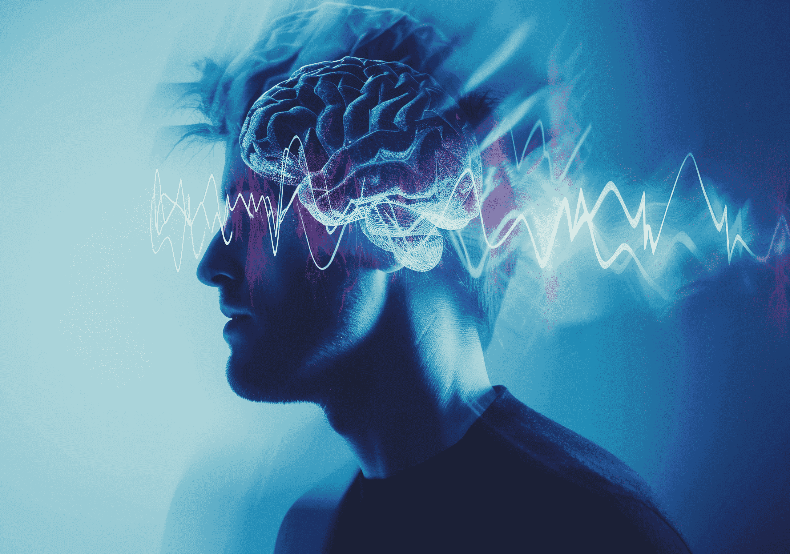
What Are Seizures?
A seizure is an abnormal electrical activity in the brain that can cause sudden changes in behavior, movements, sensations, or consciousness. Seizures can affect anyone at any age, and while they are often associated with epilepsy, they can also occur due to various other factors. It is important to seek medical attention to understand the underlying cause of seizures and determine the most appropriate treatment.
Types of Seizures
There are several different types of seizures, each with unique characteristics. Seizures are primarily categorized into two broad categories: focal seizures and generalized seizures.
1. Focal Seizures (Partial Seizures)
These seizures originate in one area of the brain and can affect only one part of the body. Focal seizures are divided into two types:
- Focal Onset Aware Seizures (Simple Partial Seizures): In these seizures, the person remains aware and conscious but may experience unusual sensations, such as tingling, visual distortions, or auditory hallucinations. The movements might involve a specific body part, like a hand or leg.
- Focal Onset Impaired Awareness Seizures (Complex Partial Seizures): These seizures affect a larger portion of the brain and cause loss of awareness. The person may appear confused, and their movements may be automatic, such as lip-smacking or repetitive gestures.
2. Generalized Seizures
Generalized seizures involve both sides of the brain and can cause a loss of consciousness. Types of generalized seizures include:
- Tonic-Clonic Seizures (Grand Mal Seizures): These are the most recognizable form of seizures. The person loses consciousness and experiences stiffening of the muscles (tonic phase) followed by violent jerking movements (clonic phase). This type of seizure can last for a few minutes and may be followed by confusion or drowsiness.
- Absence Seizures (Petit Mal Seizures): These seizures typically occur in children and involve brief episodes of staring or “zoning out,” lasting only a few seconds. The person may seem unaware of their surroundings but usually recovers quickly.
- Atonic Seizures: Also known as “drop attacks,” these seizures cause sudden loss of muscle tone, leading to a fall or collapse. Atonic seizures can be dangerous as they often result in injury due to sudden loss of control.
- Myoclonic Seizures: These involve sudden, brief jerks or twitches of the muscles, often in the arms or legs. These jerks can occur in clusters and may be mistaken for muscle spasms or tics.
3. Status Epilepticus
This is a medical emergency where seizures last for more than five minutes or occur in rapid succession without the person regaining consciousness in between. It can lead to serious complications and requires immediate medical intervention.
Causes of Seizures
Seizures can be caused by various factors. In some cases, no specific cause is identified, but here are some common triggers:
- Epilepsy: A neurological disorder characterized by recurrent seizures.
- Head injuries: Trauma to the brain can increase the risk of seizures.
- Stroke: A stroke can damage brain tissue, leading to seizures.
- Infections: Infections like meningitis or encephalitis can cause inflammation in the brain, triggering seizures.
- Brain tumors: Abnormal growth in the brain can disrupt normal brain function, leading to seizures.
- Genetic factors: Inherited conditions can make a person more susceptible to seizures.
- Alcohol withdrawal or substance abuse: Sudden withdrawal from alcohol or drugs can provoke seizures.
- High fever (in children): Febrile seizures are common in young children and are usually triggered by a high fever.
- Metabolic imbalances: Low blood sugar, electrolyte imbalances, or dehydration can also cause seizures.
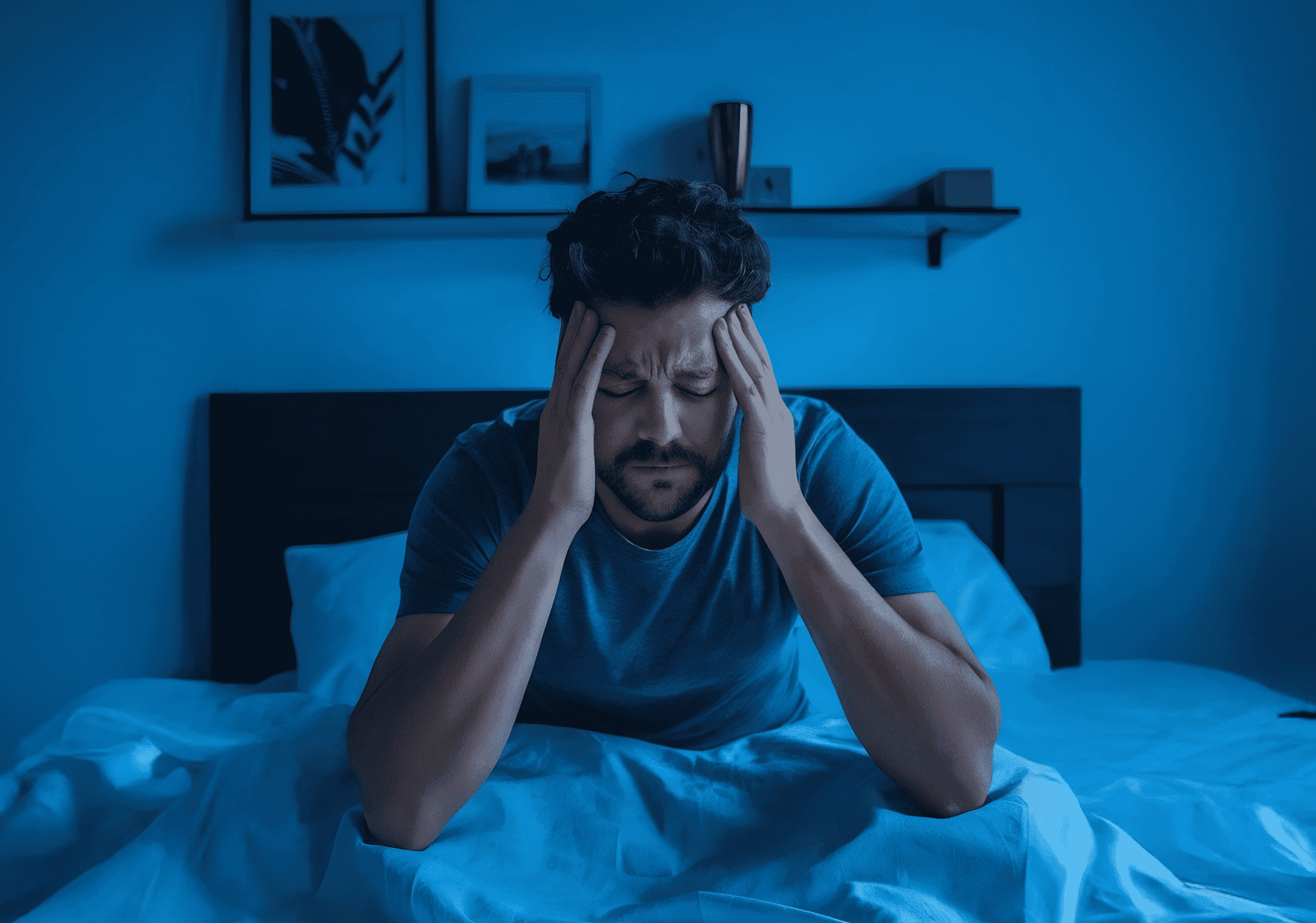
Symptoms of Seizures
The symptoms of a seizure vary depending on the type and the area of the brain involved. Common signs include:
- Sudden loss of consciousness or awareness
- Uncontrolled movements, such as jerking or twitching
- Unusual sensations, including tingling or numbness
- Visual or auditory hallucinations
- Staring spells or “zoning out” for a few seconds
- Loss of muscle control, resulting in falls or limpness
- Confusion or drowsiness following the seizure
It is important to remember that not all seizures involve convulsions. Some people may experience only subtle symptoms, such as confusion or strange sensations.
Treatment of Seizures
The treatment for seizures depends on the type, frequency, and underlying cause. At Burjeel Specialty Hospital, Sharjah, we offer personalized care and a multidisciplinary approach to treating seizures. Here are some common treatment options:
1. Medications
Anti-epileptic drugs (AEDs) are the most common treatment for controlling seizures. These medications help stabilize the brain’s electrical activity and reduce the occurrence of seizures. It may take some time to find the right medication and dosage for each individual, and regular follow-up with your neurologist is essential.
2. Surgical Treatments
In cases where seizures are difficult to control with medications, surgery may be considered. Procedures like lobectomy (removal of a small portion of the brain) or responsive neurostimulation can help manage seizures, especially in patients with focal seizures.
3. Vagus Nerve Stimulation (VNS)
VNS involves implanting a small device that stimulates the vagus nerve in the neck. This treatment is often used when medications fail to control seizures effectively.
4. Ketogenic Diet
For some patients, especially children, a high-fat, low-carbohydrate ketogenic diet can help reduce seizure frequency. This diet alters the brain’s metabolism and can provide relief for patients with intractable epilepsy.
5. Lifestyle Changes and Supportive Care
Managing stress, improving sleep hygiene, and avoiding seizure triggers are essential in minimizing the frequency of seizures. At Burjeel Specialty Hospital, we also provide comprehensive care that includes psychological support for patients and their families to manage the emotional challenges associated with seizures.
Why Choose Burjeel Specialty Hospital, Sharjah?
At Burjeel Specialty Hospital, Sharjah, we are proud to have a team of highly skilled neurologists, neurosurgeons, and allied specialists who work together to provide the best care for patients with seizure disorders. Our hospital is equipped with the latest diagnostic tools and treatment options to offer personalized care tailored to each patient’s needs.
FAQs
1. Can seizures be completely cured?
While many seizures can be controlled with medications or treatments, not all patients can be completely cured. However, with the right treatment plan, many people lead normal, active lives.
2. How do I know if I am having a seizure?
If you experience any of the symptoms mentioned above, such as unexplained jerking, confusion, or loss of consciousness, it’s important to seek medical advice.
3. Is it safe for someone with seizures to drive?
Driving laws vary by region, but generally, people with uncontrolled seizures may be restricted from driving. It’s essential to follow your doctor’s advice.
4. How can I help someone during a seizure?
If you witness someone having a seizure, stay calm, protect them from injury, and ensure their safety. Never put anything in their mouth, and if the seizure lasts for more than 5 minutes, call for emergency help.
Conclusion
Seizures are a serious medical condition that requires expert care. If you or a loved one is experiencing seizures, don’t hesitate to consult with our neurologists and neurosurgeons at Burjeel Specialty Hospital, Sharjah. With early diagnosis, appropriate treatment, and ongoing support, we can help you manage seizures and improve your quality of life. Schedule a consultation with us today!