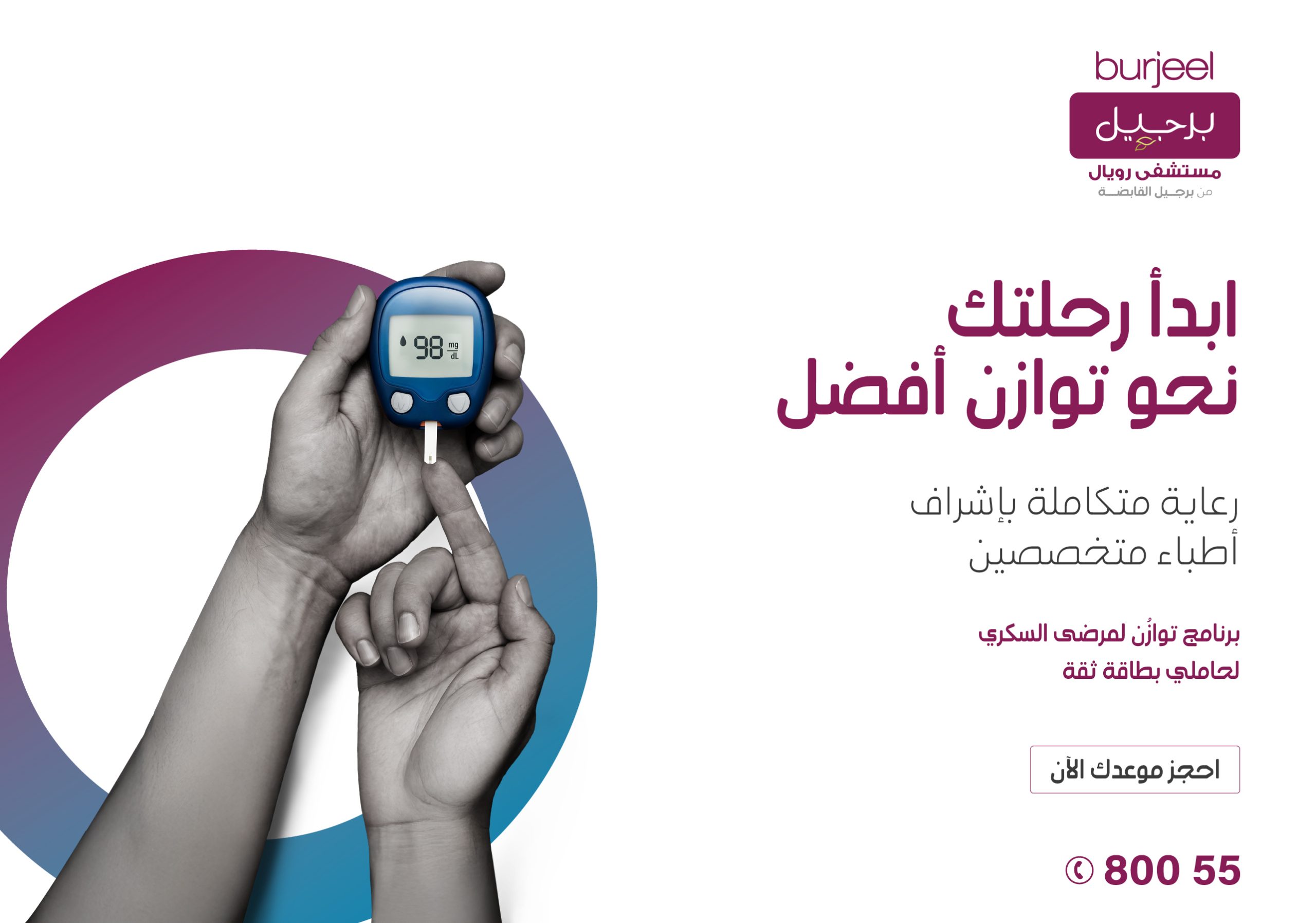
Digestive discomfort is more common than many realize — but when symptoms become persistent, they may point to Irritable Bowel Syndrome (IBS). Affecting millions worldwide, IBS is a functional gastrointestinal disorder that can significantly impact daily life.
Understanding IBS symptoms, causes, and diet management is key to improving gut health and overall well-being. At Burjeel Medical Centre, Al Marina, our specialists provide personalized care plans to help you manage IBS effectively and regain control of your digestive health.
What is IBS?
Irritable Bowel Syndrome (IBS) is a chronic condition affecting the digestive system, particularly the large intestine. Unlike structural gastrointestinal disorders, IBS does not cause visible damage — but it leads to recurring bowel movement issues, bloating, and abdominal discomfort.
IBS is often linked to the gut-brain connection, where stress and emotional health influence digestive function. While IBS is not life-threatening, it requires structured IBS management to reduce symptoms and improve quality of life.
IBS Symptoms
Recognizing IBS symptoms early can help in timely diagnosis and treatment. Symptoms vary from person to person but commonly include:
Abdominal pain or cramping
Bloating and excessive gas
Diarrhea, constipation, or alternating patterns
Mucus in stool
Feeling of incomplete bowel movement
Urgency or difficulty passing stool
👉 These irritable bowel syndrome symptoms often worsen after meals or during periods of stress.
If you experience recurring symptoms, a clinical evaluation is essential for accurate IBS diagnosis and symptoms management.
IBS Causes and Triggers
The exact IBS causes are not fully understood, but several factors contribute to the condition:
1. Gut-Brain Interaction
The gut-brain connection plays a significant role. Stress and anxiety can disrupt normal bowel function.
2. Food Intolerance
Certain foods can trigger IBS symptoms, highlighting the importance of identifying IBS triggers.
3. Abnormal Gut Motility
Irregular muscle contractions in the intestines may cause diarrhea or constipation.
4. Gut Microbiome Imbalance
Changes in gut bacteria affect digestion and sensitivity.
5. Hormonal Changes
IBS is more common in women, often influenced by hormonal fluctuations.
👉 Understanding irritable bowel syndrome causes helps in creating effective IBS treatment and diet management strategies.
IBS Diet Plan: What to Eat and Avoid
Diet plays a crucial role in IBS management. A well-structured IBS diet plan can significantly reduce symptoms.
Foods to Eat (IBS-Friendly Diet)
Low FODMAP foods (rice, oats, bananas)
Lean proteins (chicken, fish, eggs)
Cooked vegetables (carrots, spinach)
Probiotic-rich foods (yogurt, kefir)
Adequate fiber (for constipation-dominant IBS)
Foods to Avoid with IBS
High FODMAP foods (onions, garlic, beans)
Dairy (for lactose intolerance)
Fried and fatty foods
Caffeine and carbonated drinks
Artificial sweeteners
👉 A low FODMAP diet is often recommended as part of IBS diet management, helping reduce bloating and discomfort.
Best Diet Plan for IBS Patients
Creating the best diet plan for IBS patients involves personalization. There is no one-size-fits-all approach.
Key Dietary Strategies
Eat smaller, frequent meals
Stay hydrated
Gradually increase fiber intake
Keep a food diary to identify triggers
Include probiotics for gut health
👉 An effective IBS meal plan for better digestion focuses on consistency, balance, and avoiding trigger foods.
IBS Treatment and Relief Methods
While IBS cannot be permanently cured, symptoms can be effectively managed with the right approach.
IBS Treatment Options
Dietary modifications
Stress management techniques
Medications for symptom relief
Probiotics and supplements
Lifestyle changes
Natural IBS Relief Methods
Regular exercise
Mindfulness and relaxation techniques
Adequate sleep
Avoiding trigger foods
👉 Patients often ask: “How to manage IBS symptoms naturally?” The answer lies in combining diet, lifestyle, and medical guidance.
IBS Symptoms in Adults and Daily Impact
IBS affects daily routines, work productivity, and emotional well-being. Persistent symptoms like bloating and abdominal pain can lead to stress, creating a cycle that worsens IBS.
Breaking this cycle requires a structured IBS management plan tailored to individual needs.
TAKE CONTROL OF YOUR GUT HEALTH
Start Your IBS Management Journey Today
At Burjeel Medical Centre, Al Marina, our gastroenterology specialists provide comprehensive evaluation and personalized IBS treatment plans — including advanced dietary guidance and lifestyle strategies.
Common IBS symptoms include abdominal pain, bloating, gas, diarrhea, constipation, and irregular bowel habits. These symptoms often fluctuate over time.
2.What causes IBS in adults?
IBS causes include gut-brain interaction issues, food intolerance, stress, hormonal changes, and imbalances in gut bacteria.
3.What foods trigger IBS?
Common triggers include dairy, fatty foods, caffeine, artificial sweeteners, and high FODMAP foods like onions and beans.
4.How can IBS be managed with diet?
IBS can be managed with a structured IBS diet plan, including low FODMAP foods, balanced fiber intake, and avoiding triggers.
5.What is the best diet for IBS?
The best diet for IBS is personalized but often includes low FODMAP foods, probiotics, lean proteins, and cooked vegetables.
6.Can IBS be cured permanently?
IBS cannot be permanently cured, but symptoms can be effectively controlled with proper treatment and lifestyle changes.
7.How do you calm IBS symptoms quickly?
Immediate relief methods include avoiding trigger foods, staying hydrated, practicing relaxation techniques, and following a consistent diet plan.
Conclusion
IBS is a manageable condition — not a limitation. With the right understanding of IBS symptoms, causes, and diet management, you can significantly improve your digestive health and quality of life.
At Burjeel Medical Centre, Al Marina, team of board-certified Gastroenterologists combines clinical expertise with personalized care to help you navigate IBS confidently — offering effective solutions tailored to your unique needs.
A structured, multidisciplinary diabetes care program at Burjeel Royal Hospital, Al Ain and Burjeel Royal Hospital, Asharej — designed to help patients in Al Ain live healthier, more balanced lives.
Introduction: Living with Diabetes
Diabetes is one of the most prevalent chronic conditions in the UAE, with the country ranking among the highest globally for diabetes incidence. In Al Ain — a city of rich heritage and a growing population — thousands of residents manage this condition daily, navigating medication schedules, dietary choices, and lifestyle demands that can feel overwhelming without the right support.
For the diabetic, structured diabetes care has never been more important. Yet, many patients find themselves caught between fragmented consultations and a lack of coordinated follow-up, leaving critical gaps in their long-term management.
This is precisely why Burjeel Royal Hospital, Al Ain and Burjeel Royal Hospital, Asharej have launched the Tawazun Diabetic Program — a comprehensive, patient-centered diabetes management program designed to address the whole person, not just their blood glucose numbers. Rooted in the Arabic word “Tawazun” (توازن) meaning balance, the program reflects a core belief: that sustainable diabetes management is about restoring harmony across every dimension of a patient’s life.
“Tawazun” — توازن — meaning balance. Because managing diabetes is not just about numbers. It’s about your energy, your health, and your future.
What is the Tawazun Diabetic Program?
The Tawazun Diabetic Program at Burjeel Al Ain is a structured, evidence-based care pathway designed specifically for individuals living with Type 1 and Type 2 diabetes. Unlike a standard outpatient visit, Tawazun provides a continuous, coordinated journey — from initial assessment through long-term lifestyle maintenance — guided by a dedicated multidisciplinary team.
The program is built on five integrated pillars of care, each of which works in concert to deliver measurable, sustainable outcomes for patients in Al Ain:
🩺 Personalized Care Plans
Every patient enters the programme with a comprehensive clinical assessment. From this, a tailored management plan is created — factoring in medical history, comorbidities, lifestyle preferences, and personal health goals.
👥 Multidisciplinary Team
Patients benefit from the expertise of endocrinologists, diabetes nurse educators, clinical dietitians, pharmacists, and specialist consultants in ophthalmology and podiatry — all collaborating within a single coordinated framework.
🥗 Culturally Appropriate Nutrition
Dietary guidance is crafted to respect Emirati and Arab culinary traditions — making healthy eating practical, enjoyable, and culturally meaningful rather than restrictive and foreign.
💊 Medication Optimisation
The team provides close monitoring of pharmacological treatments, including the latest therapeutic options such as GLP-1 receptor agonists and SGLT-2 inhibitors, ensuring each patient’s regimen is both effective and safe.
📚 Continuous Patient Education
Knowledge is power. Through structured education sessions, workshops, and one-on-one coaching, patients develop the skills and confidence to take ownership of their condition — reducing dependence and improving long-term outcomes.
Who is the Tawazun Program For?
The Tawazun Diabetic Program is open to all adults living with diabetes who seek a more structured, supported approach to their care. It is particularly suited for:
Patients with Type 2 diabetes who have struggled to achieve target HbA1c levels despite medication
Individuals recently diagnosed with Type 1 or Type 2 diabetes who require structured onboarding and education
Patients with pre-diabetes who wish to take proactive steps to prevent progression
Those with obesity-related diabetes seeking integrated weight management support
Emirate patients in Al Ain looking for a coordinated, multi-specialist diabetes service
Patients experiencing early signs of diabetes-related complications — including peripheral neuropathy, diabetic eye disease, or kidney involvement
With fast-track access, patients receive their lab results in as little as 90 minutes — and their medications, without ever leaving home.
Whether you are newly diagnosed or have been living with diabetes for years, the Tawazun Program meets you where you are — and works with you to achieve meaningful progress.
Why Tawazun Diabetic Program Matters?
For all Emaraties covered under Thiqa insurance, managing a chronic condition like diabetes should not come with financial barriers. The Tawazun Diabetic Program is designed to align with Thiqa coverage, giving insured patients access to the full spectrum of specialist services without undue out-of-pocket burden.
Beyond insurance coverage, patients in Al Ain face a unique healthcare landscape. With a mix of public and private providers, navigating the right pathway for structured diabetes care can be complex. The Tawazun Diabetic Program simplifies this journey by bringing together all essential services — from endocrinology to dietetics to complication screening — under one coordinated program at Burjeel Royal Hospital, Al Ain and Burjeel Royal Hospital, Asharej.
Tawazun is designed with the Al Ain community in mind — offering Arabic-language support, culturally sensitive dietary guidance, and care pathways aligned with Thiqa insurance. Your health is our priority, and accessing excellent diabetes care should never be complicated.
What can Patients Achieve Through Tawazun?
The Tawazun Diabetic Program is outcomes driven. By enrolling in a structured, continuous care model, patients can expect to achieve measurable improvements across multiple dimensions of their health:
Clinical Outcomes
Reduction in HbA1c levels towards individualized target ranges
Improved fasting and post-meal blood glucose control
Optimized blood pressure and lipid profiles — key comorbidities in diabetes
Early identification and management of diabetes-related complications
Reduced risk of hypoglycemic episodes through medication review and education
Lifestyle & Behavioral Outcomes
Adoption of sustainable, culturally appropriate eating habits
Safe integration of physical activity tailored to each patient’s capacity
Effective weight management strategies aligned with diabetes goals
Improved sleep quality and stress management — both significantly impacting glucose control
Psychological & Empowerment Outcomes
Greater confidence in self-monitoring and interpreting blood glucose readings
Reduced diabetes distress and anxiety through education and peer support
A sense of control and agency over a condition that can feel overwhelming
Long-term independence in managing day-to-day diabetes decisions
Patients who complete structured diabetes management programs show significantly better glycemic control, lower rates of hospitalization, and improved quality of life compared to those receiving standard episodic care. Tawazun is built to deliver exactly these outcomes.
Diabetes is a metabolic condition characterized by elevated blood glucose levels resulting from insufficient insulin production (Type 1 diabetes), impaired insulin action (Type 2 diabetes), or both. Left unmanaged, persistently elevated glucose damages blood vessels and nerves throughout the body — leading to serious, often preventable, complications.
The Burden of Uncontrolled Diabetes
Without structured management, people living with diabetes face an elevated risk of:
Cardiovascular disease — the leading cause of mortality among people with diabetes
Diabetic nephropathy — kidney damage that may progress to renal failure
Diabetic retinopathy — a leading cause of preventable blindness
Diabetic foot ulcers — which, if untreated, can lead to amputation
Stroke and cerebrovascular disease
In the UAE, where dietary patterns, sedentary lifestyles, and genetic predisposition converge to create high prevalence rates, proactive and structured diabetes management is not optional — it is essential.
The Case for a Structured Program
Research consistently demonstrates that structured diabetes programs — those combining medical management, education, dietary counselling, and regular follow-up — outperform standard care in achieving glycemic targets, reducing complications, and improving patient quality of life. The Tawazun Diabetic Program operationalizes this evidence in a culturally adapted, locally accessible format for patients in Al Ain.
Your Tawazun Journey: Step by Step
From the moment you arrive, the Tawazun program is designed to feel seamless, supportive, and patient-centered. Here is what to expect:
Step 1: Comprehensive Initial Assessment
Your journey begins with a thorough clinical evaluation — including HbA1c testing, lipid profile, kidney function, BMI assessment, and complication screening. This forms the clinical foundation of your personalized care plan.
Step 2: Personalized Care Plan Development
Based on your assessment, your multidisciplinary team collaborates to design a care plan tailored to your specific goals, medical profile, and lifestyle preferences. You are an active participant in this process.
Step 3: Structured Education Program
You will participate in a series of diabetes education sessions covering self-monitoring techniques, medication adherence, carbohydrate awareness, physical activity guidance, and complication prevention.
Step 4: Nutritional Counselling
Our clinical dietitian will work with you to develop a personalized eating plan that reflects your tastes, cultural food preferences, and health objectives — practical, sustainable, and enjoyable.
Step 5: Regular Follow-Up & Monitoring
Ongoing monitoring is central to the program. Scheduled follow-up appointments track your clinical progress, address emerging concerns, and allow for timely adjustments to your management plan.
Step 6: Long-Term Lifestyle Maintenance
As you progress, the focus shifts to empowering you to sustain your achievements independently — with periodic check-ins and access to the Tawazun team whenever you need support.
Why Choose Burjeel for Your Diabetes Care in Al Ain?
Burjeel Royal Hospital, Al Ain and Burjeel Royal Hospital, Asharej are among the most trusted private healthcare institutions in Al Ain, offering internationally trained specialists, state-of-the-art diagnostics, and a patient-first philosophy that places your well-being at the center of every decision.
JCI-accredited quality standards ensuring patient safety and clinical excellence
Specialist endocrinologists with expertise in complex diabetes management
On-site laboratory and diagnostic services for rapid, accurate testing
Arabic and English-speaking clinical and administrative teams
Conveniently located across Al Ain for accessible, community-based care
At Burjeel, we believe that exceptional healthcare is a right, not a privilege. The Tawazun Diabetic Program brings that belief to life — delivering world-class diabetes care with a deeply human touch.
Frequently Asked Questions
1. What is the Tawazun Diabetes Program?
Tawazun is a structured, multidisciplinary diabetes management program at Burjeel Al Ain — including Burjeel Royal Hospital, Al Ain and Burjeel Royal Hospital, Asharej — designed to support patients with Type 1 and Type 2 diabetes through personalized care plans, continuous education, medication management, and lifestyle coaching.
2. Is the Tawazun Program covered under Thiqa insurance?
Yes. The Tawazun Diabetic Program accepts Thiqa insurance, making it accessible to Al Ain. We recommend confirming your specific coverage details with our patient services team.
3. How long does the program last?
The program is tailored to each individual’s needs. Most patients begin with intensive sessions over the first three months, followed by ongoing quarterly follow-ups. The timeline is adjusted based on health goals and clinical progress.
4. What specialists are involved in the program?
Our multidisciplinary team includes endocrinologists, diabetes nurse educators, clinical dietitians, pharmacists, and ophthalmology and podiatry specialists for comprehensive complication screening.
5. How do I join the Tawazun Program?
You can book your initial consultation directly through Burjeel Royal Hospital, Al Ain and Burjeel Royal Hospital, Asharej. Our team will assess your condition and enroll you into the program within a single visit.
6. Does the program offer Arabic-language support?
Absolutely. All educational materials, dietary guidance, and patient communications are available in Arabic to ensure cultural sensitivity and clarity for our local community.
Start Your Journey Today
Your health deserves balance — and the Tawazun Diabetic Program is here to help you find it. Whether you have been living with diabetes for years or are newly diagnosed, our team at Burjeel Royal Hospital, Al Ain and Burjeel Royal Hospital, Asharejis ready to walk alongside you on this journey.
Take the first step. Book your initial consultation today with our experienced endocrinologists in Al Ain and discover what structured, personalized diabetes care in Al Ain can truly achieve for your health, your energy, and your life.
Colorectal cancer is among the most prevalent cancers globally, yet it is also one of the most preventable with the right lifestyle habits and early screening. Research shows that factors such as diet, physical activity, body weight, and daily routines can significantly influence the risk of developing colorectal cancer.
Understanding how to prevent colon cancer and adopting healthier habits can help lower the chances of developing this disease. From choosing the right foods to maintaining an active lifestyle, small changes can make a meaningful difference in long-term health.
Preventive measures combined with regular medical checkups can help detect potential issues early and support overall digestive health.
1. Follow a Colorectal Cancer Prevention Diet
Diets play a crucial role in maintaining colon health. A balanced colorectal cancer prevention diet can help reduce inflammation in the digestive system and support healthy gut function.
Foods that support colon health include:
Fresh fruits and vegetables
Whole grains such as oats, brown rice, and quinoa
Legumes like lentils and beans
Nuts and seeds
Foods rich in fiber and antioxidants
Fiber-rich foods help promote regular bowel movements and may lower the risk of abnormal cell growth in the colon.
2. Reduce Processed and Red Meat Consumption
Studies suggest that consuming large amounts of processed or red meat may increase the risk of colorectal cancer.
Examples include:
Bacon and sausages
Processed deli meats
High quantities of beef, lamb, or pork
Replacing these foods with healthier protein options such as fish, poultry, or plant-based proteins may help reduce colon cancer risk naturally.
3. Stay Physically Active
Regular physical activity supports overall health and plays an important role in how to prevent colon cancer.
Exercise helps:
Improve digestion
Maintain a healthy weight
Reduce inflammation
Support immune function
Activities such as walking, cycling, swimming, or light strength training for at least 30 minutes daily can significantly benefit digestive health.
4. Maintain a Healthy Body Weight
Excess body weight has been associated with an increased risk of colorectal cancer. Maintaining a healthy weight helps regulate hormones and reduce chronic inflammation that may contribute to cancer development.
Healthy weight management can be achieved through:
Balanced nutrition
Regular exercise
Limiting sugary beverages and processed foods
Maintaining consistent eating habits
These habits contribute to long-term digestive and metabolic health.
5. Limit Alcohol Consumption
Excessive alcohol consumption can increase the risk of colorectal cancer by damaging the cells lining the colon.
To help lower cancer risk:
Limit alcohol intake
Avoid excessive drinking
Choose healthier beverage options when possible
Moderation is an important factor in reducing colon cancer risk naturally.
6. Avoid Smoking
Smoking is linked to several types of cancer, including colorectal cancer. Harmful chemicals in tobacco smoke can damage DNA and contribute to abnormal cell growth in the colon.
Quitting smoking can significantly improve overall health and lower the risk of many cancers.
Support from healthcare professionals can help individuals successfully quit smoking.
7. Get Regular Screening and Medical Checkups
One of the most effective strategies for preventing colorectal cancer is regular screening.
Screening tests such as colonoscopy can detect and remove precancerous polyps before they develop into cancer.
Most medical guidelines recommend starting colorectal cancer screening at age 45, or earlier for individuals with higher risk factors such as family history.
Early detection greatly improves treatment outcomes and survival rates.
Colorectal Cancer Care at Burjeel Specialty Hospital, Sharjah
Burjeel Specialty Hospital, Sharjah offers comprehensive colorectal cancer screening, prevention, and treatment services through its specialized Oncology Department.
A multidisciplinary team of experienced medical oncologists and surgical oncologists provides personalized care tailored to each patient’s condition.
Services include:
Advanced colorectal cancer screening programs
Diagnostic imaging and laboratory tests
Minimally invasive surgical treatments
Chemotherapy and targeted therapies
Personalized treatment plans
This coordinated approach ensures patients receive accurate diagnosis, effective treatment, and continuous support throughout their care journey.
Conclusion
Adopting healthy lifestyle habits is one of the most effective ways to lower the risk of colorectal cancer. By understanding how to prevent colon cancer and following a balanced colorectal cancer prevention diet, individuals can take proactive steps toward protecting their digestive health.
Regular physical activity, maintaining a healthy weight, limiting alcohol consumption, and avoiding smoking can all help reduce colon cancer risk naturally.
Combined with routine screening and early medical consultation, these preventive measures can play a major role in reducing the impact of colorectal cancer.
Burjeel Specialty Hospital, Sharjah provides comprehensive colorectal cancer screening and treatment through its Oncology Department, supported by experienced gastroenterologists,medical oncologists and surgical oncologists who focus on early detection and personalized care.
Frequently Asked Questions (FAQs)
1. Can lifestyle changes really help prevent colon cancer?
Yes. Research shows that healthy habits such as balanced nutrition, regular exercise, maintaining a healthy weight, and avoiding smoking can significantly reduce colorectal cancer risk.
2. What foods help prevent colorectal cancer?
Foods rich in fiber and antioxidants such as fruits, vegetables, whole grains, legumes, and nuts support digestive health and may reduce the risk of colorectal cancer.
3. At what age should colorectal cancer screening begin?
Most medical guidelines recommend starting screening at age 45 for individuals at average risk.
4. Does exercise reduce colon cancer risk?
Yes. Regular physical activity supports digestive health and helps maintain a healthy weight, which can reduce the risk of colorectal cancer.
5. Is colorectal cancer completely preventable?
Not all cases can be prevented, but many risk factors can be reduced through healthy lifestyle choices and regular screening.
Book a Consultation
If you would like to learn more about how to prevent colon cancer or discuss screening options, consult the specialists at Burjeel Specialty Hospital, Sharjah.
Experienced gastroenterologists, medical oncologists and surgical oncologists provide expert guidance, advanced screening services, and personalized treatment plans for colorectal cancer prevention and care.
Book your appointment today and take an important step toward protecting your long-term health.
Colorectal cancer is one of the most common cancers worldwide, yet it is also among the most preventable when detected early through proper screening. Coloscopy is widely considered the most effective method for identifying abnormal growths in the colon and detecting cancer at an early stage.
Many individuals are unsure about the right colonoscopy screening age or when to get a colonoscopy. Understanding the latest colon cancer screening guidelines 2025 can help people make informed decisions about their health and take proactive steps to prevent colorectal cancer.
Regular screening plays a key role in identifying potential issues early, improving treatment outcomes, and reducing the risk of advanced disease.
What is a Colonoscopy?
A colonoscopy is a medical procedure used to examine the inner lining of the colon and rectum using a thin, flexible tube equipped with a small camera called a colonoscope.
The procedure allows doctors to detect:
Precancerous polyps
Early-stage colorectal cancer
Inflammation or ulcers in the colon
Sources of unexplained gastrointestinal bleeding
Other digestive tract abnormalities
One of the major benefits of colonoscopy is that doctors can remove polyps during the procedure, helping prevent them from developing colorectal cancer.
Why Colonoscopy Screening is Important
Colorectal cancer often develops slowly over time. Many cases begin as small polyps in the colon that may gradually grow and become cancerous if not removed.
Screening helps doctors:
Detect colorectal cancer in its early stages
Identify and remove precancerous polyps
Monitor patients with higher risk factors
Reduce the likelihood of advanced-stage disease
Following the recommended colon cancer screening guidelines, 2025 significantly improves the chances of early detection and successful treatment.
Colonoscopy Screening Age: When Should You Start?
Knowing the appropriate colonoscopy screening age is essential for preventing colorectal cancer.
According to current colon cancer screening guidelines 2025, individuals with average risk should begin screening at:
Age 45
This updated recommendation reflects the rising number of colorectal cancer cases diagnosed in younger adults.
If the initial colonoscopy results are normal, the next screening is typically recommended every 10 years. However, the screening schedule may vary depending on individual health conditions and risk factors.
Who Should Get a Colonoscopy Earlier?
Some individuals may require colonoscopy screening before age 45 due to higher risk factors.
Earlier screening may be recommended for people who have:
A family history of colorectal cancer
A personal history of colon polyps
Inflammatory bowel diseases such as Crohn’s disease or ulcerative colitis
Genetic conditions like Lynch syndrome
Familial adenomatous polyposis (FAP)
In such cases, doctors may advise starting screening 10 years earlier than the age at which a close relative was diagnosed with colorectal cancer.
Symptoms That May Require Immediate Colonoscopy
Although screening is often recommended even before symptoms appear, certain warning signs may require immediate medical evaluation.
These symptoms include:
Blood in the stool
Persistent changes in bowel habits
Chronic constipation or diarrhea
Unexplained weight loss
Ongoing abdominal pain or cramping
Persistent fatigue caused by anemia
If any of these symptoms occur, it is important to seek medical advice promptly.
What Happens During a Colonoscopy?
Coloscopy is a safe and routine procedure performed by trained specialists.
Before the Procedure
Patients follow a bowel preparation process, which includes dietary adjustments and medications to cleanse the colon.
During the Procedure
The patient is given sedation for comfort
A colonoscope is inserted through the rectum
The doctor examines the colon using a camera
Polyps or abnormal tissue may be removed or biopsied
After the Procedure
Most patients recover quickly and can return home the same day.
Benefits of Colonoscopy Screening
Regular colonoscopy screening offers several important health benefits:
Early detection of colorectal cancer
Prevention through removal of precancerous polyps
Diagnosis of digestive conditions
Monitoring for individuals at higher risk
Routine screening is one of the most effective ways to reduce the burden of colorectal cancer.
Comprehensive Colorectal Care at Burjeel Specialty Hospital, Sharjah
Burjeel Specialty Hospital, Sharjah provides comprehensive screening, diagnosis, and treatment for colorectal conditions through its Gastroenterology Department and Oncology Department.
The hospital follows a multidisciplinary care approach involving experienced gastroenterologists, colorectal surgeons, medical oncologists, and surgical oncologists who work together to provide accurate diagnosis and personalized treatment.
The Gastroenterology Department plays a vital role in performing advanced diagnostic procedures such as colonoscopy and identifying early signs of colorectal disease. When necessary, colorectal surgeons and oncology specialists collaborate to deliver advanced surgical and medical treatments.
Services include:
Advanced colonoscopy screening and diagnostics
Early detection programs for colorectal cancer
Minimally invasive colorectal surgery
Chemotherapy and targeted therapy
Personalized treatment plans tailored to each patient’s needs
This coordinated approach ensures patients receive comprehensive care from screening through treatment and recovery.
When Should You Speak to a Doctor About Screening?
You should consult a doctor about colonoscopy screening if:
You are 45 years or older
You have a family history of colorectal cancer
You experience persistent digestive symptoms
You have a history of colon polyps or inflammatory bowel disease
A healthcare professional can assess your risk factors and recommend the appropriate screening schedule.
Conclusion
Colonoscopy remains one of the most effective tools for detecting and preventing colorectal cancer. Understanding the recommended colonoscopy screening age, knowing when to get a colonoscopy, and following the colon cancer screening guidelines 2025 can help individuals take proactive steps toward protecting their digestive health.
Routine screening allows doctors to detect abnormalities early and remove precancerous polyps before they develop into cancer.
Burjeel Specialty Hospital, Sharjah provides comprehensive colorectal care through its Gastroenterology Department and Oncology Department, supported by experienced gastroenterologists, colorectal surgeons, medical oncologists, and surgical oncologists who work together to deliver advanced screening and personalized treatment.
Frequently Asked Questions (FAQs)
1. At what age should colonoscopy screening begin?
For individuals at average risk, colonoscopy screening is generally recommended starting at age 45.
2. How often should a colonoscopy be performed?
If the initial colonoscopy is normal, it is typically recommended every 10 years, unless additional risk factors require more frequent monitoring.
3. Is colonoscopy painful?
Most colonoscopies are performed under sedation, making the procedure comfortable and painless for most patients.
4. Can colonoscopy help prevent colon cancer?
Yes. Colonoscopy can identify and remove precancerous polyps, helping prevent the development of colorectal cancer.
5. Are there other screening options besides colonoscopy?
Other screening options include stool-based tests and CT colonography, but colonoscopy remains the most comprehensive screening method.
Schedule Your Colonoscopy Screening
If you are approaching the recommended colonoscopy screening age or wondering when to get a colonoscopy, early screening can help protect your long-term health.
Consult the specialists at Burjeel Specialty Hospital, Sharjah, where experienced gastroenterologists, colorectal surgeons, medical oncologists, and surgical oncologists provide expert evaluation, advanced colonoscopy screening, and personalized colorectal cancer care.
Book your appointment today and take an important step toward early detection and prevention.
March is recognized worldwide as Colorectal Cancer Awareness Month, making it an important time to talk about early detection and prevention. Colorectal cancer is one of the most common cancers globally, yet it is also one of the most preventable and treatable cancers when detected early. Unfortunately, many people ignore the early warning signs, assuming the symptoms are related to minor digestive issues.
Understanding colorectal cancer symptoms, including the early signs of colon cancer and rectal cancer warning signs, can help individuals seek medical attention at the right time. Early awareness plays a crucial role in improving treatment outcomes and survival rates.
What is Colorectal Cancer?
Colorectal cancer develops in the colon or rectum, which are parts of the large intestine. It typically begins as small, noncancerous growths called polyps, which can gradually develop into cancer over time.
Early detection through awareness of early signs of colon cancer and regular screening can significantly increase survival rates. Many cases progress silently, but the body often provides warning signs that should not be ignored.
Common Colorectal Cancer Symptoms
Recognizing colorectal cancer symptoms early can help detect the disease before it spreads. Some symptoms may appear mild initially but should always be evaluated if they persist.
1. Persistent Changes in Bowel Habits
One of the earliest rectal cancer warning signs is a noticeable change in bowel habits that lasts for more than a few days.
These changes may include:
Frequent diarrhea or constipation
Narrow or thin stools
Feeling that the bowel does not empty completely
If these symptoms continue for several weeks, it is important to consult a doctor.
2. Blood in the Stool
Seeing blood in the stool can be alarming, but it is also one of the most important early signs of colon cancer.
Blood may appear as:
Bright red blood in the stool
Dark or tar-like stools
Blood detected through laboratory testing
While conditions like hemorrhoids can also cause bleeding, any unexplained bleeding should be medically evaluated.
3. Abdominal Pain or Cramping
Persistent abdominal discomfort can sometimes indicate a deeper digestive issue.
Symptoms may include:
Frequent stomach cramps
Gas or bloating
Ongoing abdominal pain
These symptoms may occur when a tumor blocks or irritates the intestine.
4. Unexplained Weight Loss
Sudden weight loss without diet or exercise changes may indicate that the body is fighting an underlying illness.
In colorectal cancer, tumors can interfere with digestion and metabolism, leading to unintentional weight loss and fatigue.
5. Weakness and Fatigue
Many people with colorectal cancer experience constant tiredness that does not improve with rest.
This can happen due to slow internal bleeding in the colon, which may lead to iron deficiency anemia.
Common symptoms include:
Low energy levels
Shortness of breath
Pale skin
Persistent fatigue
6. Feeling That the Bowel Is Not Completely Empty
Another important rectal cancer warning sign is the sensation that you still need to pass stool even after using the restroom.
This symptom can occur when a tumor in the rectum interferes with normal bowel movement.
Who is at Risk for Colorectal Cancer?
Certain factors can increase the risk of developing colorectal cancer, including:
Age over 45
Family history of colon or rectal cancer
Inflammatory bowel disease (Crohn’s disease or ulcerative colitis)
Diet high in processed or red meat
Obesity
Smoking and alcohol use
Lack of physical activity
People with these risk factors should consider regular screening and early medical consultation.
Importance of Early Screening
One of the most effective ways to detect colorectal cancer early is through routine screening tests.
Common screening methods include:
Colonoscopy
Stool-based tests
CT colonography
Screening can detect precancerous polyps before they become cancerous, making prevention possible.
At Burjeel Specialty Hospital, Sharjah, our specialists use advanced diagnostic tools and evidence-based treatment approaches to ensure accurate detection and personalized care.
Multidisciplinary Colorectal Cancer Care at Burjeel Specialty Hospital, Sharjah
The Oncology Department at Burjeel Specialty Hospital, Sharjah offers comprehensive cancer care supported by a team of gastroenterologists, medical oncologists, surgical oncologists, and multidisciplinary specialists.
Our approach includes:
Early detection and screening programs
Advanced diagnostic imaging
Minimally invasive surgical procedures
Chemotherapy and targeted therapy
Personalized treatment planning
By combining expertise and technology, our specialists provide patient-centered cancer care focused on better outcomes and quality of life.
When Should You See a Doctor?
If you notice any of the following symptoms lasting more than two weeks, it is important to seek medical advice:
Blood in stool
Persistent changes in bowel habits
Unexplained weight loss
Ongoing abdominal pain
Constant fatigue
Early medical evaluation can help detect colorectal cancer at a stage when treatment is most effective.
Conclusion
Colorectal cancer often develops quietly, but the body frequently provides warning signs. Recognizing colorectal cancer symptoms, including early signs of colon cancer and rectal cancer warning signs, can lead to timely diagnosis and life-saving treatment.
Awareness, regular screening, and prompt medical attention are key to reducing the impact of this disease.
1. What are the earliest colorectal cancer symptoms?
The earliest symptoms often include changes in bowel habits, blood in the stool, abdominal discomfort, and fatigue. However, some patients may have no symptoms initially, which is why screening is important.
2. Is colorectal cancer common in younger adults?
Although colorectal cancer is more common after age 45, cases among younger adults are increasing, making awareness of early symptoms essential.
3. How can colorectal cancer be prevented?
Prevention strategies include:
Regular screening
Maintaining a healthy diet rich in fiber
Regular physical activity
Limiting alcohol and processed foods
Avoiding smoking
4. When should I get screened for colorectal cancer?
Most people should start screening at age 45, but individuals with a family history may need screening earlier.
5. Are colorectal cancer symptoms always serious?
Some symptoms may be caused by minor digestive issues, but persistent symptoms should never be ignored, as early diagnosis greatly improves treatment success.
If you are experiencing colorectal cancer symptoms or early signs of colon cancer, early medical evaluation can make a critical difference.
Schedule a consultation with the Oncology specialists at Burjeel Specialty Hospital, Sharjah today. Our experienced gastroenterologists, medical oncologists and surgical oncologists are here to guide you through screening, diagnosis, and personalized treatment options.
Your health matters — early detection can save lives.
Colorectal cancer remains one of the most common cancers worldwide and continues to be a major public health concern. The latest colorectal cancer statistics 2025 highlight important trends in diagnosis, survival, and prevention. Understanding these numbers helps individuals, healthcare providers, and policymakers identify risk factors, improve screening programs, and strengthen awareness efforts.
Data from global cancer registries and health organizations show that colorectal cancer cases are increasing in certain populations, particularly among younger adults. At the same time, improvements in screening and treatment have helped improve the colon cancer survival rate when the disease is detected early.
Global Colorectal Cancer Statistics in 2025
According to recent global health data, colorectal cancer remains one of the most frequently diagnosed cancers worldwide.
Key colorectal cancer statistics 2025 include:
Colorectal cancer is the third most commonly diagnosed cancer globally
It is among the leading causes of cancer-related deaths worldwide
Millions of new cases are diagnosed each year
Early detection significantly improves survival outcomes
These statistics emphasize the importance of screening, awareness, and preventive lifestyle choices.
Rising Colorectal Cancer Incidence Rate
One of the most concerning trends in recent years is the increasing colorectal cancer incidence rate among younger adults.
Traditionally, colorectal cancer was considered more common in people over the age of 50. However, recent studies indicate a gradual rise in cases among individuals aged under 50.
Possible contributing factors include:
Sedentary lifestyles
Diets high in processed foods
Obesity
Reduced physical activity
Environmental and genetic influences
Because of these trends, many medical organizations now recommend beginning colorectal cancer screening at age 45 for individuals at average risk.
Colon Cancer Survival Rate: The Impact of Early Detection
The colon cancer survival rate varies significantly depending on how early the cancer is diagnosed.
When colorectal cancer is detected at an early stage before spreading to other organs, the survival rate can be significantly higher. Early-stage cancers are often easier to treat with surgery or minimally invasive procedures.
Survival outcomes generally fall into three stages:
Localized cancer: When the cancer is confined to the colon or rectum, survival rates are the highest.
Regional spread: When cancer spreads to nearby lymph nodes, treatment becomes more complex but still manageable.
Distant spread: When cancer spreads to distant organs such as the liver or lungs, survival rates decrease, making early diagnosis critical.
These statistics highlight the importance of screening programs and regular medical checkups.
Risk Factors Contributing to Colorectal Cancer
Understanding risk factors helps explain trends in the colorectal cancer incidence rate.
Common risk factors include:
Age above 45
Family history of colorectal cancer
Inflammatory bowel diseases such as Crohn’s disease or ulcerative colitis
Diet high in red and processed meat
Obesity
Smoking and excessive alcohol consumption
Physical inactivity
While some risk factors cannot be changed, lifestyle modifications and screening can help lower the risk.
Importance of Screening in Reducing Colorectal Cancer Rates
Screening is one of the most effective ways to reduce colorectal cancer cases and improve the colon cancer survival rate.
Colorectal Cancer Screening tests can detect:
Early-stage cancer
Precancerous polyps that can be removed before they become cancerous
Common screening methods include:
Colonoscopy
Stool-based tests
CT colonography
Most healthcare guidelines recommend starting screening at age 45, especially for individuals with average risk.
Early detection allows doctors to treat colorectal cancer before it progresses to advanced stages.
Advances in Colorectal Cancer Treatment
Medical advancements have improved treatment outcomes for colorectal cancer patients.
Modern Colorectal Cancer treatments include:
Minimally invasive colorectal surgery
Targeted therapies
Immunotherapy
Advanced chemotherapy protocols
Personalized treatment plans based on genetic testing
These developments have contributed to improving survival rates and patient outcomes in recent years.
Colorectal Cancer Care at Burjeel Specialty Hospital, Sharjah
Burjeel Specialty Hospital, Sharjah provides comprehensive colorectal cancer screening, diagnosis, and treatment services through its specialized Oncology Department.
A multidisciplinary team of experienced medical oncologists and surgical oncologists works closely with other specialists to provide personalized care for patients.
Services include:
Advanced colorectal cancer screening and diagnostic services
Minimally invasive colorectal surgery
Chemotherapy and targeted therapies
Personalized cancer treatment plans
Ongoing patient monitoring and supportive care
This coordinated approach ensures patients receive comprehensive care from early detection through treatment and recovery.
Conclusion
The latest colorectal cancer statistics 2025 highlight the growing importance of awareness, screening, and prevention. While the colorectal cancer incidence rate continues to rise in certain populations, improved screening programs and modern treatment options have helped increase the colon cancer survival rate, particularly when the disease is detected early.
Understanding the latest data empowers individuals to take proactive steps toward protecting their health through regular screening, healthy lifestyle choices, and timely medical consultation.
Burjeel Specialty Hospital, Sharjah provides comprehensive colorectal cancer care through its gastroenterology and oncology departments, supported by experienced gastroenterologists, medical oncologists and surgical oncologists dedicated to early detection and advanced treatment.
Frequently Asked Questions (FAQs)
1. How common is colorectal cancer in 2025?
According to recent colorectal cancer statistics 2025, colorectal cancer remains one of the most commonly diagnosed cancers worldwide and a leading cause of cancer-related deaths.
2. What is the colon cancer survival rate?
The colon cancer survival rate depends largely on the stage at diagnosis. Early-stage colorectal cancer has significantly higher survival rates compared to advanced stages.
3. Why is colorectal cancer increasing in younger adults?
The rise in the colorectal cancer incidence rate among younger adults may be linked to lifestyle factors such as diet, obesity, and reduced physical activity, although research is ongoing.
4. At what age should colorectal cancer screening begin?
Most health guidelines recommend starting colorectal cancer screening at age 45 for individuals with average risk.
5. Can colorectal cancer be prevented?
While not all cases can be prevented, lifestyle changes such as maintaining a healthy diet, exercising regularly, avoiding smoking, and undergoing regular screening can significantly reduce risk.
If you would like to learn more about colorectal cancer screening or discuss prevention and treatment options, consult the specialists at Burjeel Specialty Hospital, Sharjah.
The hospital’s gastroenterology and oncology departments, supported by experienced medical oncologists and surgical oncologists, offers advanced screening programs and personalized cancer care.
Schedule your consultation today and take an important step toward early detection and better health.
An ACL (Anterior Cruciate Ligament) tear is one of the most common and serious knee injuries, especially among athletes and physically active individuals. Whether caused by sudden twisting, pivoting, or impact, this injury can significantly affect mobility, stability, and quality of life. Understanding the ACL tear recovery process — including surgical and non-surgical options — helps patients make informed decisions about their knee injury treatment.
At Burjeel Hospital Sharjah, orthopedic specialists provide personalized treatment plans based on injury severity, activity level, and long-term goals.
What is an ACL Tear?
The ACL is a key ligament that stabilizes the knee joint by connecting the thigh bone (femur) to the shin bone (tibia). It prevents excessive forward movement and rotation of the knee.
Common Causes
Sudden stops or direction changes
Pivoting movements
Jumping and landing incorrectly
Direct blows to the knee (sports or accidents)
ACL tears are classified as:
Grade 1: Mild stretching
Grade 2: Partial tear
Grade 3: Complete rupture
Symptoms of an ACL Tear
Typical signs include:
A “popping” sensation at the time of injury
Immediate pain
Rapid swelling
Knee instability or “giving way”
Reduced range of motion
Difficulty bearing weight
Prompt medical evaluation is essential to confirm diagnosis and prevent further damage.
Non-Surgical Treatment: Who Is It For?
Not all ACL injuries require surgery. Conservative management may be appropriate for:
Partial tears
Individuals with low physical demands
Older adults
Patients willing to avoid high-impact activities
Non-Surgical Knee Injury Treatment Options
Physical therapy to strengthen surrounding muscles
Knee bracing for stability
Activity modification
Pain management and anti-inflammatory medication
Non-Surgical ACL Tear Recovery Timeline
Weeks 1–2: Reduce swelling and pain, begin gentle movement
Weeks 3–6: Strengthening exercises and improved mobility
Weeks 7–12: Advanced rehabilitation and balance training
3–6 months: Gradual return to normal daily activities
However, instability may persist, especially during sports or strenuous activities.
ACL Surgery: When Is It Recommended?
Surgical reconstruction is often advised for:
Complete ACL tears
Athletes or highly active individuals
Knee instability affecting daily life
Associated injuries (meniscus or cartilage damage)
Failure of conservative treatment
ACL surgery typically involves replacing the torn ligament with a graft from the patient’s own tissue or a donor.
ACL Surgery Timeline and Recovery
Recovery after reconstruction is longer but often restores stability and function more effectively.
ACL Surgery Timeline
First 2 Weeks
Pain control and swelling reduction
Use of crutches and knee brace
Gentle range-of-motion exercises
Weeks 3–6
Gradual weight-bearing
Physical therapy focusing on mobility and strength
Weeks 7–12
Increased strengthening and balance training
Improved walking pattern
3–6 Months
Return to light sports or physical activities
Advanced rehabilitation
6–12 Months
Full return to competitive sports (in most cases)
Strict adherence to rehabilitation protocols is essential for successful recovery.
Surgery vs. Non-Surgical Treatment: Key Differences
Aspect
Non-Surgical Treatment
ACL Surgery
Recovery Time
3–6 months
6–12 months
Knee Stability
May remain limited
Usually restored
Return to Sports
Often restricted
Typically possible
Risk of Re-Injury
Higher
Lower with proper rehab
Invasiveness
Non-invasive
Surgical procedure
Your orthopedic specialist will help determine the most appropriate approach based on your goals and lifestyle.
Long-Term Outlook
With proper treatment and rehabilitation, many individuals regain excellent knee function. However, untreated instability may increase the risk of additional injuries or early osteoarthritis.
FAQs
1. Can an ACL tear heal without surgery? Partial tears may heal with rehabilitation, but complete tears usually do not heal on their own.
2. Is ACL surgery painful? Post-operative discomfort is expected but manageable with medication and therapy.
3. When can I walk after ACL surgery? Most patients begin walking with support within a few days to weeks, depending on progress.
4. Can I return to sports after ACL reconstruction? Yes, many patients return to sports within 6–12 months with proper rehabilitation.
5. What happens if I don’t treat an ACL tear? Untreated tears can lead to chronic instability, meniscus damage, and long-term joint problems.
Conclusion
An ACL tear can be a life-altering injury, but effective treatment options are available. Understanding the ACL tear recovery process — whether through conservative management or surgical reconstruction — helps patients choose the best path for their lifestyle and goals. While non-surgical approaches may suit less active individuals, surgery often provides superior stability for those wishing to return to high-level physical activity.
Early evaluation and personalized care are key to achieving the best outcome.
Comprehensive Knee Care at Burjeel Hospital Sharjah
At Burjeel Hospital Sharjah, our experienced orthopedic specialists offer advanced diagnostics, individualized treatment plans, and state-of-the-art rehabilitation for ACL injuries and other knee conditions.
Don’t let a knee injury hold you back.
Book your consultation today to explore the most effective treatment for your ACL tear. Call now or schedule an appointment online and take the first step toward recovery and confident movement.
Back pain is one of the most common health complaints worldwide, and a herniated disc is a frequent underlying cause. While many cases improve non-surgical care, some patients continue to experience severe pain, weakness, or nerve symptoms that interfere with daily life. In such situations, herniated disc surgery may be recommended to relieve pressure on nerves and restore function.
What is a Herniated Disc?
The spine is made up of bones (vertebrae) cushioned by soft discs that act as shock absorbers. Each disc has a tough outer layer and a gel-like center. A herniated disc occurs when the inner material pushes through a tear in the outer layer, potentially pressing on nearby nerves.
Common Causes
Age-related degeneration
Sudden lifting or twisting injuries
Poor posture
Repetitive strain
Obesity
Sedentary lifestyle
Herniated discs most often occur in the lower back (lumbar spine) but can also affect the neck (cervical spine).
Symptoms That May Indicate a Herniated Disc
Symptoms depend on the location and severity of nerve compression.
Persistent back or neck pain
Pain radiating to arms or legs (sciatica)
Numbness or tingling
Muscle weakness
Difficulty walking or standing
Reduced range of motion
In severe cases, nerve damage can lead to loss of bowel or bladder control, which requires immediate medical attention.
When Conservative Treatment Isn’t Enough
Most patients improve with non-surgical treatments such as:
Physical therapy
Pain medications
Anti-inflammatory drugs
Epidural steroid injections
Activity modification
However, surgery may be considered if symptoms persist despite several weeks to months of treatment.
Indications for Surgery
Severe or worsening pain
Progressive muscle weakness
Significant nerve compression
Loss of function or mobility
Symptoms lasting longer than 6–12 weeks
Emergency conditions such as cauda equina syndrome
Modern spine surgery techniques are designed to relieve nerve pressure while preserving spinal stability.
Microdiscectomy
This is the most common procedure for lumbar herniated discs.
Removes the portion of the disc pressing on the nerve
Minimally invasive approach
Small incision
Faster recovery
High success rate for leg pain relief
Laminectomy
Part of the vertebral bone (lamina) is removed to create more space for nerves.
Used when spinal canal narrowing is present
Relieves pressure from multiple sources
Discectomy
Removal of the damaged disc portion or, in some cases, the entire disc.
Spinal Fusion
Two or more vertebrae are permanently joined to stabilize the spine.
Used when instability is present
May follow disc removal
Artificial Disc Replacement
The damaged disc is replaced with a synthetic one, preserving motion in selected patients.
Your surgeon will recommend the most appropriate back pain surgery option based on your condition, overall health, and lifestyle.
Benefits of Surgical Treatment
When indicated, surgery can provide significant relief.
Reduction or elimination of nerve pain
Improved mobility and function
Restoration of strength
Better quality of life
Prevention of long-term nerve damage
Many patients experience rapid improvement in leg or arm pain soon after surgery.
Recovery After Spine Surgery
Recovery time varies depending on the procedure and individual health.
Typical recovery expectations:
Hospital stay of 1–3 days (sometimes outpatient)
Gradual return to normal activities within weeks
Physical therapy for strengthening and mobility
Avoidance of heavy lifting during healing
Full recovery may take several weeks to months
Following post-operative instructions is crucial for optimal results.
FAQs
1. Is herniated disc surgery safe? Yes. Modern minimally invasive techniques have high success rates and low complication risks when performed by experienced surgeons.
2. Will surgery completely eliminate back pain? Surgery is most effective for nerve-related pain (such as sciatica). Some residual back discomfort may persist.
3. How long does the surgery take? Most procedures take 1–2 hours, depending on complexity.
4. Can a herniated disc recur after surgery? Yes, though recurrence is relatively uncommon. Proper rehabilitation reduces risk.
5. When can I return to work? Many patients resume light work within 2–6 weeks, depending on job demands.
Conclusion
While many herniated disc cases respond to conservative care, persistent or severe symptoms may require herniated disc surgery to prevent long-term nerve damage and restore function. Advances in spine surgery have made procedures safer, less invasive, and highly effective. Early evaluation by a specialist ensures timely treatment and the best possible outcome.
If chronic back pain or nerve symptoms are affecting your daily life, professional assessment is essential.
Book your consultation today to explore the most effective treatment for your condition. Call now or schedule an appointment online with our spine specialists for expert guidance and lasting relief.
Tonsillectomy — the surgical removal of the tonsils — is often associated with childhood, but many adults also require this procedure. Recurrent throat infections, breathing problems, or chronic tonsillitis can significantly impact quality of life, making adult tonsillectomy a recommended treatment in certain cases. Understanding recovery, risks, and expectations can help patients prepare for a smoother healing journey.
At Burjeel Hospital Sharjah, ENT specialists provide comprehensive evaluation and advanced surgical care for adults considering tonsil removal.
Why Adults May Need a Tonsillectomy
Tonsils are part of the immune system, but they can become a source of repeated infection or obstruction.
Common reasons for tonsil removal in adults include:
Chronic tonsillitis that does not respond to medication
Enlarged tonsils causing breathing or swallowing problems
Obstructive sleep apnea
Peritonsillar abscess (quinsy)
Suspicion of abnormal growth or malignancy
Although tonsillectomy is less common in adults than children, it can provide significant long-term relief when symptoms are severe.
Tonsillectomy Age: Is It Safe for Adults?
There is no strict upper limit for tonsillectomy age. Adults of various ages can safely undergo the procedure if they are medically fit. However, recovery tends to be more challenging compared to children.
Pre-surgical assessment typically includes:
Medical history review
Physical examination
Blood tests
Evaluation of anesthesia risks
Your ENT specialist will determine whether surgery is appropriate based on your overall health and symptom severity.
What Happens During the Procedure?
Adult tonsillectomy is usually performed under general anesthesia and takes about 30–60 minutes.
Surgical Steps
Patient is placed under anesthesia
Tonsils are removed using specialized instruments or energy devices
Bleeding is controlled
No external incisions are required
Most patients go home the same day unless monitoring is needed.
Tonsil Removal Recovery: What to Expect
Recovery after adult tonsillectomy typically takes longer than in children — usually about 10 to 14 days, though full recovery may take up to three weeks.
Common Recovery Symptoms
Significant throat pain
Difficulty swallowing
Ear pain (referred pain from throat)
White coating in the throat (normal healing tissue)
Mild fever
Bad breath
Fatigue
Pain often peaks around days 5–7 as scabs begin to separate.
Tips for a Smooth Recovery
Proper care can reduce complications and speed healing.
Refrain from strenuous activity for at least two weeks
Following your doctor’s instructions is essential for safe recovery.
Risks and Possible Complications
Tonsillectomy is generally safe, but like any surgery, it carries some risks.
Potential Risks
Bleeding (most significant concern in adults)
Infection
Reaction to anesthesia
Severe pain
Dehydration due to difficulty swallowing
Rare changes in voice
Immediate medical attention is required if significant bleeding occurs after surgery.
Benefits of Adult Tonsillectomy
Despite a longer recovery, many adults experience substantial improvements after surgery.
Benefits may include:
Fewer throat infections
Better sleep and breathing
Reduced antibiotic use
Improved quality of life
Relief from chronic discomfort
For patients with severe or recurrent symptoms, these benefits often outweigh the temporary recovery challenges.
FAQs
1. How painful is tonsillectomy in adults? Pain can be significant, especially during the first week, but it is manageable with medication and proper care.
2. When can I return to work after surgery? Most adults need 10–14 days off work, depending on job demands and recovery progress.
3. Is bleeding normal after tonsillectomy? Small streaks of blood may occur, but active bleeding requires immediate medical attention.
4. Can tonsils grow back after removal? It is extremely rare for tonsils to regrow after complete removal.
5. Will my immunity weaken after tonsillectomy? No. The immune system has many other tissues that compensate for the absence of tonsils.
Conclusion
An adult tonsillectomy can be life-changing for individuals suffering from recurrent infections or breathing issues. Although tonsil removal recovery may be more demanding in adults than in children, the long-term benefits often include improved health, sleep, and comfort. Understanding the procedure, risks, and healing process helps patients prepare confidently for surgery and recovery.
If persistent throat problems interfere with your daily life, consultation with an ENT specialist is the first step toward lasting relief.
Expert ENT Care at Burjeel Hospital Sharjah
At Burjeel Hospital Sharjah, our experienced ENT specialists team provides advanced evaluation, safe surgical techniques, and comprehensive post-operative care for adult tonsillectomy.
Take the first step toward a healthier throat and better quality of life.
Cholesterol is often discussed during routine health checkups, yet many people remain unsure what their numbers mean. Understanding your cholesterol profile is essential for protecting long-term heart health and preventing serious conditions such as heart attack and stroke. This guide provides a clear overview of cholesterol levels explained, including LDL, HDL, and triglycerides — and why each matter.
At Burjeel Hospital Sharjah, our cardiology specialists emphasize that knowing your numbers is the first step toward preventing cardiovascular disease.
What is Cholesterol?
Cholesterol is a waxy, fat-like substance found in your blood. Your body needs it to build cells, produce hormones, and aid digestion. However, too much cholesterol can lead to plaque buildup in arteries, restricting blood flow to the heart and brain.
Cholesterol travels through the bloodstream in particles called lipoproteins, mainly LDL and HDL.
LDL Cholesterol: The “Bad” Cholesterol
LDL (Low-Density Lipoprotein) is commonly referred to as “bad cholesterol” because high levels contribute to plaque formation in arteries — a process known as atherosclerosis.
Why High LDL Is Dangerous
Narrows and hardens arteries
Increases risk of heart attack and stroke
Reduces oxygen supply to vital organs
Often shows no symptoms until complications occur
Ideal LDL Levels
Optimal: Less than 100 mg/dL
Near optimal: 100–129 mg/dL
High: 160 mg/dL or above
Lower LDL levels are generally better, especially for individuals with diabetes, hypertension, or existing heart disease.
HDL Cholesterol: The “Good” Cholesterol
HDL (High-Density Lipoprotein) is known as good cholesterol because it helps remove excess cholesterol from the bloodstream and transports it back to the liver for disposal.
What is Good Cholesterol and Why It Matters
HDL protects arteries by reducing plaque buildup and lowering cardiovascular risk.
Ideal HDL Levels
Men: 40 mg/dL or higher
Women: 50 mg/dL or higher
Protective level: 60 mg/dL or above
Higher HDL levels are associated with better heart protection.
Triglycerides: The Overlooked Risk Factor
Triglycerides are a type of fat in the blood that stores excess calories from food. While not cholesterol, they play a major role in cardiovascular risk.
Causes of High Triglycerides
High-sugar or high-carbohydrate diets
Obesity
Physical inactivity
Diabetes
Excess alcohol intake
Smoking
Healthy Triglyceride Levels
Normal: Less than 150 mg/dL
Borderline high: 150–199 mg/dL
High: 200 mg/dL or above
Elevated triglycerides combined with low HDL and high LDL significantly increase heart disease risk.
Why Your Cholesterol Numbers Matter for Heart Health
An abnormal lipid profile often develops silently, without noticeable symptoms. Over time, plaque buildup can lead to coronary artery disease, stroke, or peripheral vascular disease.
Risk factors that make cholesterol control especially important include:
Family history of heart disease
Diabetes or hypertension
Smoking
Sedentary lifestyle
Obesity
Age over 40
Regular screening helps detect problems early, allowing for lifestyle changes or medical treatment before complications occur.
How to Improve Your Cholesterol Profile
Lifestyle Changes
Eat a heart-healthy diet rich in fruits, vegetables, whole grains, and healthy fats
Reduce saturated and trans fats
Exercise regularly (at least 150 minutes per week)
Maintain a healthy weight
Quit smoking
Limit alcohol intake
Medical Management
If lifestyle changes are insufficient, doctors may prescribe medications such as statins or other lipid-lowering therapies to reduce cardiovascular risk.
FAQs
1. How often should I check my cholesterol levels? Adults should have their cholesterol checked every 4–6 years, or more frequently if risk factors are present.
2. Can young people have high cholesterol? Yes. Genetics, diet, and lifestyle can cause high cholesterol even in young adults.
3. Is high HDL always protective? Generally yes, but extremely high levels may not always provide additional benefit. A balanced profile is key.
4. Can cholesterol be controlled without medication? Many people can improve their numbers through diet, exercise, and weight management, though some may still require medication.
5. Do cholesterol problems cause symptoms? Usually no. Most people feel normal until serious complications develop, which is why routine screening is crucial.
Conclusion
Understanding your cholesterol profile is vital for maintaining lifelong heart health. LDL, HDL, and triglycerides each play a distinct role in cardiovascular risk, and maintaining balanced levels can significantly reduce the chances of heart disease and stroke. Regular testing, healthy lifestyle habits, and timely medical care are the cornerstones of prevention.
Take Charge of Your Heart Health Today
At Burjeel Hospital Sharjah, our team of experienced cardiology specialists offers comprehensive cholesterol screening, personalized risk assessment, and advanced treatment plans tailored to your needs.
Schedule your heart health checkup today and take a proactive step toward a healthier future.
This website uses cookies. We use cookies to make sure you get the best experience on our website. Please read our privacy policy and Terms & Conditions.
Burjeel Holdings Cookie Policy
Our website uses cookies and similar tracking technologies to enhance website functionality and analyze site usage. Our aim is to improve the experience for visitors to our website.
What is a cookie?
Cookies are small text files downloaded to your device when you visit a website. Cookies are also sent back to the original website or another website that recognises the cookie and act as a memory for a website, allowing the site to remember details on subsequent visits. For instance, cookies can remember end user preferences, improve user experience as well as adjust content to items most relevant to end users
Burjeel Holdings uses cookies for the following purposes:
Providing website functionality;
Analyzing site usage
Generic type cookies
Session and persistent cookies
When a site is launched, these cookies attempt to remember information about you, such as your consent to view YouTube videos or to use investor tools
Session cookies exist only until you close your web browser
Persistent cookies exist longer but for a specified period of time.
Third party cookies
Third-party cookies are created by domains that are not the website (or domain) that you are visiting. These cookies are used to understand your browsing of the website, for example, which page you visit or how long you stay on each page.
These types of cookies are set by Burjeel Holdings affiliates and/or vendors we are working with to enhance end user experience
The Cookie Preferences Panel
You may utilize the Cookie Preferences Panel at any time to change or withdraw your consent for each cookie category - except strictly necessary cookies - by clicking the “Cookie preferences” button in the bottom of the screen or clicking the “Cookie Policy“ link, located in the website footer. You may select “Allow all cookies” on the panel which permits the download of all available cookies for use.
The panel identifies categories of cookies and enables you to control and change your cookie preferences. Detailed information about the individual cookies is provided on Cookie Policy page, it is delivered in an open and transparent way, providing complete visibility into how cookies are used to enrich your visitor experience and make an informed choice to allow their usage.
You may use the Toggle bar next to each category of cookies to set acceptance or denial of cookies being placed on your device. Be aware however, that restricting cookies may impact the functionality of a website. Many of the interactive functions offered by websites are dependent on cookies and disabling or blocking cookies can prevent these services from working and reduce their usefulness. Once your selections are made, click the “Confirm your preferences” button to confirm your cookie usage choices.
Cookie Category Description
Mandatory Cookies
The strictly necessary cookies help make a website usable by enabling basic functions like page navigation and access to secure areas of the website. These cookies are necessary for the website to function and cannot be switched off in our systems. They are usually only set in response to actions made by you which amount to a request for services, such as setting your privacy preferences, logging in or filling in forms. You may be able to decline these cookies through your browser, but necessary parts of the site will not then work. These cookies do not store any personally identifiable information.
Operational Cookies
These cookies allow us to count visits and traffic sources, perform customer surveys and other web analytics, so we can measure and improve the performance of our site. They help us know which pages are the most and least popular and see how visitors move around the site. The information these cookies collect is aggregated and in some instances limited identifiable data may be collected.
Functional Cookies
These cookies enable the website to provide enhanced functionality, aid site personalisation, maintain user-selected options and site navigation aids. They may be set by us or by third party providers whose services we have added to our pages. If you do not allow these cookies then some or all of these services may not function properly.
Marketing and targeted advertising cookies
They are not used on this website.
Individual Cookies Descriptions
You can find more information about the individual cookies we use and the purposes for which we use them in the table below:
Name
Provider
Purpose
Expiry
Type
Operational
_ga
Google
Registers a unique ID that is used to generate statistical data on how the visitor uses the website.
2 years
HTTP Cookie
_ga_#
Google
Used by Google Analytics to collect data on the number of times a user has visited the website as well as dates for the first and most recent visit.
2 years
HTTP Cookie
Functional
cookies_policy
Burjeel Holdings
Stores the user’s cookie consent state for the current domain.
1 year
HTTP Cookie
SERVERID
Closir
Session
HTTP Cookie
_hjSessionUser_#
Closir
1 year
HTTP Cookie
_hjSession_#
Closir
1 day
HTTP Cookie
CLID
Euroland
351 day
HTTP Cookie
MUID
Euroland
371 day
HTTP Cookie
Marketing
VISITOR_INFO1_LIVE
YouTube
Tries to estimate the users’ bandwidth on pages with integrated YouTube videos.
180 days
HTTP Cookie
YSC
YouTube
Registers a unique ID to keep statistics of what videos from YouTube the user has seen.
Session
HTTP Cookie
We only use necessary cookies that help make a website usable by enabling basic functions like page navigation and access to secure areas of the website. The website cannot function properly without these cookies.We and do not store any PII data on the websites.
Azure Cookies
Azure TuPMix
Description : The TiPMix cookie is set by Azure to determine which web server the users must be directed to.
Duration : 1 hour
Azure x-ms-routing-name
Description : Azure sets this cookie for routing production traffic by specifying the production slot.
Duration : 1 hour
Azure ARRAfinity
Description : ARRAffinity cookie is set by Azure app service, and allows the service to choose the right instance established by a user to deliver subsequent requests made by that user.
Duration : Session
Azure ARRAffinitySamSite
Description : This cookie is set by Windows Azure cloud, and is used for load balancing to make sure the visitor page requests are routed to the same server in any browsing session
Duration : Session
Analytics cookies
Google Tag Manager _gcl_au
Description : Google Tag Manager sets the cookie to experiment advertisement efficiency of websites using their services.
Duration : 3 months
Google Analytics _ga_*
Description : Google Analytics sets this cookie to store and count page views.
Duration : 1 year 1 month 4 days
Google Analytics _ga
Description : Google Analytics sets this cookie to calculate visitor, session and campaign data and track site usage for the site's analytics report. The cookie stores information anonymously and assigns a randomly generated number to recognise unique visitors.
Duration : 1 year 1 month 4 days
Google Analytics ga*
Description : Google Analytics sets this cookie to store and count page views.
Duration : 1 year 1 month 4 days
Facebook _fbp
Description : Facebook sets this cookie to display advertisements when either on Facebook or on a digital platform powered by Facebook advertising after visiting the website.
Duration : 3 months
Functional Cookies
Polylang pll_language
Description : These are used to remember the language the user selects when returning to the website and get the language information when unavailable in another way.
Duration : 1 year
We are currently using only the mandatory cookies, and the websites cannot function properly without these. None of the cookies are storing any user data [like -- login information, items in shopping cart, users tracking for advertising purposes, user preferences and activities].
Along with the Cookie Preferences Panel, you can also change your browser and/or mobile device settings so that cookies from this website cannot be placed on your computer or mobile device. You can usually change the browser’s settings to refuse new cookies, disable existing ones or simply let you know when new ones are sent to your device. In order to set your browser to reject cookies, refer to the help instructions supplied by the browser provider (usually located within the “Help”, “Tools” or “Edit” menu) or the settings of your mobile device.
Please be aware that, if you refuse or disable cookies, some of the website’s functionality may be lost.
Learn more about who we are, how you can contact us and how we process personal data in our Privacy Policy If you have any queries in relation to this notice, please contact us at dataprotectionofficer@burjeelholdings.com