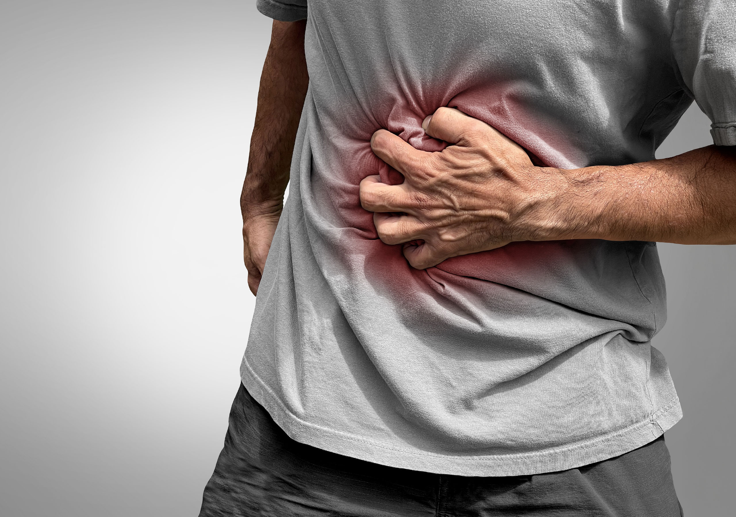
Many people notice veins on their legs becoming more visible over time. At first, it may seem like a cosmetic issue, just a few twisted blue lines beneath the skin. But for many individuals, these veins can gradually bring discomfort, heaviness, swelling, and sometimes even pain.
Varicose veins are one of the most common vein conditions worldwide. The good news is that today, modern medical treatments make it possible to manage and treat them safely and effectively, often without surgery. But before discussing treatment, it’s important to understand what your body might be trying to tell you.
“Doctor, Why Are These Veins Appearing on My Legs?”
This is one of the most common questions patients ask when they first visit a clinic.
Varicose veins develop when the tiny valves inside the veins stop working properly. These valves normally help blood flow upward toward the heart. When they weaken, blood begins to pool inside the veins instead of moving smoothly through them.
Over time, this pressure causes the veins to stretch and enlarge, making them visible under the skin.
Because the veins in the legs must constantly work against gravity, this is where varicose veins most often appear.
“How Do I Know If What I’m Feeling Is Varicose Veins?”
Many people live with symptoms for years without realizing they may be related to vein problems.
In the early stages, you might notice small changes such as:
- Blue or purple veins becoming more visible on your legs
- A feeling of heaviness in the legs by the end of the day
- Legs that feel unusually tired after standing or sitting for long periods
- Mild swelling around the ankles
- Occasional burning or throbbing in the legs
- Night-time leg cramps
At first, these symptoms can be easy to ignore. Many people assume they are simply tired or overworked. But over time, the symptoms can slowly become more noticeable.
When Does Varicose Veins Symptoms Start to Progress?
If varicose veins remain untreated, the body may start showing more visible signs that circulation is being affected.
Some people notice:
- Persistent swelling in the lower legs
- Darkening or discoloration of the skin around the ankles
- Dry, itchy skin near the veins
- Tightening or thickening of the skin
- Increasing discomfort while standing for long periods
- In more advanced cases, small wounds or ulcers near the ankles
These symptoms may indicate chronic venous insufficiency, a condition where the veins are no longer able to effectively return blood to the heart.
“Can Varicose Veins Cause Blood Clots?”
This is another concern many patients have—and it’s an important question.
Sometimes, varicose veins can lead to a condition called superficial thrombophlebitis, which occurs when a small blood clot forms in a surface vein.
If this happens, you may notice:
- Redness along the vein
- Tenderness or warmth in the area
- A firm or painful vein
- Swelling in the leg
In rare situations, clots can develop deeper in the leg veins, a condition known as deep vein thrombosis (DVT). This requires immediate medical attention.
The important thing to remember is that most complications can be prevented with early evaluation and treatment.
“What Should I Do If I Notice These Symptoms?”
If your legs have been feeling heavy, swollen, or uncomfortable, the best first step is simply to speak with a vein specialist.
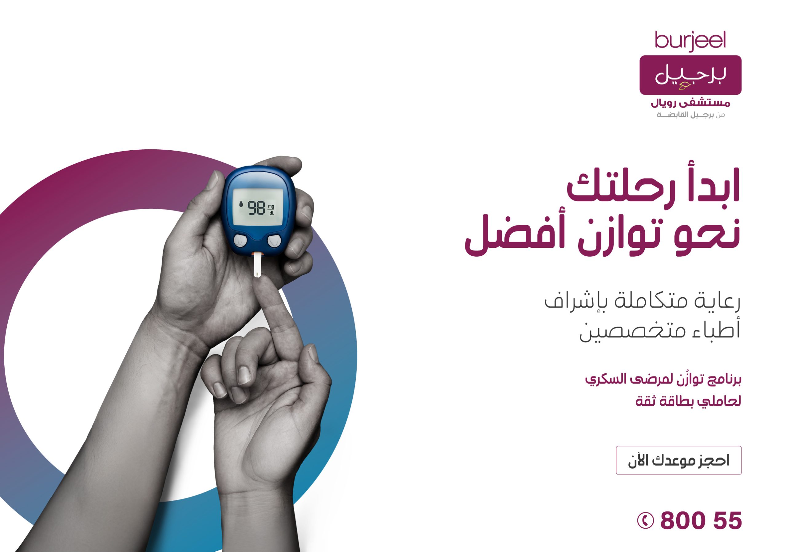
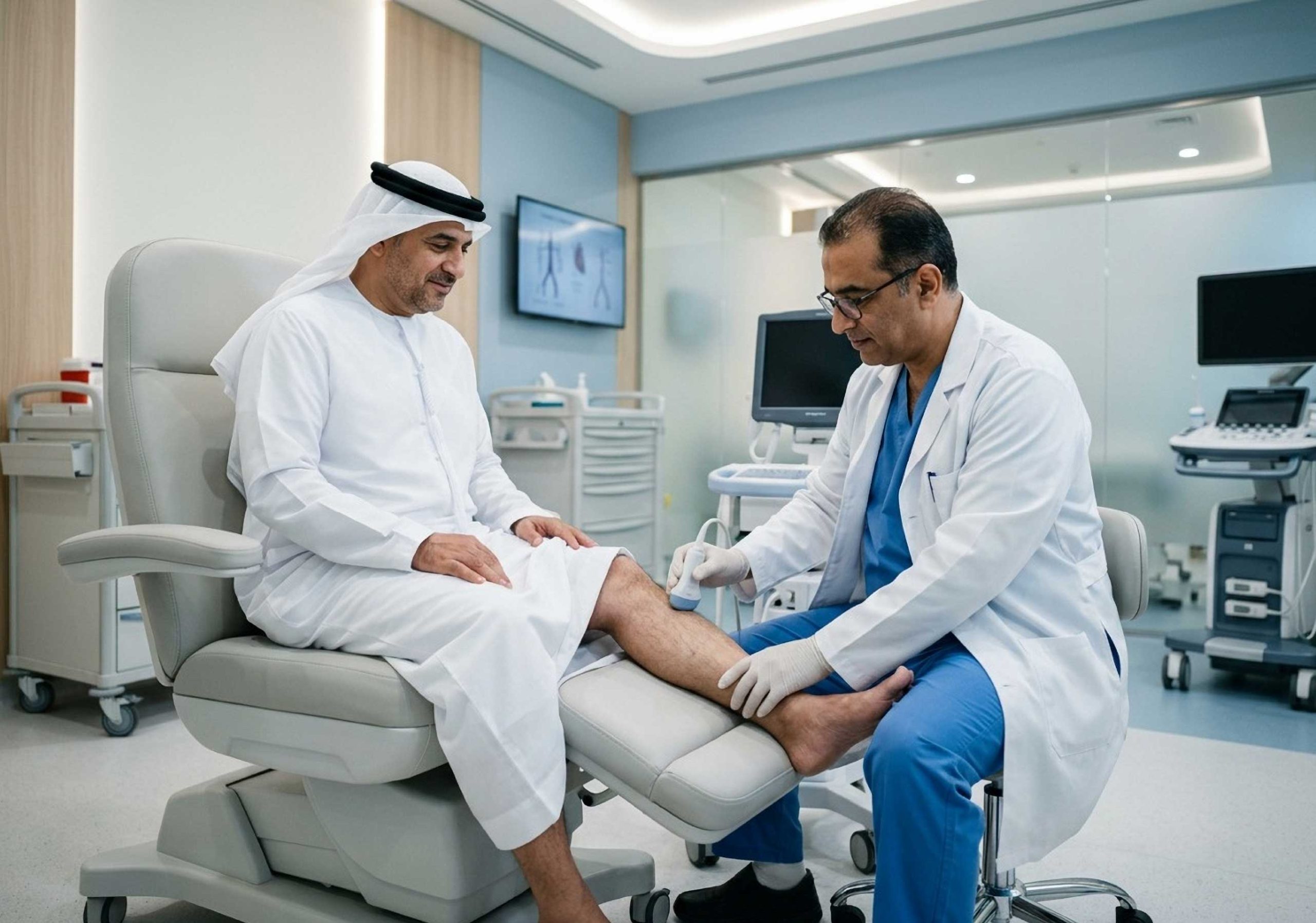
Many patients feel relieved when they learn that diagnosing vein problems is straightforward. Doctors usually begin with a duplex ultrasound, a painless imaging test that shows how blood flows through the veins and whether the valves are functioning properly.
This test helps determine:
- Whether the veins are allowing blood to flow correctly
- If the valves are weakened
- Whether any clots are present
Once the underlying cause is identified, the doctor can recommend the most appropriate treatment.
“Why Do Some People Get Varicose Veins While Others Don’t?”
Varicose veins can develop for several reasons, and often it is a combination of factors.
Some of the most common include:
- Family history
If your parents or close relatives have varicose veins, you may be more likely to develop them.
- Long hours standing or sitting
Jobs that keep you on your feet or seated for long periods can slow circulation in the legs.
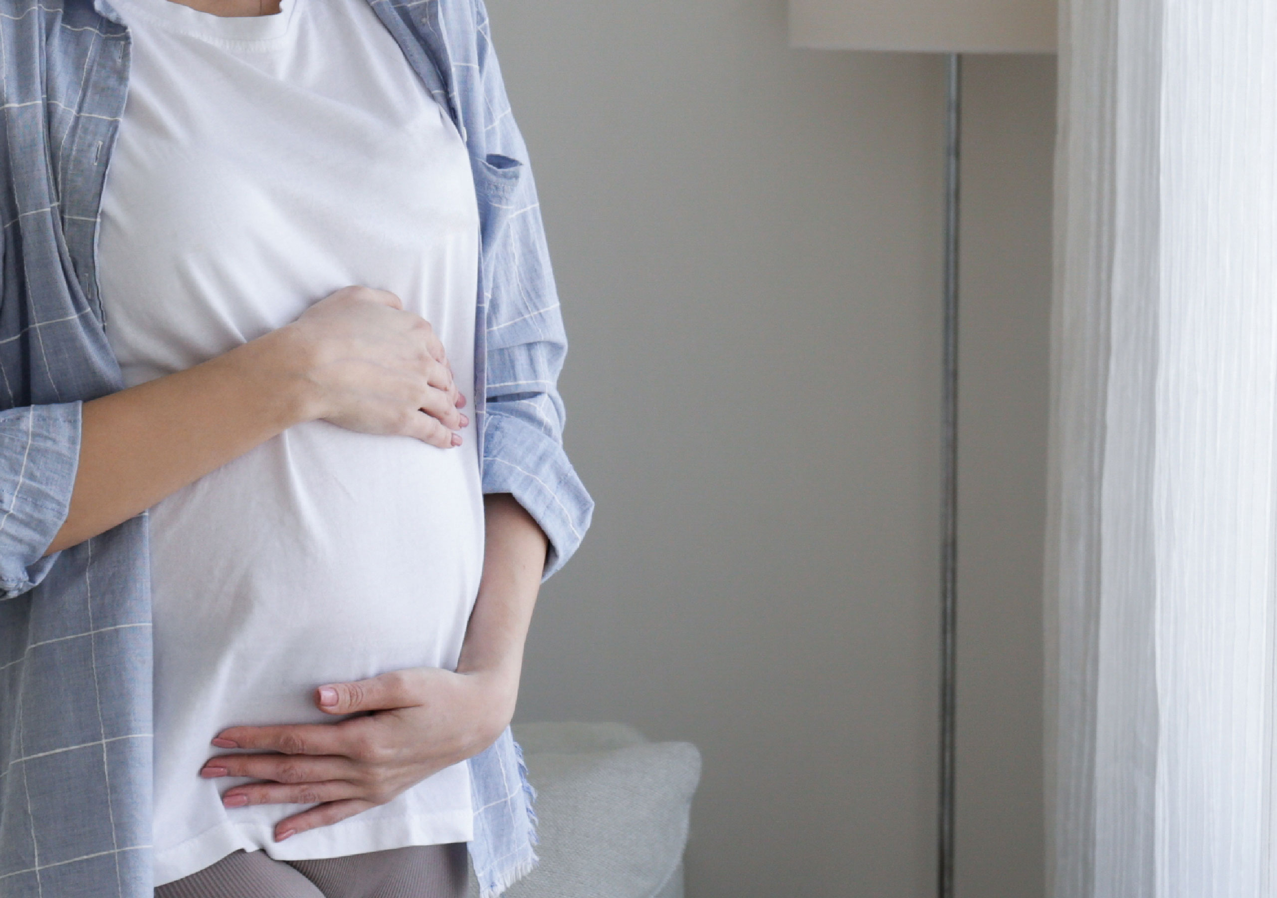
- Pregnancy
Hormonal changes and increased blood volume during pregnancy can put extra pressure on the veins.
- Age
Over time, vein valves naturally become less efficient.
- Excess body weight
Additional pressure on the legs can strain the veins.
- Limited physical activity
Movement helps blood circulate. A sedentary lifestyle can contribute to poor venous circulation.
The Good News: Modern Treatments Are Highly Effective
For many people, hearing the word “vein treatment” suggests surgery and long recovery periods. Fortunately, that is no longer the case.
Today, most treatments for varicose veins are minimally invasive procedures performed in outpatient clinics, allowing patients to return home the same day.
Lifestyle Changes That Can Help
In the early stages, small lifestyle adjustments can significantly reduce symptoms:
- Regular walking or gentle exercise
- Elevating the legs when resting
- Avoiding long periods of standing or sitting
- Maintaining a healthy weight
- Wearing compression stockings recommended by your doctor
These steps help improve circulation and relieve discomfort.
Varicose Veins Treatments
Endovenous Laser Treatment (EVLT)
One of the most widely used modern treatments involves inserting a thin laser fiber into the affected vein. The heat from the laser gently seals the vein closed, allowing blood to flow through healthier veins instead.
Patients appreciate that this treatment:
- Is minimally invasive
- Requires local or general anesthesia
- Allows quick recovery
- Has a very high success rate
Radiofrequency Ablation (RFA)
Another advanced treatment uses radiofrequency energy to close the damaged vein.
Many patients prefer this option because it:
- Causes minimal discomfort
- Does not require surgical incisions
- Allows a quick return to daily activities
Sclerotherapy
For smaller veins or spider veins, doctors may inject a medical solution that causes the vein to gradually collapse and fade away.
This treatment is commonly used for:
- Spider veins
- Smaller varicose veins
- Cosmetic improvements
Microphlebectomy
When larger veins are visible beneath the skin, they can sometimes be removed through very small incisions. The procedure leaves minimal scarring and is often combined with other treatments for the best results.
Why Early Treatment Makes a Difference
Many patients delay seeking help because they assume varicose veins are simply a cosmetic issue. But early treatment can make a significant difference.
Addressing the problem sooner can help:
- Relieve leg pain and swelling
- Improve blood circulation
- Prevent blood clots
- Avoid skin complications or ulcers
- Restore comfort and confidence
Most importantly, modern treatments allow patients to return to normal life quickly with minimal downtime.
When Should You See a Vascular Surgeon?
If you have been experiencing any of the following symptoms, it may be time to consult a vascular surgeon:
- Persistent leg heaviness or pain
- Swelling in the ankles or feet
- Visible, bulging veins on the legs
- Skin changes near the ankles
- Warm or painful veins
- Sudden swelling or redness that may suggest a clot
Early diagnosis often leads to simpler treatments and better long-term outcomes.
Taking the First Step Toward Healthier Legs
If you have noticed changes in your legs whether it’s visible veins, discomfort, or swelling it’s important to listen to your body.
Varicose veins are very common, and you are certainly not alone in experiencing them. The reassuring news is that modern vein care has advanced tremendously, and effective treatment options are available.
Speaking with a Vascular Specialist or a Vascular Surgeon at Burjeel Hospital Abu Dhabi, can help you understand what is happening in your body and guide you toward the right solution so you can move forward with comfort, confidence, and healthier legs