Brain tumors can affect people of all ages and may develop silently before symptoms become noticeable. Early recognition of brain tumor symptoms is crucial, as timely diagnosis can significantly improve treatment outcomes and quality of life. Many early signs are subtle and may be mistaken for common conditions like stress, migraines, or fatigue. Knowing the warning signs brain tumor patients often experience helps ensure prompt medical evaluation.
At Burjeel Hospital Sharjah, neurologists and neuro specialists emphasize that early detection plays a vital role in successful management.
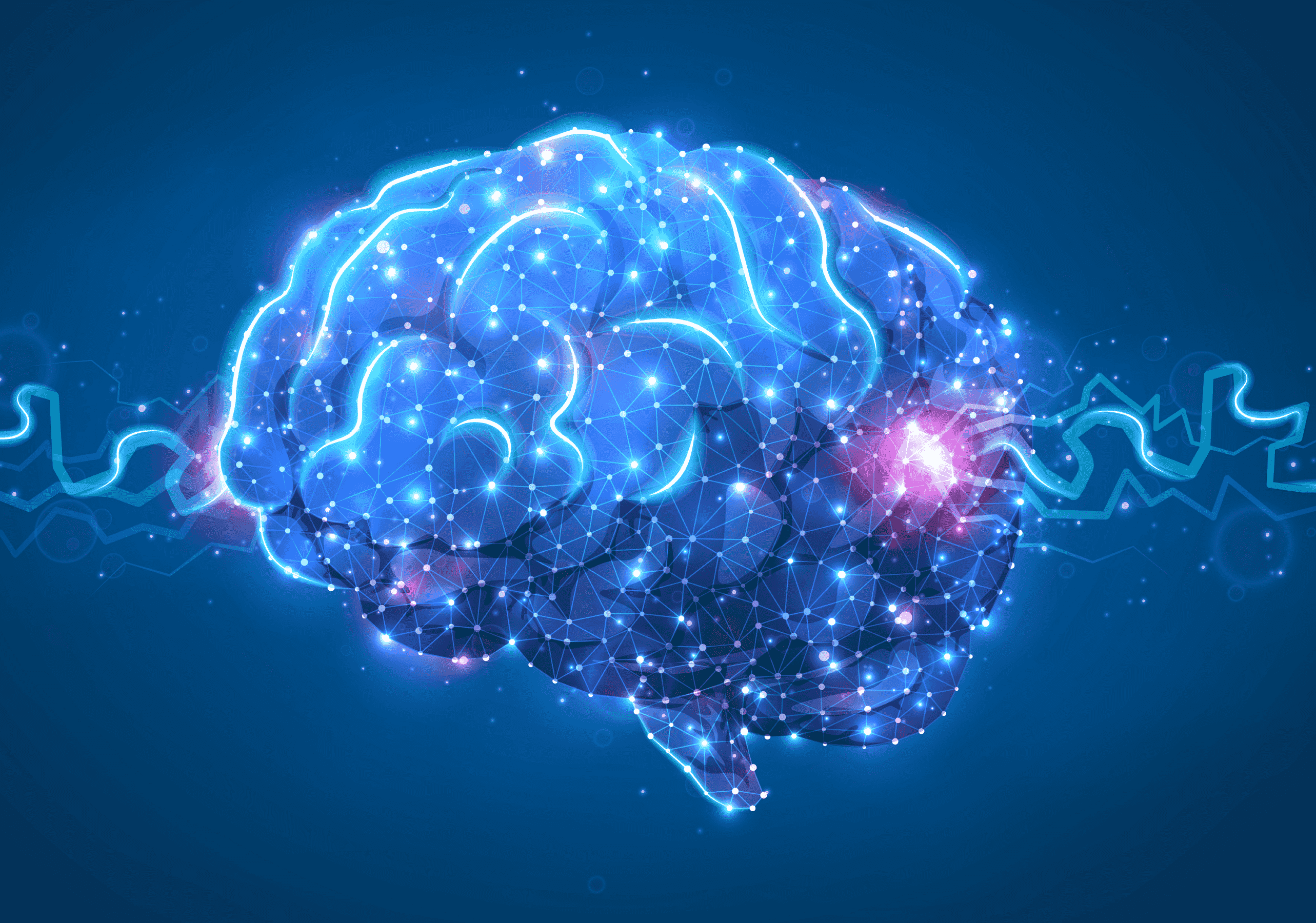
What Is a Brain Tumor?
A brain tumor is an abnormal growth of cells within the brain or surrounding tissues. Tumors may be:
- Benign (non-cancerous) — slow-growing and less likely to spread
- Malignant (cancerous) — aggressive and capable of invading nearby structures
Regardless of type, tumors can disrupt brain function by increasing pressure inside the skull or interfering with vital areas that control movement, speech, memory, and vision.
Common Brain Tumor Symptoms
Symptoms vary depending on tumor size, location, and growth rate.
Persistent or Worsening Headaches
Headaches are among the most frequent symptoms. Unlike typical headaches, they may:
- Be worse in the morning
- Intensify over time
- Not respond to usual pain medications
- Be accompanied by nausea or vomiting
Any new or unusual headache pattern should be evaluated.
Seizures
Seizures in someone without a prior history are a major red flag. They may involve:
- Convulsions
- Sudden loss of awareness
- Uncontrolled movements
- Sensory disturbances
Seizures often prompt immediate medical investigation.
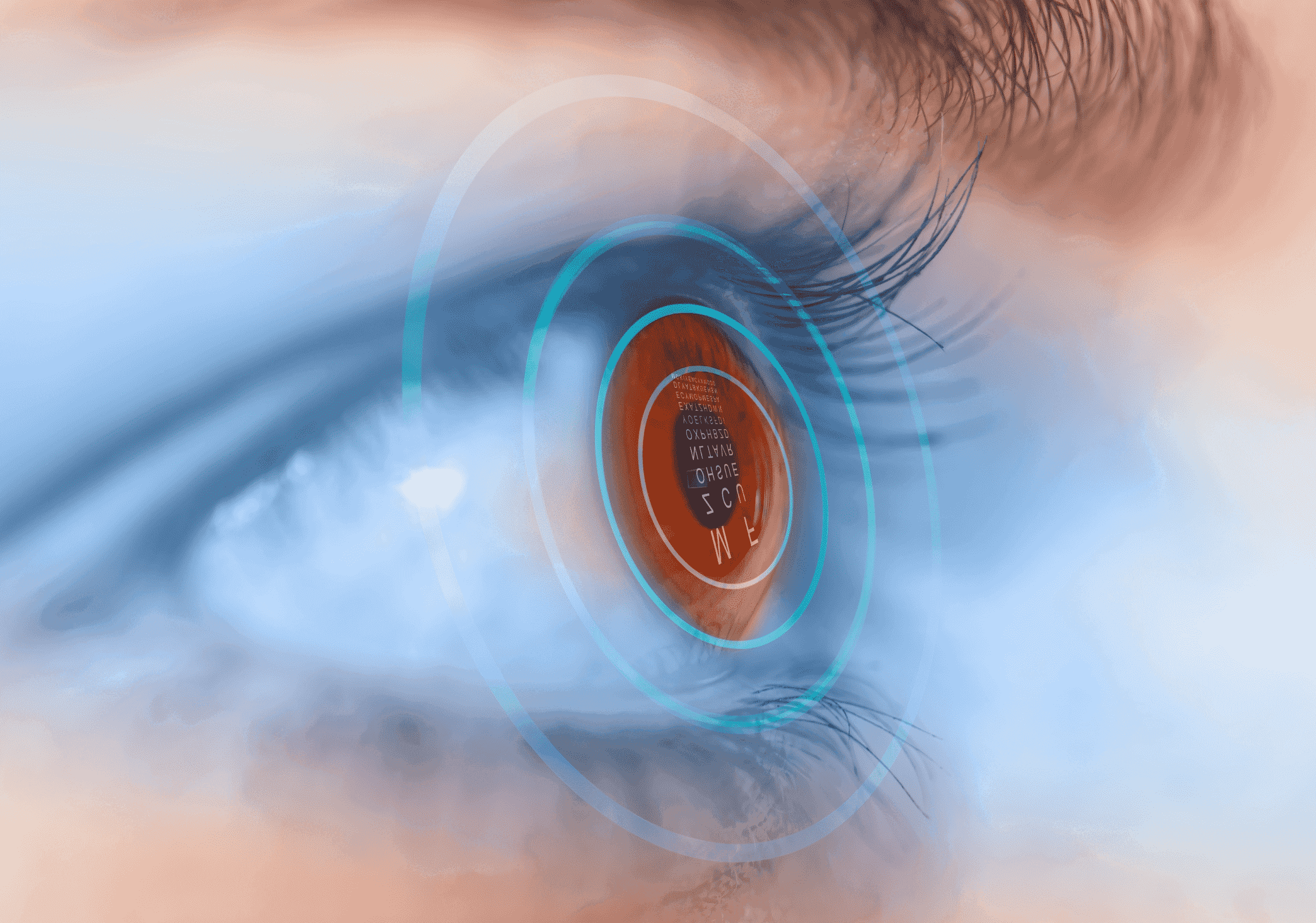
Vision Problems
Tumors affecting visual pathways may cause:
- Blurred or double vision
- Loss of peripheral vision
- Difficulty focusing
- Unexplained vision changes
These symptoms can sometimes be mistaken for eye problems.
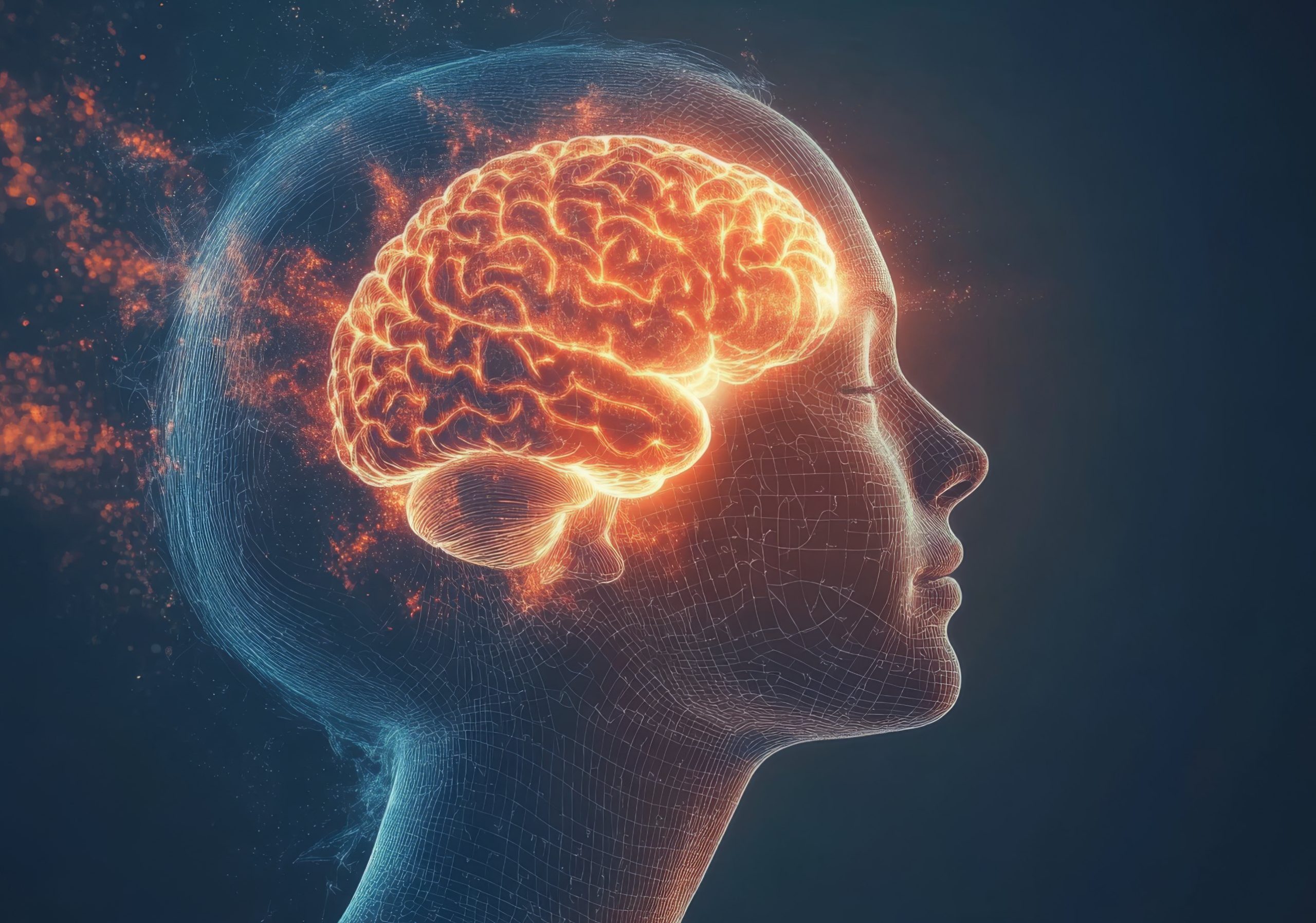
Cognitive and Personality Changes
Brain tumors can alter mental function and behavior.
Possible changes include:
- Memory problems
- Confusion
- Difficulty concentrating
- Mood swings
- Personality changes
- Poor judgment
Family members often notice these changes first.
Weakness or Numbness
Depending on tumor location, patients may experience:
- Weakness on one side of the body
- Numbness or tingling
- Difficulty with coordination
- Trouble walking or maintaining balance
Speech or Hearing Difficulties
Tumors in specific brain regions may affect communication.
Symptoms may include:
- Slurred speech
- Trouble finding words
- Difficulty understanding language
- Hearing loss or ringing in the ears
Warning Signs Brain Tumor Patients Should Not Ignore
Seek urgent medical attention if symptoms are severe, progressive, or accompanied by:
- Persistent vomiting
- Sudden neurological deficits
- New seizures
- Severe drowsiness
- Loss of consciousness
Early evaluation allows doctors to determine the cause and initiate appropriate treatment.
When to See a Neurologist
Understanding when to see neurologist services is essential for timely diagnosis.
You should consult a neurologist if you experience:
- Persistent unexplained headaches
- New neurological symptoms
- Seizures
- Changes in memory or behavior
- Vision or coordination problems
- Symptoms that worsen over time
Neurologists use advanced imaging tests such as MRI or CT scans to identify abnormalities in the brain.
Risk Factors for Brain Tumors
While many brain tumors have no clear cause, certain factors may increase risk:
- Exposure to radiation
- Family history of brain tumors
- Genetic conditions
- Increasing age (for some tumor types)
However, tumors can occur even without identifiable risk factors.
FAQs
1. Are headaches always a sign of a brain tumor?
No. Most headaches are not related to tumors, but persistent or unusual headaches should be evaluated.
2. Can brain tumors develop without symptoms?
Yes. Small tumors may remain silent until they grow large enough to affect brain function.
3. Are brain tumors curable?
Treatment of success depends on tumor type, location, and stage. Many benign tumors can be effectively treated.
4. Do brain tumors always cause seizures?
No, but seizures are a common presenting symptom in many cases.
5. How are brain tumors diagnosed?
Diagnosis typically involves neurological examination and imaging studies such as MRI or CT scans, sometimes followed by biopsy.
Conclusion
Recognizing early brain tumor symptoms can be lifesaving. Because symptoms often mimic common conditions, they are easy to overlook. However, persistent or progressive neurological changes should never be ignored. Understanding the warning signs brain tumor patients experience — and knowing when to see neurologist care — ensures timely diagnosis and the best possible treatment outcomes.
If you or a loved one experiences concerning symptoms, seeking medical evaluation without delay is essential.
Expert Neurological Care at Burjeel Hospital Sharjah
At Burjeel Hospital Sharjah, our highly skilled neurologists and neurosurgeons in the Department of Neurosciences provide advanced diagnostic imaging, expert neurological evaluation, and comprehensive treatment for brain tumors and other neurological conditions.
Don’t ignore warning signs — early detection saves lives.
Book a consultation today or call to schedule an appointment with our neurology specialists. Your brain health deserves expert care and attention.