Thyroid cancer is one of the more treatable cancers when found early, but outcomes and treatment plans can change significantly depending on the stage at diagnosis. Understanding staging helps patients make sense of what comes next—from imaging and biopsy to surgery, radioactive iodine, and advanced thyroid cancer treatment options for disease that has spread or returned.
Thyroid cancer symptoms to watch for
Many people notice no early warning signs, but these are common thyroid cancer symptoms:
- A new lump or swelling in the front of the neck
- Hoarseness or voice changes that don’t improve
- Trouble swallowing or a persistent “something stuck” feeling
- Neck discomfort or enlarged lymph nodes
Symptoms of thyroid cancer in females can look similar, but women sometimes report symptoms being mistaken for “thyroid imbalance” or fatigue-related issues. Any new neck lump or persistent voice/swallowing symptoms should be checked by a specialist.
For late-stage thyroid cancer symptoms, people may experience more pronounced lymph node swelling, breathing/swallowing difficulty, persistent cough not linked to infection, or symptoms related to spread (for example, bone pain if bones are involved). (Symptoms vary by cancer type and where it spreads.)
What are the thyroid cancer stages?
Doctors most commonly use the TNM staging system (Tumor size/extent, lymph Nodes, Metastasis) and group it into stages (I–IV). In thyroid cancer, staging can be influenced by factors like age and cancer type.
In simple terms:
- Early-stage disease is usually confined to the thyroid (and sometimes nearby lymph nodes).
- Advanced thyroid cancer typically refers to cancer that is widely invasive, has spread to distant organs, or is radioactive iodine–refractory (not responding to radioactive iodine), requiring systemic therapies.
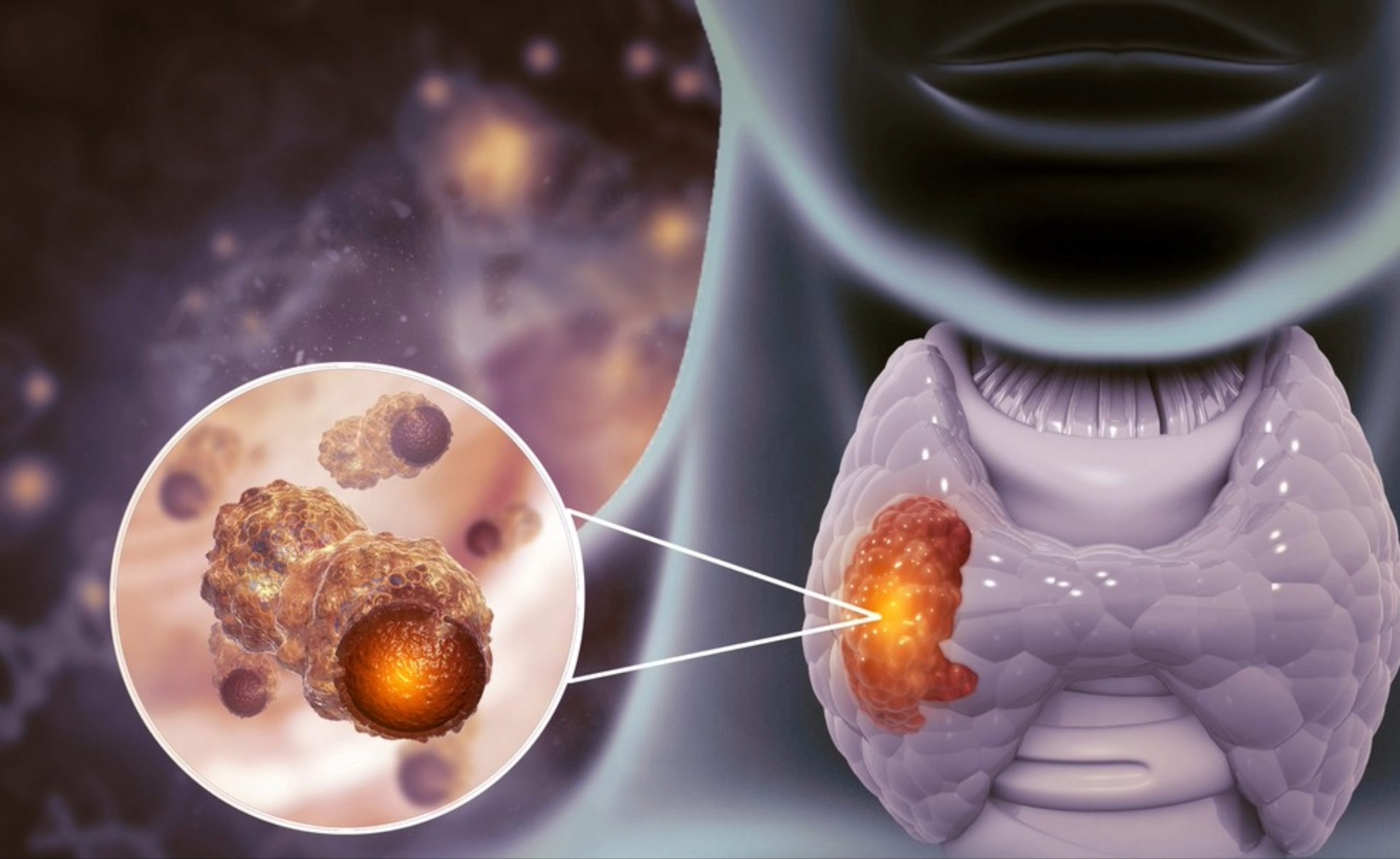
Papillary thyroid carcinoma and staging
Papillary thyroid carcinoma is the most common type of thyroid cancer and often has an excellent prognosis, especially when detected early and treated appropriately. Staging rules for differentiated cancers like papillary/follicular thyroid cancer follow AJCC (8th edition) criteria.
Thyroid cancer diagnosis: tests that confirm stage and type
A thorough thyroid cancer diagnosis usually includes:
- Clinical exam + ultrasound
Ultrasound evaluates thyroid nodules and cervical lymph nodes and guides next steps.
- Fine-needle aspiration biopsy (FNA)
This is the key test to confirm cancer cells in a suspicious nodule.
- Imaging for staging (when indicated)
CT/MRI, PET/CT, or whole-body scans may be used depending on risk and suspected spread.
- Molecular testing (in selected cases)
Gene testing can guide targeted therapy choices for advanced thyroid cancer treatment, especially in metastatic or recurrent disease. Updated professional guidance increasingly emphasizes the role of molecular/genetic testing in the patient journey.
Does thyroid cancer show up in blood tests?
Blood tests rarely “diagnose” thyroid cancer by themselves. They are used to:
- Check thyroid function (TSH, T3/T4) before/after treatment
- Track tumor markers in specific cancers (for example, thyroglobulin for many differentiated thyroid cancers after thyroid removal, and calcitonin for medullary thyroid cancer)
So, does thyroid cancer show up in blood tests? Not reliably as a first diagnostic tool—biopsy and imaging are usually what confirm it.
Thyroid cancer treatment by stage: what patients can expect
Treatment is individualized, based on stage, pathology, risk of recurrence, and patient factors.
1) Early-stage thyroid cancer (often Stage I–II)
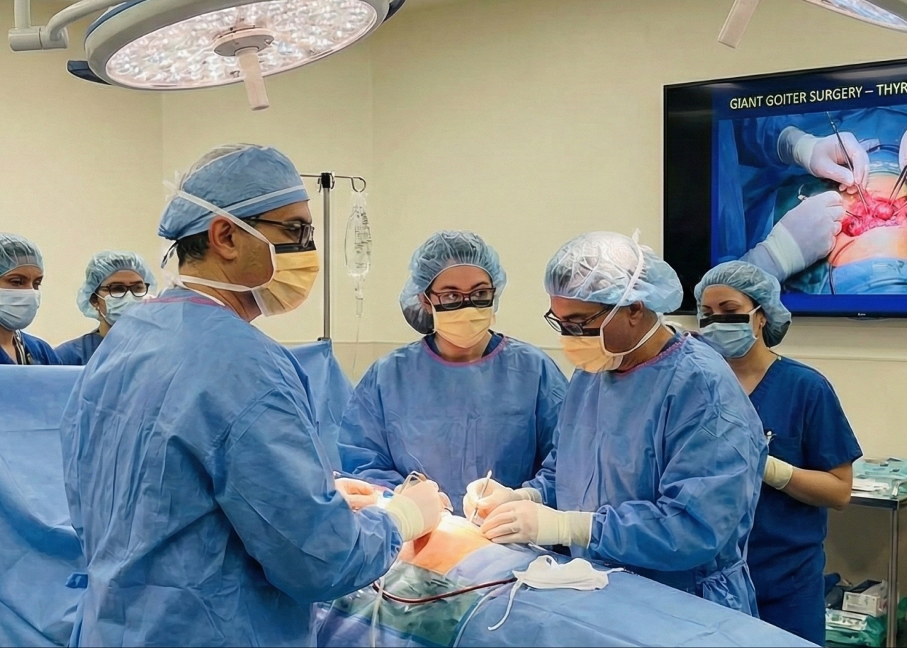
Typical approach
- Surgery: lobectomy or total thyroidectomy depending on tumor size/risk
- Radioactive iodine (RAI) may be recommended for higher risk differentiated cancers, but is not used for all cases.
- TSH suppression therapy (thyroid hormone replacement) to reduce stimulation of cancer cells
2) Locally advanced disease (higher risk or regional spread)
Treatment may include
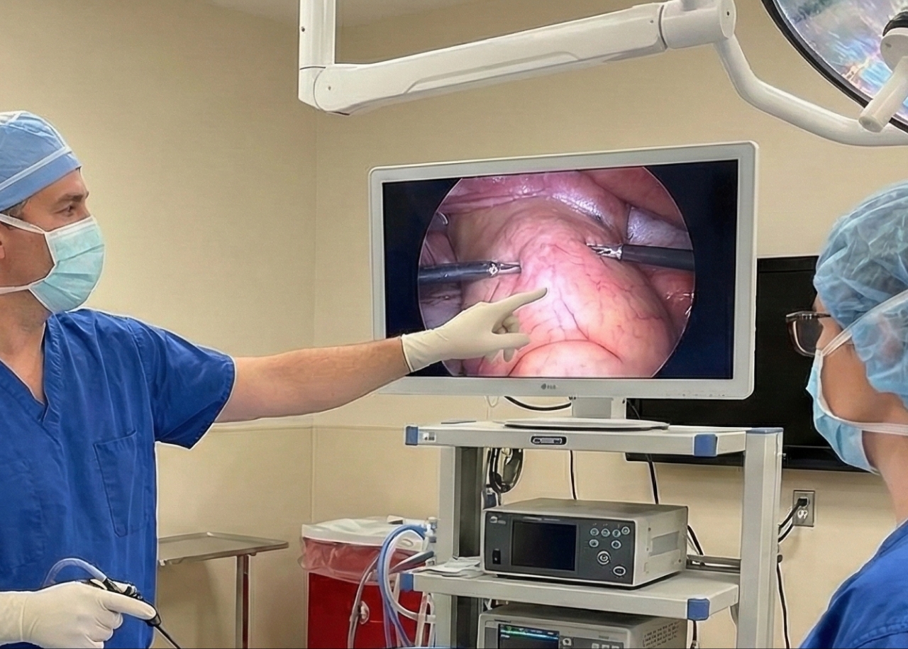
- More extensive surgery (including lymph node dissection if nodes are involved)
- RAI for appropriate differentiated cancers
- External beam radiotherapy in selected scenarios (for residual disease or unresectable local invasion)
3) Advanced thyroid cancer (metastatic or RAI-refractory)
When thyroid cancer spreads or stops responding to RAI, doctors consider systemic therapy and targeted treatments. NCCN guidance and peer-reviewed updates describe a broadened systemic therapy landscape for advanced disease.
Advanced thyroid cancer treatment options can include:
- Multi-kinase inhibitors (MKIs) such as lenvatinib or sorafenib for progressive, RAI-refractory differentiated thyroid cancer.
- Targeted therapies based on mutations/fusions, for example:
- RET inhibitors (e.g., selpercatinib, pralsetinib) for RET-altered thyroid cancers
- NTRK inhibitors (e.g., larotrectinib, entrectinib) for NTRK fusion–positive cancers (when present)
- Redifferentiation strategies (in select patients) to potentially restore iodine uptake before RAI—an emerging approach discussed in recent treatment reviews.
- Immunotherapy in specific settings (often based on tumor features and prior treatments), typically under specialist guidance and sometimes as part of clinical trials.
Key point: “Advanced thyroid cancer” does not mean there are no options—today, molecular testing can unlock therapies tailored to the tumor’s biology.
Latest treatments and what’s changed recently
In the last few years, thyroid cancer care has become more precise due to:
- Routine consideration of molecular profiling in advanced/recurrent cases to match patients to targeted therapy
- Stronger integration of RET-targeted drugs and other mutation-driven therapies in treatment pathways
- Expanded systemic options for radioiodine-refractory disease, improving disease control for some patients
FAQs
1) Is papillary thyroid cancer deadly?
Most papillary thyroid carcinoma cases are highly treatable, and many patients do very well long-term—especially when diagnosed early. Risk depends on stage, tumor behavior, spread, and response to treatment.
2) What are late stage thyroid cancer symptoms?
They can include more significant neck swelling, persistent swallowing/breathing difficulty, and symptoms related to spread (like bone pain or respiratory symptoms). Always get persistent or worsening symptoms evaluated.
3) Does thyroid cancer show up in blood tests?
Blood tests can support evaluation and monitoring, but they don’t reliably detect thyroid cancer on their own. Diagnosis usually relies on ultrasound + biopsy, with blood markers used mainly for follow-up in specific thyroid cancer types.
4) What is the most common thyroid cancer treatment?
For many patients, treatment starts with surgery, sometimes followed by radioactive iodine and thyroid hormone therapy depending on risk.
5) What does “advanced thyroid cancer” mean?
It commonly refers to cancer that has spread, returned repeatedly, invaded vital structures, or become RAI-refractory, where systemic therapy (including targeted treatments) may be needed.
Worried About a Thyroid Lump or Symptoms?
If you notice a neck lump, persistent hoarseness, swallowing difficulty, or you’ve been told you have a suspicious thyroid nodule, early assessment matters. At Burjeel Royal Hospital, Al Ain, our endocrinologists and thyroid specialists can guide you through thyroid cancer diagnosis, staging, and a personalized care plan—ranging from surgery and endocrine follow-up to advanced thyroid cancer treatment using modern targeted approaches when appropriate.
Book an appointment with us now!