UAE’s First Totally Robotic Three-Stage Esophagectomy with Pharyngo-Gastric Reconstruction: A Surgical Milestone

A Groundbreaking Surgical Procedure in the UAE

Burjeel Medical City (BMC) has successfully performed the UAE’s first totally robotic three-stage esophagectomy with pharyngo-gastric reconstruction, marking a significant milestone in the country’s advanced surgical capabilities. This innovative procedure, conducted by Dr. Ali Ayoob, HOD & Consultant Gastrointestinal Surgery at Burjeel Medical City, demonstrates the hospital’s commitment to providing cutting-edge surgical solutions for complex cases.

The Patient’s Journey

The case involved a 34-year-old woman who had undergone total radical thyroidectomy and lymph node dissection for locally advanced papillary carcinoma thyroid at another hospital. During the initial surgery, she experienced an intraoperative esophageal injury on the right lateral wall at the level of the pharynx, which was immediately addressed with primary suturing.

However, complications arose when the patient developed leakage of oral fluids through the drain site on the fourth day after surgery. Despite conservative management with nasogastric tube (NGT) aspiration, nothing by mouth (NPO) protocol, and antibiotic therapy, the esophageal leak persisted. The patient also developed mediastinitis and pleural effusion, further complicating her condition.

With evidence of a significant and persistent esophageal leak confirmed by contrast CT of the neck, she was transferred to Burjeel Medical City for specialized surgical care. Additionally, the patient was dealing with right vocal cord palsy and hypocalcemia as a result of her previous surgery.

Initial Surgical Intervention

Upon arrival at BMC, the patient underwent neck exploration surgery on October 28, 2023. The procedure included:

  • Upper GI endoscopy
  • Laparoscopy-assisted retrograde endoscopy
  • Neck wound exploration
  • Closure of the pharyngeal defect
  • Closure of the distal cut margin of the esophagus
  • Laparoscopic feeding jejunostomy

Following this intervention, the patient was discharged in stable condition with a plan for definitive surgery to reconstruct her swallowing pathway. During this recovery period, she received treatment with calcium supplements and Eltroxin (levothyroxine).

A subsequent CT scan of the neck and laryngoscopy revealed a completely stenotic esophageal inlet, requiring a high pharyngeal anastomosis—a particularly challenging surgical scenario.

The Groundbreaking Robotic Procedure

After thorough discussion in a multidisciplinary team meeting, the decision was made to proceed with definitive surgery. On January 3, 2024, the patient underwent the landmark procedure, which included:

  1. Robotic mobilization of the intrathoracic esophagus
  2. Robotic mobilization of the gastric tube in the abdomen
  3. Formation of a gastric conduit, which was transported to the neck through the posterior mediastinum
  4. Neck exploration from the right side with resection of the previously stenosed esophagus
  5. Exposure of the pharyngeal wall on the right posterolateral aspect (right pyriform fossa)
  6. Creation of a pharyngo-gastric end-to-side handsewn anastomosis
  7. Placement of a nasogastric tube through the anastomosis to the stomach

The Surgical Challenge

The procedure represented a significant technical challenge for several reasons:

“We have been performing esophageal reconstruction in the neck for a long time, but reconstruction of the swallowing pathway high up in the neck onto the pharynx is a real challenging situation,” explains Dr. Ali Ayoob. “High reconstruction is usually done with removal of the larynx, but here surgery is done sparing the larynx.”

What makes this case particularly noteworthy is that while the surgical team had extensive experience with video-assisted thoracoscopic (VAT) esophagectomy, this marked the first time that the da Vinci Robot was used for esophagectomy via the thorax and gastric conduit formation in the UAE.

Benefits of Robotic Surgery for Complex Procedures

The use of robotic technology in such a complex procedure offers several advantages:

  1. Enhanced precision: The robotic system provides surgeons with greater dexterity and precision, particularly valuable when operating in anatomically challenging areas like the neck and thorax.
  2. Improved visualization: The high-definition, 3D visualization allows for better identification of critical structures during the procedure.
  3. Minimally invasive approach: Despite the complexity of the surgery, the robotic approach typically results in smaller incisions, potentially leading to faster recovery.
  4. Better outcomes for challenging reconstructions: The robotic system’s capabilities are particularly beneficial for complex reconstructions such as the pharyngo-gastric anastomosis performed in this case.

A Testament to Advanced Surgical Capabilities

This groundbreaking procedure demonstrates the advanced level of surgical expertise and technological capabilities available at Burjeel Medical City. It also highlights the importance of multidisciplinary collaboration in managing complex cases, as evidenced by the comprehensive care provided to this patient from initial evaluation through to definitive treatment.

The successful performance of the UAE’s first totally robotic three-stage esophagectomy with pharyngo-gastric reconstruction represents not just a milestone for the hospital, but for the entire healthcare landscape of the UAE.

Experts

Advanced Oral Cavity Cancer: Complex Surgical Management with Microvascular Free Fibula Flap Reconstruction

Introduction

Oral cavity cancer represents a significant health challenge worldwide, with over 350,000 new cases diagnosed annually. Management of advanced cases—particularly those involving the mandible (lower jaw) and adjacent soft tissues—presents formidable surgical challenges that require sophisticated techniques to both eradicate the cancer and restore form and function. Achieving these dual goals demands a delicate balance between oncological radicality and functional-aesthetic reconstruction.

This case study examines the successful surgical management of a locally advanced oral cavity cancer at Burjeel Specialty Hospital in Sharjah, highlighting the complex decision-making process and advanced surgical techniques that enabled both effective cancer treatment and optimal functional reconstruction. The case demonstrates the importance of multidisciplinary collaboration and the application of state-of-the-art microvascular surgical techniques in achieving favorable outcomes for patients with advanced head and neck malignancies.

Understanding Oral Cavity Cancer

Epidemiology and Risk Factors

Oral cavity cancer constitutes a significant proportion of head and neck malignancies globally, with particularly high incidence rates in regions where tobacco chewing, smoking, and alcohol consumption are prevalent. Key risk factors include:

  • Tobacco use (smoking and smokeless forms)
  • Alcohol consumption
  • Chronic irritation from ill-fitting dentures or sharp teeth
  • Human papillomavirus (HPV) infection
  • Poor oral hygiene
  • Genetic predisposition

In the UAE and surrounding regions, the practice of tobacco chewing—often mixed with betel nut, lime, and other substances—remains a significant risk factor for oral cancer, particularly among expatriate populations from South and Southeast Asia.

Pathology and Progression

The vast majority (over 90%) of oral cavity cancers are squamous cell carcinomas (SCCs) that arise from the mucosal lining. These malignancies typically progress through several stages:

  1. Early mucosal changes: Often appearing as leukoplakia (white patches) or erythroplakia (red patches)
  2. Carcinoma in situ: Malignant cells confined to the epithelium
  3. Invasive carcinoma: Penetration beyond the basement membrane
  4. Local advancement: Invasion into adjacent structures (muscle, bone, skin)
  5. Regional spread: Metastasis to regional lymph nodes
  6. Distant metastasis: Spread to distant organs (less common in oral SCC)

When oral SCCs involve the lower alveolus (tooth-bearing portion of the mandible), they frequently invade the underlying bone, necessitating segmental mandibulectomy as part of the surgical treatment.

Case Presentation

Patient Profile

Mr. Sarwar Ali Khan, a 57-year-old male with a significant history of tobacco chewing, presented to Dr. Prasanta Kumar Dash, Medical Oncologist at Burjeel Specialty Hospital, with a chief complaint of a non-healing ulcer on the left side of his cheek that had been present for approximately 12 months. The patient worked as a driver for a government organization in Dubai.
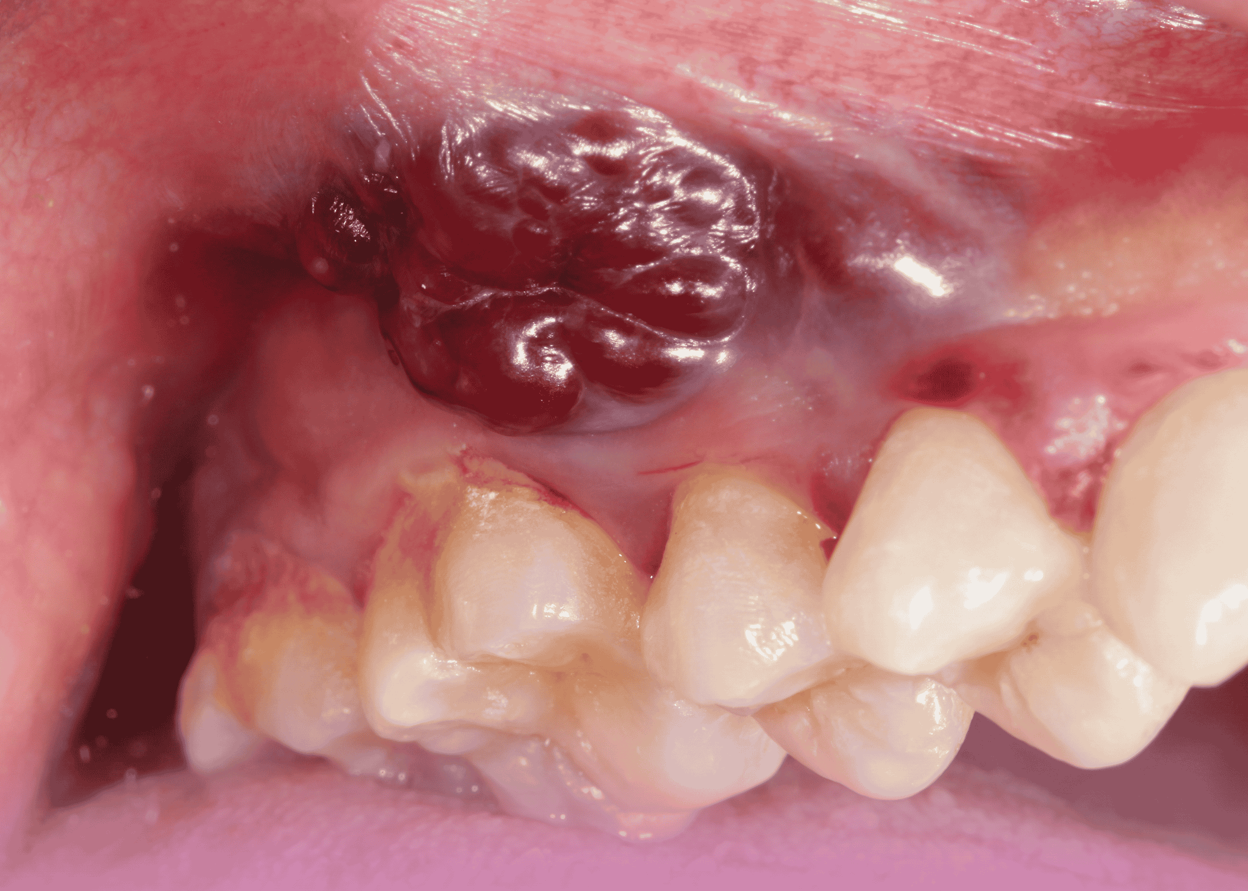
Clinical Findings

Physical examination revealed:

  • A locally advanced cancerous lesion invading the lower jaw bone and involving the overlying skin
  • The lesion measured approximately 3×3 cm
  • A discharging sinus was present on the left side of the cheek
  • No clinically palpable significant lymphadenopathy

Diagnostic Workup

The following investigations were performed:

  1. Biopsy: Revealed moderately differentiated squamous cell carcinoma
  2. Contrast-Enhanced CT (CECT) scan of the neck: Confirmed the locally advanced nature of the malignancy
  3. PET-CT scan: Corroborated the findings of the CECT and helped rule out distant metastasis
  4. Previous imaging review: A CT scan performed in India three months earlier (September) showed a significantly smaller lesion, indicating rapid disease progression

Based on these findings, the patient was diagnosed with locally advanced left-sided oral cavity cancer (cT4a N1 M0) with invasion of the mandible and overlying skin.

Multidisciplinary Approach

Tumor Board Discussion

The case was presented and discussed in a multidisciplinary tumor board involving physicians and oncologists from across the Burjeel network. After thorough deliberation of the diagnostic findings and consideration of various treatment options, the board unanimously recommended proceeding with surgical resection as the primary treatment modality.

This decision was based on several factors:

  • Locally advanced disease amenable to surgical resection
  • Absence of distant metastasis
  • The aggressive progression of the disease over a short period
  • The involvement of critical structures (mandible and skin) necessitating en bloc resection

Treatment Planning

Based on the tumor board recommendation, a comprehensive surgical plan was developed that included:

  1. Composite resection: Involving a segment of the left mandible with wide mucosal and skin margins
  2. Modified radical neck dissection: On the left side to address regional lymphatics
  3. Microvascular free fibula flap: For bone and soft tissue reconstruction
  4. Consideration for adjuvant therapy: Based on final histopathological findings

This complex surgical plan required careful coordination between multiple surgical specialists, including head and neck surgeons, plastic surgeons, and anesthesiologists.

Surgical Management

Procedure Details

The surgery was performed on January 6, 2024, at Burjeel Specialty Hospital in Sharjah by a multidisciplinary team led by Dr. Mohammed Basheeruddin Inamdar (Surgical Oncologist) and Dr. Rajkumar CBD (Plastic Surgeon), with support from Dr. Satish V. (ENT Surgeon) and Dr. Mohammed Eid Ali (Anesthesiologist).

The procedure consisted of several key components:

1. Tumor Resection

A complex composite resection was performed, including:

  • Segmental mandibulectomy from the left first premolar to the retromolar trigone
  • Wide excision of oral mucosa with appropriate margins
  • Excision of involved overlying skin
  • Modified radical neck dissection on the left side

2. Microvascular Free Flap Harvest and Preparation

Simultaneously, a fibula free flap was harvested from the patient’s leg:

  • The fibula bone was contoured to match the resected mandibular segment
  • A skin paddle was included with the flap to replace the resected oral mucosa and external skin
  • The peroneal vessels (artery and vena comitantes) were meticulously dissected for microvascular anastomosis

3. Microvascular Reconstruction

The harvested free flap was positioned and secured in the defect:

  • The fibula bone was shaped and fixated to recreate the mandibular contour
  • Microvascular anastomosis was performed between:
    • The facial artery and peroneal artery
    • Two vena comitantes and branches of the jugular veins
  • These anastomoses were completed using an operating microscope with precise suturing techniques
  • The soft tissue components of the flap were inset to replace the resected oral mucosa and skin

Despite the complexity of the procedure, the operation was completed successfully with a duration of approximately 11 hours and minimal blood loss (300ml). No blood transfusion was required.

Technical Challenges and Considerations

The surgical management of this case involved several significant challenges:

1. Three-Dimensional Planning

The resection and reconstruction required meticulous three-dimensional planning to ensure:

  • Complete tumor removal with clear margins in all dimensions (bone, mucosa, and skin)
  • Precise shaping of the fibula to match the native mandibular contour
  • Appropriate positioning of the skin paddle to replace both intraoral and external defects

2. Microvascular Expertise

The success of free flap reconstruction depends critically on the technical expertise of the microsurgical team:

  • Vessel selection and preparation for anastomosis
  • Precise microvascular suturing under high magnification
  • Ensuring adequate blood flow to the transferred tissue
  • Careful positioning to prevent vessel kinking or compression

3. Aesthetic and Functional Considerations

Beyond oncological clearance, the surgical team had to address:

  • Mandibular continuity for chewing function
  • Oral competence for speech and swallowing
  • External facial appearance
  • Minimal donor site morbidity

Outcomes and Follow-up

Immediate Postoperative Course

The patient’s immediate postoperative course was uneventful, with:

  • Stable vital signs
  • Good flap perfusion
  • Appropriate pain control
  • No evidence of complications

Histopathological Findings

The final histopathology report confirmed:

  • Moderately differentiated squamous cell carcinoma
  • Clear surgical margins
  • Lymph node status consistent with preoperative staging

Long-term Prognosis

The complete resection with clear margins provides the patient with the best chance for long-term survival and disease control. Following this type of comprehensive surgical management, patients typically undergo:

  • Regular clinical follow-up to monitor for recurrence
  • Adjuvant therapy if indicated by pathological findings
  • Rehabilitation for speech and swallowing as needed
  • Dental rehabilitation in preparation for potential implants

Discussion

Significance of the Case

This case exemplifies several important aspects of contemporary head and neck cancer management:

1. Integrated Multidisciplinary Care

The successful outcome resulted from seamless collaboration between:

  • Medical oncology for initial assessment and staging
  • Surgical oncology for tumor resection
  • Plastic surgery for complex reconstruction
  • Anesthesiology for perioperative management
  • Pathology for diagnostic confirmation and margin assessment

This integrated approach ensures comprehensive patient care from diagnosis through treatment and rehabilitation.

2. Advanced Reconstructive Techniques

The use of microvascular free tissue transfer represents the gold standard for complex head and neck reconstruction:

  • Fibula free flaps provide both bone for mandibular reconstruction and soft tissue for mucosal/skin replacement
  • Vascularized bone has superior healing properties compared to non-vascularized grafts
  • The robust blood supply allows for better healing in previously irradiated fields
  • Long-term outcomes show better functional and aesthetic results compared to other reconstructive methods

3. Evolving Standards of Care

This case reflects current best practices in advanced oral cancer management:

  • Primary surgical resection for operable disease
  • Immediate reconstruction to restore form and function
  • Consideration of quality of life alongside oncological outcomes
  • Risk-adapted adjuvant therapy based on pathological findings

Broader Clinical Implications

This case has several important implications for the broader field of head and neck oncology:

1. Importance of Early Detection

The rapid progression of this patient’s disease over just three months underscores the critical importance of early detection and intervention in oral cavity cancers. Public health initiatives focused on:

  • Awareness of oral cancer signs and symptoms
  • Regular dental examinations with oral cancer screening
  • Education about risk factors, particularly tobacco cessation
  • Prompt biopsy of suspicious oral lesions

Could significantly improve outcomes by facilitating diagnosis at earlier, more treatable stages.

2. Accessibility of Advanced Care

The successful execution of this complex procedure at Burjeel Specialty Hospital in Sharjah demonstrates the growing accessibility of advanced surgical techniques in the region. This has important implications for:

  • Patient retention within regional healthcare systems
  • Reduction in medical tourism for complex care
  • Development of local surgical expertise and training opportunities
  • Overall improvement in regional cancer care standards

3. Technical Advancement in Reconstructive Surgery

The integration of microvascular techniques into head and neck reconstruction represents a significant technical advancement that has revolutionized outcomes for patients with advanced disease:

  • Before free flap techniques, reconstruction options were limited and often yielded suboptimal functional and aesthetic results
  • Microvascular techniques have expanded the scope of resectable disease by providing reliable reconstruction for even the most complex defects
  • Continued refinement of these techniques promises even better outcomes in the future

Future Directions

Technological Advancements

Several emerging technologies may further enhance the management of complex oral cavity cancers:

1. Virtual Surgical Planning

Computer-aided design and manufacturing (CAD/CAM) technology allows for:

  • Precise preoperative planning of resection and reconstruction
  • Custom cutting guides for both mandible resection and fibula osteotomies
  • Patient-specific reconstruction plates that perfectly match the planned reconstruction
  • Reduced operating time and improved accuracy

2. Intraoperative Navigation

Similar to GPS systems in vehicles, surgical navigation enables:

  • Real-time tracking of instruments relative to the patient’s anatomy
  • Enhanced precision in tumor resection
  • Verification of reconstruction accuracy
  • Especially valuable in complex three-dimensional reconstructions

3. Advances in Flap Monitoring

Newer technologies for postoperative flap monitoring include:

  • Implantable Doppler probes for continuous blood flow assessment
  • Near-infrared fluorescence angiography for real-time perfusion evaluation
  • Smartphone-based photoplethysmography for nurse-driven monitoring
  • These technologies can enable earlier detection of vascular compromise and improve flap salvage rates

Treatment Paradigms

Evolving treatment paradigms in oral cancer management include:

1. De-intensification Strategies

For appropriately selected patients:

  • Reduction in the extent of surgery when possible
  • Selective neck dissection approaches
  • Risk-stratified adjuvant therapy
  • These approaches aim to maintain oncological outcomes while reducing treatment-related morbidity

2. Immunotherapy Integration

The role of immunotherapy in head and neck cancer continues to evolve:

  • Currently established in recurrent/metastatic disease
  • Emerging data for neoadjuvant and adjuvant applications
  • Potential for combination with conventional treatments
  • May ultimately change the treatment landscape for advanced oral cancers

Conclusion

This case of locally advanced oral cavity cancer successfully managed at Burjeel Specialty Hospital in Sharjah exemplifies the power of multidisciplinary collaboration and advanced surgical techniques in addressing complex oncological challenges. The combination of radical tumor resection and sophisticated microvascular reconstruction enabled both cancer control and functional rehabilitation for a patient with aggressive disease.

The case highlights the importance of timely intervention, comprehensive treatment planning, and technical expertise in achieving optimal outcomes. It also demonstrates the growing accessibility of world-class cancer care within the UAE, reducing the need for patients to travel abroad for complex procedures.

For healthcare providers, this case underscores the value of specialized training in advanced surgical techniques and the benefits of multidisciplinary tumor boards in treatment planning. For patients with advanced oral cancers, it offers hope that even extensive disease can be effectively treated with approaches that prioritize both oncological clearance and functional reconstruction.

As technologies and treatment paradigms continue to evolve, the management of complex head and neck cancers will likely become even more refined, potentially leading to improved survival rates and quality of life for affected patients.

Post-Traumatic Diaphragmatic Hernia: Surgical Management of a Complex Case with Thoracoabdominal Approach

Introduction

Diaphragmatic hernias represent a significant surgical challenge, particularly when they are large, contain multiple organs, and result from previous trauma. These uncommon conditions occur when abdominal contents herniate into the thoracic cavity through a defect in the diaphragm, potentially compromising both respiratory and digestive functions. While congenital forms like Bochdalek and Morgagni hernias are well-recognized, traumatic diaphragmatic hernias (TDH) are less common and may present with delayed symptoms years after the initial injury.

This case study explores the successful surgical management of a complex post-traumatic diaphragmatic hernia at Burjeel Hospital in Sharjah, highlighting the multidisciplinary approach required to address this challenging condition. The case is particularly notable for the large size of the defect, the extensive organ herniation, and the rare associated finding of absent pericardium.

Understanding Traumatic Diaphragmatic Hernias

Etiology and Pathophysiology

Traumatic diaphragmatic injuries occur in approximately 0.8-8% of patients with blunt thoracoabdominal trauma, most commonly from motor vehicle accidents. The injury typically results from sudden increases in intra-abdominal pressure during trauma, causing the diaphragm to rupture at its weakest points. Left-sided ruptures are more common (approximately 75% of cases) due to the protective effect of the liver on the right hemidiaphragm.

The natural history of undiagnosed diaphragmatic injuries often follows three distinct phases:

  1. Acute phase: Immediately following injury, marked by respiratory distress and other trauma-related symptoms
  2. Latent phase: A period of minimal or absent symptoms that may last for months to years
  3. Obstructive or strangulation phase: When herniated abdominal contents become incarcerated or strangulated, leading to acute symptoms

The progressive nature of these hernias results from the pressure gradient between the abdominal and thoracic cavities, which gradually enlarges the defect and promotes herniation of abdominal contents into the thorax.

Clinical Implications

Untreated diaphragmatic hernias can lead to serious complications:

  • Respiratory compromise: Due to compression of lung tissue and mediastinal shift
  • Gastrointestinal obstruction: When herniated bowel becomes obstructed
  • Strangulation: Compromised blood supply to herniated organs
  • Pleural effusion: Accumulation of fluid in the pleural space
  • Cardiac dysfunction: From compression or displacement of the heart

Early diagnosis and surgical repair are essential to prevent these potentially life-threatening complications.

Case Presentation

Patient Profile

A 29-year-old male presented to Burjeel Hospital in Sharjah with complaints of abdominal pain and difficulty breathing that significantly affected his daily activities. These symptoms had persisted for approximately two years without improvement, despite various treatments. Notably, the patient reported a history of blunt trauma to the chest and abdomen from a motor vehicle accident, which had occurred prior to the onset of his symptoms.

Clinical Presentation

The patient described progressive shortness of breath, particularly with exertion, and intermittent abdominal discomfort. Physical examination revealed decreased breath sounds over the left hemithorax and abdominal tenderness, raising suspicion for a thoracoabdominal pathology.

Diagnostic Workup

Initial investigations included:

  1. Chest X-ray: Revealed elevation of the left hemidiaphragm with abnormal gas shadows in the left thoracic cavity
  2. CT scan of the abdomen and chest: Demonstrated a large defect in the left hemidiaphragm with herniation of multiple abdominal organs into the left thoracic cavity, including:
    • Spleen
    • Stomach
    • Large portions of the colon
  3. Additional findings: The CT scan also showed significant atelectasis (collapse) of the left lung due to compression by the herniated abdominal contents

Based on these findings and the patient’s history of trauma, a diagnosis of post-traumatic diaphragmatic hernia was established.

Surgical Management

Preoperative Planning

After thorough discussion of the case with the patient and his family, the decision was made to proceed with surgical repair. Given the complexity of the case, a multidisciplinary surgical team was assembled, including:

  • General surgeons
  • Thoracic surgeons
  • Anesthesiologists

The surgical approach was carefully planned, taking into consideration the chronicity of the hernia, the size of the defect, and the multiple organs involved.

Surgical Approach

The surgical team, led by Dr. Mohamed Eraki (Consultant of General and Laparoscopic Surgery), Dr. Imtiaz Ahmed and Dr. Taj Mohamed (Consultant Thorax Surgeons), opted for a combined thoracoabdominal approach to ensure optimal exposure and management of both abdominal and thoracic components of the hernia.

The procedure consisted of:

  1. Initial abdominal laparotomy: To access the abdominal cavity and assess the condition of the herniated organs
  2. Left lateral thoracotomy: To provide direct access to the diaphragmatic defect and thoracic cavity
  3. Comprehensive exploration: Of both cavities to identify all anatomical abnormalities

Intraoperative Findings

The surgery revealed several significant findings:

  1. Large diaphragmatic defect: Confirming the preoperative imaging
  2. Extensive herniation: The spleen, stomach, and large portions of the colon were found within the left thoracic cavity
  3. Redundant diaphragm: The remaining diaphragmatic tissue was abnormally loose and stretched
  4. Absent pericardium: A rare associated congenital anomaly was discovered, with absence of the pericardial sac
  5. Compressed left lung: The lung was significantly atelectatic but otherwise viable

Surgical Technique

The surgical team performed several key steps:

  1. Organ reduction: Carefully reducing the herniated abdominal organs (spleen, stomach, and colon) back into the abdominal cavity
  2. Assessment of viability: All herniated organs were thoroughly examined and found to be viable with no signs of ischemia or strangulation
  3. Diaphragmatic repair: Multiple rows of plication (folding and suturing) of the redundant diaphragm were performed from both the thoracic and abdominal approaches
  4. Chest drainage: Placement of thoracic drains to evacuate air and fluid
  5. Layered closure: Of both the thoracotomy and laparotomy incisions

Postoperative Care

Following the procedure, the patient was transferred to the Intensive Care Unit for close monitoring. Key aspects of postoperative management included:

  • Respiratory support and monitoring
  • Pain management
  • Early mobilization
  • Gradual resumption of oral intake

The patient’s recovery was uneventful, with progressive improvement in respiratory function and resolution of abdominal symptoms. After an appropriate period of monitoring, the patient was discharged from the hospital in good health.

Discussion

Surgical Considerations

This case highlights several important surgical considerations in the management of complex diaphragmatic hernias:

1. Approach Selection

The choice of surgical approach is critical in complex diaphragmatic hernias:

  • Transthoracic approach: Provides excellent exposure of the diaphragm and thoracic cavity, allowing direct visualization of the hernia and any adhesions to thoracic structures
  • Transabdominal approach: Offers better access to abdominal organs and facilitates their reduction and assessment
  • Combined approach: As used in this case, provides comprehensive access to both cavities in complex cases

For large, chronic hernias with multiple herniated organs, the combined approach offers significant advantages, despite increased surgical trauma.

2. Repair Technique

Several options exist for diaphragmatic reconstruction:

  • Primary repair: Direct suturing of the defect, possible when there is sufficient healthy tissue
  • Plication: Folding and suturing of redundant diaphragm to restore tension and function
  • Mesh reinforcement: Use of prosthetic or biologic materials for large defects with insufficient tissue
  • Muscle flap reconstruction: In cases of extensive tissue loss

In this case, the redundant nature of the diaphragm allowed for effective plication without the need for prosthetic materials, which minimizes the risk of infection and erosion into adjacent structures.

3. Management of Associated Anomalies

The finding of an absent pericardium in this case represents a rare association that required no specific intervention but demonstrates the importance of thorough intraoperative exploration and readiness to address unexpected findings.

Clinical Relevance

This case underscores several clinically relevant points:

1. Delayed Presentation

The two-year interval between the traumatic event and definitive diagnosis highlights the often indolent course of traumatic diaphragmatic injuries. Healthcare providers should maintain a high index of suspicion in patients with a history of thoracoabdominal trauma who present with respiratory or gastrointestinal symptoms, even years after the initial injury.

2. Diagnostic Challenges

Diaphragmatic hernias can be challenging to diagnose, particularly when symptoms are nonspecific. CT imaging has emerged as the gold standard for diagnosis, providing detailed information about the defect size, location, and herniated contents.

3. Multidisciplinary Approach

The successful management of this complex case required collaboration between general surgeons, thoracic surgeons, and anesthesiologists. This multidisciplinary approach ensured comprehensive assessment and treatment of both thoracic and abdominal components of the hernia.

4. Long-term Outcomes

Complete resolution of the patient’s symptoms following surgical repair confirms that even long-standing diaphragmatic hernias can be successfully treated with appropriate surgical intervention. Early diagnosis and repair remain preferable to prevent complications, but even delayed repair can provide excellent outcomes.

Conclusion

This case illustrates the successful surgical management of a complex post-traumatic diaphragmatic hernia with multiple herniated organs and an associated congenital anomaly. Through a carefully planned combined thoracoabdominal approach, the surgical team at Burjeel Hospital in Sharjah was able to reduce the herniated contents, repair the diaphragmatic defect, and restore normal anatomy and function.

For clinicians, this case emphasizes the importance of maintaining awareness of diaphragmatic injuries in patients with a history of thoracoabdominal trauma, even when presentation is delayed. The case also highlights the value of advanced imaging in diagnosis and the benefits of a multidisciplinary surgical approach in complex cases.

For patients suffering from undiagnosed diaphragmatic hernias, this case offers hope that even long-standing, complex hernias can be successfully treated with appropriate surgical intervention, resulting in significant improvement in symptoms and quality of life.

Experts

Chemical Radiculopathy: A Novel Approach to Understanding and Treating Non-Compressive Sciatica

Introduction

Sciatica—characterized by radiating pain from the lower back down the leg along the sciatic nerve distribution—affects millions of people worldwide and is a common cause of disability and decreased quality of life. The traditional understanding of sciatica has centered primarily around structural causes, particularly herniated discs and spinal stenosis mechanically compressing nerve roots. However, emerging evidence suggests that in some cases, sciatic pain may occur in the absence of visible structural compression on imaging studies, pointing to alternative mechanisms that are biochemical rather than mechanical in nature.

This case study explores the successful diagnosis and treatment of chemical radiculopathy, a fascinating yet underrecognized cause of sciatica, highlighting how advanced interventional pain management techniques can provide significant relief even when conventional imaging suggests no obvious mechanical compression.

Understanding Chemical Radiculopathy

Beyond Mechanical Compression

The conventional wisdom in spine medicine has long held that sciatic pain results primarily from physical compression of nerve roots, typically by herniated disc material or bony overgrowth. However, research has increasingly revealed that inflammation and biochemical irritation of nerve roots can produce identical symptoms even without visible compression on imaging studies.

Chemical radiculopathy occurs when inflammatory mediators from degenerated discs or surrounding structures leak through annular tears (fissures in the disc’s outer fibrous ring) and irritate nearby nerve roots. This represents an early stage of disc degeneration that precedes the development of frank herniation visible on imaging.

The Inflammatory Cascade

Several biochemical substances have been implicated in this inflammatory process:

  1. Phospholipase A2: An enzyme found in high concentrations in herniated disc material
  2. Cytokines: Particularly tumor necrosis factor-alpha (TNF-α) and interleukins
  3. Matrix metalloproteinases: Enzymes involved in disc degeneration
  4. Prostaglandins: Inflammatory mediators that sensitize nerve roots to pain
  5. Substance P: A neuropeptide involved in pain transmission

These inflammatory mediators can diffuse through annular tears and reach nerve roots without any significant structural displacement of disc material, explaining why some patients exhibit classic radicular symptoms despite minimal findings on imaging studies.

Case Presentation

Patient Profile

The patient was a female athlete in her 40s who was a long-distance runner. She presented with severe lower extremity pain that was more pronounced than her axial (back) pain, suggesting a predominantly radicular component to her symptoms.

Clinical Examination

The physical examination revealed several significant findings:

  • Bilateral straight leg raise (SLR) was limited to 30 degrees due to pain
  • Inability to bear weight on toes and heels because of pain
  • No detectable neurological deficit (normal strength, sensation, and reflexes)
  • Clinical pattern strongly suggestive of L5 and S1 nerve root involvement

These findings were consistent with bilateral radiculopathy affecting the L5 and S1 nerve roots, despite the absence of neurological deficits.

Diagnostic Imaging

Magnetic Resonance Imaging (MRI) of the lumbar spine revealed:

  • Degenerated and desiccated L5-S1 disc (loss of normal hydration and height)
  • Importantly, axial views showed the absence of mechanical compression
  • Both exiting nerve roots appeared free from impingement

This created a clinical paradox: the patient had clear symptoms of nerve root irritation, but imaging showed no structural cause for these symptoms.

Interventional Management

Diagnostic-Therapeutic Procedure

Based on the clinical presentation and imaging findings, Dr. Shailendra Chauhan, Consultant in Anesthesia and Pain Management at Burjeel Day Surgery Center, performed a bilateral L5-S1 selective nerve root block under fluoroscopic guidance.

The procedure involved:

  1. Precise needle placement at the L5 and S1 nerve roots bilaterally
  2. Injection of non-ionic contrast to confirm appropriate spread and the absence of significant mechanical compression
  3. Administration of a therapeutic mixture containing lidocaine (a local anesthetic) and Depo-Medrol (a corticosteroid)

Immediate Outcome

The results were dramatic and immediate:

  • Complete resolution of pain immediately following the procedure
  • Bilateral straight leg raise improved from 30 degrees to 80 degrees
  • Restored ability to bear weight on toes and heels
  • Significant improvement in overall mobility

This dramatic response to selective nerve root block with anti-inflammatory medication strongly supported the diagnosis of chemical radiculopathy as the underlying cause of the patient’s sciatica.

Discussion

Diagnostic Challenges

Chemical radiculopathy presents several diagnostic challenges:

  1. Imaging-Clinical Mismatch: Patients exhibit classic radicular symptoms despite minimal or no compression on imaging.
  2. Overlooked Diagnosis: Many practitioners continue to focus exclusively on structural causes visible on imaging, potentially missing chemical causes.
  3. Early Intervention: Chemical radiculopathy may represent an earlier stage of disc pathology before structural herniation occurs, offering a window for preventive interventions.

Treatment Implications

Understanding chemical radiculopathy expands the treatment options available to patients with sciatica:

  1. Targeted Anti-inflammatory Therapy: Selective nerve root blocks deliver potent anti-inflammatory medications directly to the site of inflammation.
  2. Beyond Decompression: In cases where imaging shows no significant compression, pursuing surgical decompression may be unnecessary and unlikely to address the true cause of pain.
  3. Annular Repair Techniques: Emerging therapies aimed at sealing annular tears may help prevent leakage of inflammatory mediators.
  4. Systemic Anti-inflammatory Approaches: Oral medications, dietary modifications, and lifestyle interventions that reduce systemic inflammation may play a supportive role.

Future Management Plan

For this particular patient, a comprehensive follow-up plan was established:

  1. Recommendation to repeat the procedure after 3-4 months if pain recurs
  2. Consideration of discography (a diagnostic procedure to identify painful discs) in case of recurrence
  3. Possible annuloplasty (a procedure to repair annular tears) if indicated by discography

Clinical Implications

This case highlights several important clinical implications:

Expanded Diagnostic Framework

The recognition of chemical radiculopathy as a cause of sciatica encourages clinicians to move beyond the traditional structure-based paradigm when evaluating patients with radicular symptoms. A more comprehensive approach should include:

  1. Thorough clinical examination: Identifying patterns of pain and dysfunction that may suggest radicular involvement even in the absence of neurological deficits
  2. Critical interpretation of imaging: Recognizing that “normal” or “minimally abnormal” imaging does not rule out significant radicular pain
  3. Diagnostic injections: Utilizing selective nerve root blocks as diagnostic tools to identify pain generators and confirm chemical causes

Therapeutic Options

Understanding chemical radiculopathy expands the therapeutic options available to patients:

  1. Targeted injections: Delivering anti-inflammatory medications directly to the site of chemical irritation
  2. Pharmacological approaches: Medications specifically targeting neuroinflammation and neuropathic pain mechanisms
  3. Avoidance of unnecessary surgery: Preventing surgical interventions that may not address the underlying chemical causes
  4. Regenerative approaches: Emerging therapies aimed at improving disc health and repairing annular integrity

Evidence-Based Application

The case underscores the growing clinical evidence that anatomical abnormalities are not always required to cause radiculopathy, and that biochemical etiologies play a significant role in many cases. This paradigm shift is supported by:

  1. Basic science research demonstrating the presence of inflammatory mediators in disc material
  2. Clinical studies showing successful treatment of radicular pain with anti-inflammatory approaches
  3. Observations of patients with clear radicular symptoms despite minimal imaging findings

Conclusion

This case illustrates the importance of considering chemical radiculopathy in the differential diagnosis of patients with sciatic symptoms, particularly when imaging studies fail to demonstrate significant mechanical compression. The dramatic response to selective nerve root block with anti-inflammatory medication in this patient serves as a powerful reminder that pain mechanisms extend beyond simple mechanical models.

For clinicians, this case encourages a broader conceptual framework when evaluating patients with radicular pain, incorporating both structural and chemical etiologies. For patients suffering from sciatica without clear imaging findings, the recognition of chemical radiculopathy offers hope through targeted interventional approaches.

The successful management of this case at Burjeel Day Surgery Center demonstrates the value of a sophisticated, mechanism-based approach to pain diagnosis and treatment, ultimately leading to improved outcomes and quality of life for patients with complex pain conditions.

Experts

Ischemic Colitis with Multiple Perforations: Surgical Management of a Life-Threatening Condition

Introduction

Ischemic colitis represents a significant challenge in gastrointestinal surgery, particularly when complicated by multiple perforations. This condition occurs when blood flow to the colon is reduced to a level insufficient to maintain normal cellular function, leading to inflammation and potentially tissue death. While ischemic colitis is more commonly seen in elderly patients with atherosclerotic disease, its occurrence in younger individuals is rare and often associated with unusual underlying conditions.

This case study presents the successful emergency management of a 32-year-old male patient with severe ischemic colitis complicated by multiple colonic perforations at Burjeel Hospital in Sharjah. The case highlights the importance of prompt recognition, appropriate diagnostic workup, and decisive surgical intervention in managing this potentially fatal condition.

Understanding Ischemic Colitis

Pathophysiology

Ischemic colitis results from inadequate blood flow to the colon, which can occur through several mechanisms:

  1. Occlusive disease: Blockage of arteries or veins by thrombi, emboli, or external compression
  2. Non-occlusive ischemia: Low flow states due to systemic hypoperfusion, vasospasm, or shunting
  3. Venous thrombosis: Impaired venous drainage leading to congestion and secondary arterial insufficiency

The colon is particularly vulnerable to ischemia due to its relatively precarious blood supply, especially at the “watershed” areas where different vascular territories meet, such as the splenic flexure and rectosigmoid junction.

Clinical Presentation

The presentation of ischemic colitis varies widely depending on the severity and extent of ischemia:

  • Mild cases: May present with crampy abdominal pain, urgency, and bloody diarrhea
  • Moderate cases: More severe pain, peritoneal signs limited to the affected area, and systemic inflammatory response
  • Severe cases: Peritonitis, septic shock, and multi-organ failure due to transmural necrosis and perforation

Etiology

While atherosclerosis is the most common underlying cause in elderly patients, other potential etiologies include:

  • Cardiac arrhythmias and low output states
  • Vasculitis and hypercoagulable states
  • Medications (vasoconstrictors, estrogens, danazol)
  • Vascular interventions and surgery
  • Coagulopathies (inherited or acquired)
  • Mechanical obstruction of mesenteric vessels

Case Presentation

Patient Profile and Initial Presentation

A 32-year-old male patient presented to the Emergency Department at Burjeel Hospital, Sharjah, with complaints of recurrent attacks of abdominal pain accompanied by vomiting. The pain was generalized but with particular tenderness in the left side of the abdomen. This was an unusual presentation for a young adult male without significant past medical history.

Initial Diagnostic Workup

The patient was admitted for further evaluation. Magnetic Resonance Imaging (MRI) of the abdomen revealed diffuse multifocal areas of wall edema with mild wall thickening and surrounding fat stranding involving the descending colon. These areas showed mild enhancement on post-contrast images. The radiological features suggested colitis, and a colonoscopy was recommended for further evaluation.

Initial Management and Clinical Course

The patient was started on medical management, including intravenous fluids, bowel rest, and appropriate antibiotics. He showed clinical improvement and was discharged after three days of inpatient treatment.

However, the improvement was short-lived. Just five days after discharge, the patient returned to the Emergency Department with severe abdominal pain and generalized tenderness, suggesting a significant deterioration in his condition.

Advanced Imaging and Preoperative Diagnosis

A contrast-enhanced CT scan of the abdomen and pelvis was performed urgently. The scan revealed a clustered loop of small bowel (jejunal loops) with bowel loops located lateral and anterior to the ascending colon, associated with a pulled branch of mesenteric vessels. There was evidence of a possible defect in the mesentery. These findings suggested the possibility of a transmesenteric (internal) hernia.

Based on the patient’s clinical deterioration and the concerning CT findings, the decision was made to proceed with emergency surgery.

Surgical Management

Operative Findings

The patient underwent emergency laparotomy, which revealed critical findings not fully appreciated on preoperative imaging:

  1. Free fluid in the peritoneal cavity, indicative of peritonitis
  2. Multiple areas of necrosis and perforation affecting several segments of the colon:
    • Hepatic flexure
    • Transverse colon
    • Part of the descending colon

These findings represented a severe case of ischemic colitis with multiple perforations, a life-threatening condition requiring immediate intervention.

Surgical Procedure

Given the extent and severity of the colonic involvement, the surgical team led by Dr. Mohamed Eraki, Consultant General and Laparoscopic Surgeon and Head of the Surgery Department at Burjeel Hospital, Sharjah, performed the following procedure:

  1. Extended Colectomy: Resection of the affected bowel segments, including the ascending colon, transverse colon, and part of the descending colon
  2. Temporary Stoma Creation: A right-sided colostomy was created to divert the fecal stream
  3. Mucous Fistula: A left-sided mucous fistula was fashioned from the distal bowel segment
  4. Thorough Lavage: The abdominal cavity was thoroughly irrigated to reduce bacterial contamination
  5. Drainage: Two large drains were placed to monitor for potential postoperative complications

Postoperative Care

Following surgery, the patient was transferred to the Intensive Care Unit for close monitoring and management of potential septic complications. The postoperative course was successful, with gradual recovery of bowel function and overall clinical improvement. The patient was eventually discharged from the hospital in stable condition.

Diagnostic Investigation and Case Conclusion

Following recovery, a comprehensive workup was initiated to identify the underlying cause of this unusual case of ischemic colitis in a young adult. After detailed investigations, it was discovered that the patient had a congenital coagulopathy (a hereditary blood clotting disorder), which was determined to be the most likely etiology for his ischemic colitis.

This finding highlighted the importance of considering uncommon causes of ischemia in younger patients without traditional risk factors for vascular disease. Congenital coagulopathies can create a hypercoagulable state, predisposing to thrombosis in the mesenteric vessels and subsequent ischemic damage to the bowel.

Discussion

Clinical Significance

This case illustrates several important clinical points:

  1. Atypical Presentation: Ischemic colitis in young adults is rare and should prompt investigation for unusual underlying causes such as hypercoagulable states, vasculitis, or vascular anomalies.
  2. Diagnostic Challenges: The initial presentation and imaging findings were suggestive of colitis but did not immediately reveal the severity of the condition. The patient’s rapid deterioration after initial improvement highlights the potentially progressive nature of ischemic bowel disease.
  3. Surgical Decision-Making: The case demonstrates the critical importance of timely surgical intervention in cases of suspected bowel ischemia. Delaying surgery when peritoneal signs are present can significantly increase morbidity and mortality.
  4. Role of Extended Colectomy: In cases of extensive colonic ischemia with multiple perforations, extended colectomy with temporary diversion is often necessary to control sepsis and remove all compromised tissue.

Congenital Coagulopathy and Ischemic Colitis

The association between congenital coagulopathies and ischemic colitis merits special attention. Various hereditary thrombophilias can predispose to arterial and venous thrombosis in the mesenteric circulation:

  • Factor V Leiden mutation
  • Prothrombin gene mutation
  • Protein C or S deficiency
  • Antithrombin III deficiency
  • Hyperhomocysteinemia

In patients with these conditions, even minor reductions in mesenteric blood flow that might be well-tolerated in individuals with normal coagulation can trigger a cascade of thrombosis, ischemia, and tissue necrosis. This underscores the importance of considering hereditary thrombophilias in the differential diagnosis of ischemic colitis, particularly in younger patients without other risk factors.

Surgical Approach to Ischemic Colitis with Perforation

The surgical management of ischemic colitis with perforation follows several key principles:

  1. Control of Contamination: Removing the perforated bowel and lavaging the peritoneal cavity to reduce bacterial load and minimize septic complications.
  2. Complete Resection of Non-Viable Tissue: Ensuring all ischemic segments are removed, often requiring assessment of tissue viability through direct inspection, assessment of bleeding at cut edges, and sometimes intraoperative Doppler assessment.
  3. Damage Control Strategy: In critically ill patients, a staged approach may be necessary, with initial resection and diversion followed by later restoration of intestinal continuity once the patient is stabilized.
  4. Temporary Fecal Diversion: Creating a stoma to protect any anastomosis or to simplify the initial procedure when the patient is unstable. This is often essential in cases of fecal peritonitis to prevent further contamination.

In this case, the extended colectomy with end stoma and mucous fistula creation represents the application of these principles, prioritizing the patient’s survival in a life-threatening situation.

Postoperative Considerations

Short-term Management

Following surgery for ischemic colitis with multiple perforations, several aspects of care are critical:

  1. Hemodynamic Stabilization: Maintaining adequate perfusion to prevent further ischemic damage to other organs
  2. Broad-Spectrum Antibiotics: Continued administration to control polymicrobial intra-abdominal infection
  3. Nutritional Support: Early initiation of nutrition to support wound healing and immune function
  4. Thromboprophylaxis: Given the underlying hypercoagulable state, appropriate anticoagulation is essential once bleeding risk is controlled
  5. Stoma Management: Early involvement of stoma care specialists to optimize adaptation and prevent complications

Long-term Planning

Once recovered from the acute event, several considerations come into play:

  1. Investigation and Management of Underlying Coagulopathy: Detailed hematological evaluation and long-term management plan, potentially including anticoagulation therapy
  2. Restoration of Intestinal Continuity: Assessment for potential stoma reversal and colocolonic or ileocolonic anastomosis, typically considered after 3-6 months
  3. Surveillance: Regular follow-up to monitor for recurrent ischemia or other complications
  4. Quality of Life Considerations: Addressing the psychological and social impact of temporary or permanent stoma

Future Perspectives

Advancements in Diagnostics

Earlier diagnosis of ischemic colitis could potentially prevent the severe complications seen in this case. Several emerging modalities may help in this regard:

  1. Biomarkers: Intestinal fatty acid binding protein (I-FABP), citrulline, and D-lactate have shown promise as early markers of intestinal ischemia
  2. Advanced Imaging: CT angiography protocols specifically optimized for mesenteric vasculature can improve detection of early ischemic changes
  3. Colonoscopic Assessment: Early endoscopic evaluation with modalities like narrow-band imaging might improve visualization of mucosal ischemic changes

Therapeutic Innovations

Evolving therapeutic approaches that may improve outcomes in ischemic colitis include:

  1. Endovascular Interventions: Balloon angioplasty or stenting of mesenteric vessels in cases of occlusive disease
  2. Novel Anticoagulants: Direct oral anticoagulants may offer advantages over traditional therapy for patients with thrombophilia
  3. Targeted Therapies: Agents specifically addressing the inflammatory cascade in ischemia-reperfusion injury

Conclusion

This case of ischemic colitis with multiple perforations in a young patient with underlying congenital coagulopathy illustrates the importance of considering uncommon etiologies in atypical presentations. The successful outcome highlights the value of prompt surgical intervention with appropriate resection and temporary diversion in managing this life-threatening condition.

For clinicians, this case reinforces several key lessons:

  1. Ischemic colitis should be included in the differential diagnosis of acute abdominal pain even in younger patients without typical risk factors
  2. Rapid deterioration can occur in ischemic colitis, necessitating close monitoring and low threshold for surgical intervention
  3. Extended colectomy with temporary diversion represents an appropriate damage-control approach in cases with multiple perforations
  4. Underlying coagulopathies should be investigated in cases of ischemic colitis without obvious etiology, particularly in younger patients

The successful management of this complex case at Burjeel Hospital Sharjah demonstrates the high level of surgical expertise and comprehensive care available at the institution, enabling effective treatment of rare and life-threatening conditions.

Experts

Retrocaval Ureter: Surgical Management of an Ultra-Rare Congenital Anomaly in a Pediatric Patient

Introduction

Retrocaval ureter, also known as circumcaval ureter, is an exceptionally rare congenital anomaly in which the ureter passes behind the inferior vena cava, creating a potential obstruction to urine flow. With a reported worldwide incidence of only 0.06-0.17% of the population, most medical professionals will never encounter this condition in their career. The anomaly is even more uncommon in children, as most cases are diagnosed in adults during the third or fourth decade of life.

This case study details the successful diagnosis and surgical management of retrocaval ureter in a 15-year-old patient at Burjeel Farha in Al Ain, United Arab Emirates. The case highlights the importance of appropriate diagnostic imaging in children with persistent, unexplained abdominal symptoms and demonstrates how targeted surgical intervention can dramatically improve quality of life for patients with this rare condition.

Understanding Retrocaval Ureter

Embryological Basis

Retrocaval ureter is fundamentally a venous anomaly rather than a ureteral one. During embryological development, the right subcardinal vein normally regresses, and the supracardinal vein forms the infrarenal portion of the inferior vena cava (IVC). In retrocaval ureter, the subcardinal vein persists and forms the infrarenal IVC, causing the ureter to course posterior to it. This creates an S-shaped or reverse J-shaped proximal ureter that winds around the IVC, potentially causing functional obstruction.

Classification

Retrocaval ureters are classified into two types based on radiographic appearance:

  1. Type I (more common): Shows moderate to severe hydronephrosis with the ureter having an S-shaped or “fishhook” deformity
  2. Type II (less common): Shows milder hydronephrosis with the ureter having a sickle-shaped curve

Clinical Presentation

The clinical manifestations of retrocaval ureter can vary widely:

  • Some patients remain asymptomatic throughout life
  • Others develop symptoms in adolescence or adulthood
  • Common symptoms include flank pain, recurrent urinary tract infections, hematuria, and renal calculi
  • Symptoms are typically related to the degree of ureteral obstruction and subsequent hydronephrosis

Diagnosis is frequently delayed due to the rarity of the condition and the nonspecific nature of symptoms, which may mimic more common disorders like urolithiasis or pyelonephritis.

Case Presentation

Patient History

A 15-year-old male patient presented to Burjeel Farha Hospital in Al Ain for the first time, accompanied by his parents. They reported that their son had been experiencing irregular abdominal pain and vomiting several times per week for several years. Despite consultations with numerous physicians who had suggested allergies and gastric disorders as potential causes, the prescribed medications had provided only temporary relief.

The parents noted a concerning progression in their son’s condition, with worsening pain and daily episodes of vomiting in recent months. This deterioration in his symptoms prompted them to seek further medical evaluation.

Initial Evaluation

The patient was initially assessed by a pediatrician at Burjeel Farha, who ordered an abdominal ultrasound—remarkably, the first ultrasound examination the child had ever undergone despite years of symptoms. The ultrasound revealed significant dilation of the right renal pelvis (hydronephrosis), prompting referral to Dr. Humam Qaraschouli, Consultant Urologist and Head of the Urology Department.

Dr. Qaraschouli performed a repeat ultrasound himself to further evaluate the findings. The examination confirmed hydronephrosis but also showed dilation of the proximal ureter, suggesting that the condition was not a typical ureteropelvic junction obstruction. To rule out urinary calculi as a cause of obstruction, a non-contrast CT scan of the abdomen was ordered. While this study confirmed the absence of stones, it revealed an unusual course of the right ureter.

Definitive Diagnosis

Based on the suspicious findings, a follow-up contrast-enhanced CT scan was performed, which clearly demonstrated the retrocaval course of the right ureter. The imaging showed the classic J-hook or reverse S appearance of the proximal ureter as it traveled behind the inferior vena cava before continuing its normal path to the bladder. The diagnosis of retrocaval ureter was established.

To complete the evaluation and better visualize the distal ureter (which was not clearly visible on the CT scan), a cystoscopy with retrograde ureterography was performed. This confirmed the diagnosis and provided additional anatomical details essential for surgical planning.

Surgical Management

Preoperative Planning

Given the patient’s young age, progressive symptoms, and confirmed anatomical abnormality, surgical correction was deemed necessary. The goals of surgery were to:

  1. Relieve the obstruction
  2. Restore normal urinary drainage
  3. Preserve renal function
  4. Alleviate the patient’s symptoms

The surgical approach chosen was an open flank incision rather than laparoscopic or robotic techniques, considering the specific anatomical details of this case.

Surgical Procedure

Under general anesthesia administered by Dr. Maitham Badawy, Specialist Anesthesia, Dr. Qaraschouli performed the following steps:

  1. A right flank incision was made to access the retroperitoneal space
  2. The right ureter was identified and carefully exposed
  3. The ureter was divided at the point where it passed behind the inferior vena cava
  4. The ureter was fully mobilized from its retrocaval position
  5. The ureter was relocated to its normal anatomical position, anterior to the inferior vena cava
  6. Spatulation of both cut ends of the ureter was performed to widen the anastomosis
  7. A double-J ureteral stent was placed to maintain patency during healing
  8. An end-to-end ureteral anastomosis was performed to reestablish urinary continuity
  9. The incision was closed in layers

Postoperative Care

Four weeks after the initial procedure, the patient underwent a second brief procedure to remove the double-J stent. At this time, ureteroscopy with ureterography was performed to assess the healed anastomosis and confirm unobstructed urinary flow. The evaluation showed excellent results with a patent ureter and no evidence of stenosis at the anastomotic site.

Clinical Outcome

The results of this intervention were dramatic and immediate. Following surgery, the patient experienced complete resolution of his long-standing symptoms. The abdominal pain and vomiting that had plagued him for years disappeared entirely. At follow-up, he was described as being in “perfect condition,” with a remarkable improvement in his quality of life.

The parents reported their satisfaction with the care provided at Burjeel Farha and expressed gratitude that they did not need to travel abroad for this specialized procedure. This highlights the importance of advanced pediatric urological expertise being available within the region.

Discussion

Clinical Significance

This case underscores several important clinical lessons:

  1. Importance of Imaging: Despite years of symptoms and multiple medical consultations, this was the first time the patient had undergone ultrasound imaging. Earlier imaging might have led to earlier diagnosis and intervention, potentially preventing years of suffering.
  2. High Index of Suspicion: The diagnosis of retrocaval ureter requires awareness of this rare entity and appropriate diagnostic workup when hydronephrosis is identified without an obvious cause.
  3. Value of Comprehensive Evaluation: The stepwise diagnostic approach—from ultrasound to non-contrast CT to contrast-enhanced CT and finally to retrograde ureterography—was essential for accurate diagnosis and surgical planning.
  4. Effective Surgical Intervention: Despite the rarity of the condition, surgical correction with ureteral reanastomosis proved highly effective, with immediate and complete symptom resolution.

Epidemiological Context

The extreme rarity of this condition cannot be overstated. With a worldwide incidence between 0.06-0.17%, retrocaval ureter is one of the least common congenital urological anomalies. Most reported cases in the literature involve adults, making this pediatric case even more unusual.

The condition shows a male predominance with a male-to-female ratio of approximately 3:1, which aligns with this case of a male patient. This gender disparity is likely related to the embryological development of the inferior vena cava.

Comparison with Alternative Approaches

While this case was managed with traditional open surgery, it’s worth noting that minimally invasive approaches have been described in the literature:

  1. Laparoscopic Repair: Can be performed with transperitoneal or retroperitoneal approaches, offering benefits of smaller incisions and faster recovery
  2. Robotic-Assisted Repair: Provides enhanced visualization and precision for the ureteral anastomosis
  3. Endourological Management: In select cases, ureteral stenting alone may provide symptom relief in patients who are poor surgical candidates

The choice of open surgery in this case was appropriate given the patient’s specific anatomy and the surgeon’s expertise. The excellent outcome validates this approach, though future similar cases might benefit from minimally invasive techniques as expertise with these approaches continues to evolve.

Conclusion

This case represents a successful diagnosis and surgical management of an exceptionally rare congenital anomaly in a pediatric patient. After years of misdiagnosis and ineffective treatments, the patient experienced complete symptom resolution following surgical correction of his retrocaval ureter.

The case highlights several important points for medical practitioners:

  1. The critical role of appropriate imaging studies in evaluating persistent, unexplained abdominal symptoms in children
  2. The need to consider rare anatomical anomalies when common diagnoses fail to explain a patient’s symptoms
  3. The dramatic improvement in quality of life that can result from correct diagnosis and appropriate surgical intervention

For the medical community in the UAE, this case demonstrates the high level of specialized pediatric urological expertise available at Burjeel Farha in Al Ain, eliminating the need for families to travel internationally for complex urological care.

Most importantly, for the young patient in this case, a rare diagnosis followed by expert surgical correction has transformed his life, freeing him from years of unexplained pain and allowing him to enjoy a normal, symptom-free adolescence.

Experts

Clival Chordoma: Advanced Endoscopic Surgical Management of a Rare Skull Base Tumor

Introduction

Skull base tumors represent some of the most challenging conditions in neurosurgery. Their deep location, proximity to vital neurovascular structures, and complex surrounding anatomy make them particularly difficult to access and remove safely. Among these rare entities, clival chordomas stand out as especially challenging lesions that require multidisciplinary expertise and advanced technological approaches.

This case study explores the successful management of a clival chordoma in a young patient at Burjeel Medical City (BMC), highlighting the integration of cutting-edge minimally invasive techniques, intraoperative imaging, and comprehensive multidisciplinary care that resulted in an optimal outcome for a potentially devastating condition.

Understanding Clival Chordomas

What are Chordomas?

Chordomas are rare, slow-growing malignant tumors that develop from remnants of the notochord, an embryonic structure involved in the development of the spine. These tumors typically arise along the axial skeleton, with approximately 35% occurring at the skull base (clivus), 50% at the sacrum, and 15% in the mobile spine.

With an incidence of approximately 8 cases per 10 million people, clival chordomas are exceedingly rare. They typically present in adults between 40-60 years of age, though they can occur in any age group.

Clinical Presentation

The clinical manifestation of clival chordomas depends on their size, exact location, and the surrounding structures they compress or invade. Common presentations include:

  1. Cranial nerve deficits: Due to compression of cranial nerves traversing the skull base
  2. Headaches: Often due to increased intracranial pressure or direct pressure on pain-sensitive structures
  3. Visual disturbances: Including double vision (diplopia), visual field defects, or decreased visual acuity
  4. Brainstem compression symptoms: May include balance problems, motor or sensory deficits
  5. Pituitary dysfunction: If the tumor extends into the sellar region

Diagnostic Challenges

Diagnosing clival chordomas presents several challenges:

  1. Rarity: Many clinicians may never encounter a case during their entire career
  2. Nonspecific symptoms: Initial symptoms like headache or double vision may be attributed to more common conditions
  3. Deep location: The tumor’s position at the skull base makes clinical examination difficult
  4. Variable radiographic appearance: Can mimic other skull base lesions

Case Presentation

Patient Profile

The patient in this case was a 35-year-old Pakistani gentleman who worked as a driver. His presenting complaint was double vision (diplopia) that had developed over a period of a few months, progressively worsening to the point where it significantly interfered with his daily activities and forced him to stop driving.

The patient initially sought medical attention at another hospital, where he was found to have paralysis of the left sixth cranial nerve (abducens nerve), which controls lateral eye movement. This explained his diplopia, as the abducens nerve palsy resulted in impaired ability to abduct the left eye.

Diagnostic Workup

The patient underwent magnetic resonance imaging (MRI) which revealed a concerning finding: an extra-axial abnormal signal intensity area in the prepontine cistern along the clivus. The lesion measured approximately 33 × 16 × 29 mm in craniocaudal, anteroposterior, and transverse dimensions.

The imaging characteristics included:

  • Low signal intensity on T1-weighted images
  • Heterogeneous signal on T2-weighted and FLAIR images
  • Heterogeneous signal on diffusion-weighted imaging
  • No significant enhancement on post-contrast images

The lesion demonstrated an infiltrative nature, invading and compressing the pons with a clear mass effect on the brainstem. Additionally, the vertebral artery was displaced toward the left. The tumor partially extended into the sella, compressing the pituitary gland, and laterally abutted the left temporal lobe and bilateral cavernous sinuses, with possible extension into the left cavernous sinus.

These radiological findings were highly suggestive of a clival chordoma, a rare skull base tumor.

Initial Diagnosis

Based on the clinical presentation and imaging findings, an initial diagnosis of a retroclival large mass with brain stem compression was made, with clival chordoma being the most likely etiology.

Comprehensive Treatment Approach

Multidisciplinary Assessment

Upon referral to Burjeel Medical City, the patient underwent a thorough evaluation in the neurosurgery clinic. The neurosurgical team explained his condition in detail and discussed various treatment options.

Additional investigations were conducted, including:

  1. Endocrine workup: To assess any impact on pituitary function
  2. Formal visual assessment: To document the extent of visual impairment

The case was then presented at the Oncology Tumor Board, headed by Prof. Humaid Obaid bin Harmal Al Shamsi, with participation from specialists in radiology, medical oncology, and radiation oncology. Following comprehensive discussion, the board recommended surgical intervention as the primary treatment modality, with subsequent adjuvant therapy to be determined based on histopathological findings.

Surgical Planning

The surgical team opted for a minimally invasive endoscopic endonasal trans-clival approach. This modern technique avoids the need for craniotomy and brain retraction by accessing the tumor through the natural corridor of the nasal cavity and sinuses. The surgical plan included:

  1. Endoscopic Trans-Nasal Trans-Clival Microsurgical Resection: A minimally invasive approach to access and remove the tumor
  2. Neuronavigation Guidance: Advanced computer-assisted technology to provide real-time surgical orientation
  3. Electrophysiological Monitoring: Continuous monitoring of neural function during surgery to minimize the risk of neurological injury
  4. Intraoperative Frozen Section: To confirm the diagnosis during surgery
  5. Skull Base Reconstruction: To repair the surgical defect and prevent postoperative cerebrospinal fluid leak

Surgical Execution

The surgery proceeded as planned, with an endoscopic approach through the nasal cavity. During the procedure, several key steps were executed:

  1. A biopsy with intraoperative frozen section analysis confirmed the diagnosis of clival chordoma
  2. The surgical team performed a meticulous resection of the tumor through a narrow corridor between critical neurovascular structures
  3. While the patient was still under anesthesia, an intraoperative MRI was performed, revealing a small residual portion of the tumor hidden behind the left carotid artery
  4. Based on this crucial information, the patient was immediately returned to surgery for further resection to achieve gross total removal of the tumor

This case highlights the significant value of intraoperative MRI technology, which allowed the surgeons to identify residual tumor that might otherwise have been missed, enabling a more complete resection in a single surgical session.

A follow-up MRI performed 24 hours after surgery confirmed gross total resection of the tumor with no visible residual.

Multidisciplinary Post-Surgical Management

Following the final histopathological confirmation of clival chordoma, the case was again presented to the Tumor Board for discussion of adjuvant therapy. Based on the tumor characteristics and extent of resection, adjuvant radiotherapy was recommended to reduce the risk of recurrence.

This comprehensive, multi-stage approach to treatment planning exemplifies the value of multidisciplinary collaboration in managing complex oncological cases.

Challenges and Considerations

The management of this case involved several significant challenges:

1. Rarity of the Condition

With an estimated incidence of only 8 cases per 10 million people, clival chordomas are exceptionally rare. Consequently, few neurosurgeons gain substantial experience in managing these tumors, making each case a significant technical challenge requiring specialized expertise.

2. Critical Anatomical Location

The tumor’s position at the skull base presented formidable anatomical challenges:

  • Located behind the clival bone and anterior to the brainstem
  • Difficult to access transcranially without significant risk of neurological morbidity
  • Situated in close proximity to critical vascular structures, including the carotid arteries anteriorly and the basilar artery posteriorly
  • Adjacent to multiple cranial nerves that could be damaged during surgery

3. Technical Surgical Challenges

The endoscopic approach, while minimally invasive, presented its own technical demands:

  • Operating through a narrow corridor with limited maneuverability
  • Navigating between vital structures with minimal margin for error
  • Working with a 4mm endoscope through the nasal passage to access a deep-seated tumor
  • Creating a surgical window through the clival bone between the carotid arteries (approximately 1cm wide by 1.5cm high) to remove a 3cm tumor

4. Oncological Considerations

Chordomas present unique oncological challenges:

  • Currently, the median survival for patients with clival chordomas is approximately 7 years
  • The overall survival rates are reported as 68% at 5 years and 40% at 10 years
  • Complete surgical resection offers the best chance for long-term survival, potentially reaching 67% at 10 years
  • The tumor’s proximity to critical structures often makes complete resection difficult to achieve safely

Technological Advantages

This case demonstrates the impact of advanced technology in modern neurosurgery:

Endoscopic Skull Base Surgery

Endoscopic approaches represent the cutting edge of skull base surgery, offering:

  • Minimally invasive access through natural corridors
  • Excellent visualization with magnified, high-definition views
  • Reduced collateral damage to surrounding structures
  • Faster recovery compared to traditional open approaches

Intraoperative MRI

The availability of intraoperative MRI proved crucial in this case:

  • Identified residual tumor that would otherwise have been missed
  • Enabled immediate return to surgery for complete resection
  • Eliminated the need for a second operation
  • Potentially improved long-term oncological outcomes

Comprehensive Cancer Center Capabilities

Burjeel Cancer Institute at BMC offered several advantages:

  • A highly trained, subspecialized team of physicians
  • State-of-the-art equipment and facilities
  • Up-to-date treatment protocols
  • Complete continuum of care from diagnosis through surgery and adjuvant therapy

Conclusion

This case illustrates the successful management of a rare and challenging clival chordoma through a state-of-the-art minimally invasive approach. The integration of advanced surgical techniques, intraoperative imaging, and multidisciplinary collaboration exemplifies modern neuro-oncological care at its best.

The comprehensive capabilities at Burjeel Medical City—including specialized surgical expertise, advanced intraoperative imaging, and multidisciplinary tumor board—were instrumental in achieving an optimal outcome in this case. For rare and complex conditions like clival chordomas, such a comprehensive approach offers patients the best opportunity for successful treatment and improved quality of life.

This case serves as an excellent example of how technological advancement and multidisciplinary collaboration can overcome even the most challenging neurosurgical conditions, providing hope and healing for patients facing rare and difficult-to-treat disorders.

Experts

PIPAC Therapy: Pioneering Treatment for Peritoneal Carcinomatosis at Burjeel Holdings

Introduction

Peritoneal carcinomatosis—the spread of cancer cells throughout the peritoneal cavity—represents one of the most challenging scenarios in oncology. Patients with malignant ascites (abnormal buildup of fluid in the abdomen containing cancer cells) often experience significant discomfort, reduced quality of life, and limited treatment options. Traditional intravenous chemotherapy frequently yields suboptimal results due to poor penetration into peritoneal tissues, while conventional intraperitoneal chemotherapy has been associated with significant complications and uneven drug distribution.

In this context, Pressurized Intraperitoneal Aerosolized Chemotherapy (PIPAC) emerges as an innovative approach, designed to overcome these limitations. This case study discusses the successful implementation of PIPAC therapy at Burjeel Holdings—a pioneering achievement as one of the earliest applications of this technique in the United Arab Emirates.

Understanding PIPAC Therapy

Mechanism of Action

PIPAC represents a significant advancement in the delivery of chemotherapy for peritoneal carcinomatosis. The procedure involves several key components that distinguish it from conventional treatments:

  1. Aerosolization: Rather than using liquid chemotherapy, PIPAC converts the anticancer drugs into a fine aerosol spray consisting of micro-droplets.
  2. Pressurization: The aerosol is delivered under pressure (approximately 12 mmHg), enhancing tissue penetration through physical principles.
  3. Minimal Access Delivery: The entire procedure is performed laparoscopically through just two small incisions, minimizing surgical trauma.
  4. Homogeneous Distribution: The aerosol format allows for more even distribution throughout the peritoneal cavity compared to liquid.
  5. Enhanced Tissue Penetration: The combination of aerosolization and pressure results in deeper penetration of chemotherapy agents into tumor nodules.

Advantages Over Conventional Approaches

PIPAC offers several potential benefits compared to traditional intravenous or intraperitoneal chemotherapy:

  • Improved Drug Delivery: Enhanced penetration into peritoneal tissues by a factor of 3-4 times compared to liquid solutions
  • Reduced Systemic Exposure: Lower doses with minimal systemic absorption, resulting in fewer side effects
  • Repeatable Procedure: Can be performed multiple times at intervals of 6-8 weeks
  • Minimally Invasive: Outpatient procedure with rapid recovery
  • Visual Assessment: Allows direct visualization and biopsy of peritoneal disease during each session
  • Quality of Life: Typically associated with less toxicity and better preservation of function

Current Applications

PIPAC is primarily indicated for patients with peritoneal carcinomatosis arising from:

  • Gastric cancer
  • Colorectal cancer
  • Ovarian cancer
  • Peritoneal mesothelioma
  • Other malignancies with peritoneal dissemination

It may be employed in various clinical scenarios:

  • As palliative treatment for symptom control
  • In combination with systemic chemotherapy
  • As neoadjuvant therapy before definitive surgery
  • For patients who have progressed on conventional treatments

Case Presentation

Patient Profile

A 72-year-old female patient presented to Burjeel Holdings on March 11, 2024, with a history of:

  • Carcinoma of the stomach with malignant ascites for several months
  • Previous emergency surgery (laparotomy with subtotal gastrectomy) for a perforated gastric ulcer due to malignancy
  • Recurrent ascites requiring multiple drainage procedures (three paracentesis procedures within two months)

Disease History

The patient’s oncological journey began with an emergency presentation of a perforated gastric ulcer that was discovered to be malignant. The initial surgical intervention on March 22, 2024, in her home country included:

  • Laparotomy
  • Subtotal gastrectomy for perforation and peritonitis
  • Roux-en-Y gastrojejunostomy performed with palliative intent

Intraoperative findings included:

  • 1×1 cm perforation in the anterior wall near the gastric antrum
  • Large neoplastic lesion along the lesser curvature
  • Suspicious desmoplastic areas on the posterior wall of the stomach
  • Enlarged lymph nodes
  • Suspicious malignant-appearing deposits on the root of mesentery

Histopathology

Pathological examination of the surgical specimen revealed:

  • Poorly differentiated adenocarcinoma of the stomach with perforation
  • Positive lymph node involvement (1 out of 10 nodes)
  • Positive immunohistochemistry markers: CK20, CDX2, CK7
  • Negative synaptophysin

Previous Treatment

Following surgery, the patient received systemic chemotherapy:

  • 12 cycles of CAPEOX regimen (capecitabine and oxaliplatin)
  • Completed the full course

Despite this comprehensive treatment approach, the patient developed progressive abdominal distension due to recurrent ascites. This necessitated therapeutic paracentesis (fluid drainage) on three separate occasions within a two-month period, with approximately 3.5 liters of fluid removed during each procedure. The frequent need for these interventions significantly impacted her quality of life and functional status.

Clinical Assessment

At presentation, the patient reported:

  • Progressive abdominal distension
  • Discomfort and limited mobility
  • Decreased quality of life due to frequent paracentesis requirements

Diagnostic Evaluation

Recent investigations included:

MRI Abdomen (February 23, 2024):

  • Persistent abdominopelvic ascites
  • Omental-peritoneal nodularity and strand changes
  • Clumped small bowel loops without frank obstruction
  • Normal-appearing liver

PET-CT Scan:

  • Moderate ascites
  • Mildly thickened peritoneal reflections
  • Stable non-avid right adrenal adenoma
  • Stable non-avid pancreatic cyst

Tumor Markers:

  • CA 125: 7.2 U/ml
  • CA 19-9: 30.75 U/ml
  • CEA: 2.39 ng/ml

Treatment Decision and Implementation

Multidisciplinary Assessment

The patient’s case was evaluated by a multidisciplinary team including:

  • Dr. Mohammed Basheeruddin Inamdar (Specialist Surgical Oncologist and Robotic Surgeon)
  • Dr. Prasanta Kumar Dash (Specialist Medical Oncology)
  • Dr. Mehdi (additional team member)

After comprehensive review of her clinical history, imaging studies, and consideration of available treatment options, the team proposed PIPAC as an innovative approach to address her recurrent malignant ascites.

Rationale for PIPAC Selection

Several factors influenced the decision to proceed with PIPAC:

  1. Failure of Conventional Therapy: Despite completing 12 cycles of systemic chemotherapy, the patient had developed progressive peritoneal disease with recurrent malignant ascites.
  2. Symptom Burden: The need for repeated paracentesis procedures (three times in two months) indicated significant symptom burden and quality-of-life impact.
  3. Localized Disease Control: The patient’s disease appeared predominantly confined to the peritoneal cavity, making a local-regional approach appropriate.
  4. Age and Performance Status: At 72 years, the patient would benefit from a less toxic, minimally invasive approach rather than aggressive systemic therapy.
  5. Therapeutic Goal: Palliative management focused on symptom control and quality-of-life improvement aligned well with PIPAC’s capabilities.

Procedure Details

The PIPAC procedure was performed under general anesthesia using minimally invasive techniques:

  1. Access: Two small keyhole incisions (5mm and 12mm) were created for laparoscopic access
  2. Assessment: Diagnostic laparoscopy was performed to visualize the extent of peritoneal disease
  3. Preparation: After evacuation of ascitic fluid, the abdomen was inflated with carbon dioxide to create a pneumoperitoneum
  4. Aerosolization: Chemotherapy drugs were aerosolized using a specialized nebulizer (CapnoPen) connected to a high-pressure injector and delivered into the peritoneal cavity
  5. Exposure: The aerosol was maintained under pressure within the peritoneal cavity for a predetermined period to allow optimal tissue penetration
  6. Evacuation: Following the treatment phase, the aerosol was safely evacuated through a closed filtration system
  7. Closure: The small incisions were closed, completing the minimally invasive procedure

The entire procedure was completed as a day case surgery, with the patient discharged the same day after recovery from anesthesia.

Significance and Innovation

Regional Context

The implementation of PIPAC therapy at Burjeel Holdings represents a significant milestone in advanced cancer care in the region:

  • Believed to be only the second case of PIPAC therapy performed in the United Arab Emirates
  • Demonstrates the institution’s commitment to adopting innovative approaches for challenging oncological scenarios
  • Expands the therapeutic options available to patients with peritoneal carcinomatosis in the region

Institutional Capabilities

The successful implementation of PIPAC therapy highlights several key institutional strengths:

  1. Advanced Facilities: The procedure requires specialized equipment and technology available only at comprehensive cancer centers
  2. Specialized Training: Surgical oncologists performing PIPAC require specific training and expertise in the technique
  3. Multidisciplinary Approach: Collaboration between surgical oncology, medical oncology, and supporting specialties is essential for patient selection and management
  4. Comprehensive Care: Ability to offer advanced palliative interventions represents an important component of complete cancer care

Clinical Implications

Patient Selection

PIPAC therapy is particularly beneficial for:

  • Patients with peritoneal carcinomatosis from gastric, ovarian, colorectal, or other GI malignancies
  • Those with recurrent malignant ascites requiring frequent paracentesis
  • Individuals who have progressed on systemic therapy
  • Patients seeking minimally invasive palliative options with low morbidity

Integration with Standard Care

PIPAC can be integrated into comprehensive cancer care in several ways:

  • Palliative Setting: For symptom control and quality-of-life improvement
  • Combination Approach: Alongside systemic chemotherapy for enhanced disease control
  • Sequential Therapy: As part of a multimodal treatment strategy
  • Clinical Trials: Within research protocols to establish optimal protocols and outcomes

Practical Advantages

From a practical perspective, PIPAC offers several important advantages:

  • Day Case Procedure: Minimal hospitalization requirement
  • Minimal Side Effects: Reduced systemic exposure compared to IV chemotherapy
  • Repeatable: Can be performed multiple times as needed (typically every 6-8 weeks)
  • Assessment Opportunity: Allows repeated visual assessment of treatment response
  • Tissue Acquisition: Facilitates biopsy for molecular and histological evaluation

Future Directions

Ongoing Research

The field of PIPAC therapy continues to evolve, with several areas of active investigation:

  • Optimization of drug selection and dosing
  • Development of novel aerosolization devices
  • Combination approaches with immunotherapy
  • Personalization based on molecular profiling
  • Expansion to additional tumor types

Institutional Development

Burjeel Holdings’ successful implementation of PIPAC therapy represents an opportunity for:

  • Development of a specialized peritoneal malignancy program
  • Participation in international collaborative research
  • Training and education of additional specialists
  • Expansion of treatment access to more patients in the region

Conclusion

The successful implementation of Pressurized Intraperitoneal Aerosolized Chemotherapy (PIPAC) at Burjeel Holdings demonstrates the institution’s commitment to advancing cancer care through innovative approaches. As one of the earliest applications of this technique in the UAE, it represents an important addition to the therapeutic armamentarium for patients with peritoneal carcinomatosis and malignant ascites.

PIPAC offers a minimally invasive, repeatable approach with enhanced drug delivery to peritoneal tissues while minimizing systemic exposure and side effects. For selected patients, particularly those with recurrent malignant ascites requiring frequent paracentesis procedures, PIPAC may provide improved symptom control and quality of life.

The multidisciplinary approach employed in this case, combining surgical oncology and medical oncology expertise, highlights the comprehensive cancer care available at Burjeel Holdings. As experience with the technique grows and research continues to refine protocols and applications, PIPAC therapy is likely to play an increasingly important role in the management of peritoneal malignancies.

Experts

Vascularized Bone Graft for Advanced Kienbock’s Disease: A Surgical Innovation

Introduction

Kienbock’s disease, a relatively rare condition affecting the lunate bone in the wrist, presents significant challenges for both patients and surgeons. This progressive disorder is characterized by avascular necrosis (death of bone tissue due to insufficient blood supply) of the lunate, one of the eight small carpal bones that make up the wrist. Left untreated, the condition leads to wrist pain, diminished grip strength, limited motion, and eventually, debilitating arthritis.

This case study explores an advanced surgical approach – the vascularized bone graft – for managing a case of advanced Kienbock’s disease in a young patient. The procedure, performed at Burjeel Hospital in Abu Dhabi, demonstrates how microsurgical techniques can offer hope for patients with conditions previously considered difficult to treat effectively.

Understanding Kienbock’s Disease

Pathophysiology

Kienbock’s disease involves the progressive collapse of the lunate bone in the wrist due to compromised blood supply. This vascular disruption leads to bone necrosis, fragmentation, and eventual collapse, resulting in altered wrist mechanics, pain, and functional limitations.

The condition typically progresses through several stages:

  • Stage I: Ischemia (reduced blood supply) without radiographic changes
  • Stage II: Sclerosis (hardening) of the lunate visible on X-rays
  • Stage III: Collapse and fragmentation of the lunate
    • Stage IIIA: Lunate collapse without scaphoid rotation
    • Stage IIIB: Lunate collapse with fixed scaphoid rotation
  • Stage IV: Advanced collapse with degenerative changes throughout the wrist

Risk Factors

Several factors may contribute to the development of Kienbock’s disease:

  • Anatomical variations: A shorter ulna relative to the radius (negative ulnar variance) may increase mechanical stress on the lunate
  • Vascular anatomy: Variations in blood supply to the lunate can predispose to ischemia
  • Trauma: History of wrist injury
  • Systemic factors: Conditions affecting blood flow or coagulation
  • Occupational factors: Activities involving repetitive wrist loading or vibration

Clinical Presentation

Patients with Kienbock’s disease typically present with:

  • Wrist pain, particularly during activities
  • Swelling and tenderness over the lunate area
  • Limited range of motion
  • Decreased grip strength
  • In advanced stages, wrist instability and crepitus

Case Presentation

Patient Profile

A 31-year-old female presented with persistent pain in her left wrist of approximately one year’s duration. Her medical history included a traumatic injury to the left wrist one year prior, which was conservatively managed at another facility. Despite this management, she continued to experience persistent wrist pain, leading to her referral to Burjeel Hospital with a diagnosis of Kienbock’s disease.

Clinical Findings

On examination, the patient exhibited:

  • Diffuse swelling around the left wrist
  • Tenderness throughout the wrist, particularly over the lunate area
  • Painful and restricted range of motion

Diagnostic Imaging

The radiographic assessment revealed:

  • X-rays showing sclerosis of the lunate
  • Loss of height of the lunate bone
  • Fragmentation of the lunate structure

Magnetic Resonance Imaging (MRI) confirmed these findings and supported the diagnosis of advanced Kienbock’s disease, classified as Stage 3A according to Lichtman’s classification system. At this stage, the lunate had undergone collapse but without scaphoid rotation or carpal instability.

Treatment Approach

Surgical Options Assessment

For Stage 3A Kienbock’s disease, several surgical options exist:

  1. Proximal Row Carpectomy: Removal of the scaphoid, lunate, and triquetrum bones
  2. Limited Wrist Fusion: Joining certain carpal bones to redistribute forces
  3. Revascularization Procedures: Techniques to restore blood supply to the lunate
  4. Total Wrist Arthrodesis: Complete fusion of the wrist in severe cases
  5. Denervation Procedures: To reduce pain while maintaining some motion

After thorough discussion with the patient regarding the risks, benefits, and long-term outcomes of each approach, a revascularization procedure was selected. This decision was based on the patient’s young age, the stage of disease, and the desire to preserve wrist function.

Surgical Technique

The patient underwent a specialized procedure combining several techniques:

  1. 4+5 Extensor Compartmental Artery-Based Vascularized Bone Graft: This microsurgical procedure involves harvesting bone with its blood supply from the dorsal distal radius and transferring it to the lunate to provide both structural support and blood supply.
  2. Posterior Interosseous Nerve (PIN) Neurectomy: Division of a nerve branch to reduce pain signals from the affected area.
  3. Scapho-capitate Pinning: Temporary stabilization of these carpal bones with Kirschner wires (K-wires) to maintain proper alignment during healing.

Technical Challenges

The surgical team faced several challenges during this procedure:

  1. Microsurgical Complexity: The harvesting of bone with its minute blood vessel supply requires advanced microsurgical expertise and specialized equipment.
  2. Anatomical Variations: The 4+5 extensor compartmental artery is small and can have variable anatomy between patients.
  3. Vascular Pedicle Preservation: Maintaining the viability of the blood vessels during transfer is critical and technically demanding.
  4. Recipient Site Preparation: Creating an appropriate cavity in the necrotic lunate while preserving its outer shell.

Surgical Implementation

The procedure was performed under general anesthesia with a tourniquet applied to the upper arm. A detailed step-by-step approach was followed:

  1. A dorsal approach to the wrist was utilized to expose both the 4th and 5th extensor compartments and the lunate.
  2. The Posterior Interosseous Nerve was identified and neurectomy performed to provide pain relief.
  3. The lunate was accessed and a cavity created after curettage of the necrotic bone while preserving its outer shell.
  4. A vascularized bone graft based on the 4+5 extensor compartmental artery was harvested from the distal radius, with careful preservation of its vascular pedicle.
  5. The vascularized bone graft was transferred and inserted into the prepared cavity in the lunate.
  6. Temporary scapho-capitate pinning was performed with K-wires to provide stability during the healing process.
  7. Meticulous closure was performed in layers.

Intraoperative photographs documented key steps of the procedure, including:

  • PIN nerve identification before neurectomy
  • Preparation of the cavity in the lunate after curettage
  • Harvesting of the bone graft with its vascular pedicle
  • Insertion of the vascularized bone graft into the lunate cavity

Post-Operative Course

The perioperative period was uneventful, with the patient discharged on the same day following appropriate pain management and wound care instructions. A below-elbow splint was applied to protect the wrist during the initial healing phase.

Post-operative radiographs demonstrated:

  • Restoration of lunate height
  • Proper positioning of the scapho-capitate K-wires
  • Maintained carpal alignment

Clinical Outcome

At the six-month follow-up assessment, the patient reported significant improvement:

  • Pain-free wrist
  • Ability to perform daily activities without discomfort
  • Progressive improvement in range of motion
  • Return to normal hand function for most activities

Radiographic evaluation at six months showed signs of lunate revascularization with maintenance of carpal height and alignment.

Discussion

Significance of Vascularized Bone Grafting

Vascularized bone grafting represents an important advancement in the management of Kienbock’s disease, particularly for younger patients in whom preservation of wrist mechanics is crucial. The technique offers several advantages over conventional approaches:

  1. Biological Reconstruction: Provides both structural support and blood supply to the necrotic lunate.
  2. Potential Disease Modification: May halt the progression of avascular necrosis and prevent further collapse.
  3. Preservation of Natural Anatomy: Maintains normal carpal relationships and biomechanics.
  4. Avoidance of Salvage Procedures: May prevent the need for more drastic surgeries like proximal row carpectomy or wrist fusion.

Contraindications and Limitations

Despite its benefits, vascularized bone grafting is not suitable for all patients with Kienbock’s disease:

  • Advanced Stages (Stage IV): When significant degenerative changes are present throughout the wrist
  • Technical Demands: Requires microsurgical expertise and equipment
  • Recovery Period: Longer rehabilitation compared to some alternative procedures
  • Limited Evidence Base: Long-term outcomes still being established through research

Comparison with Alternative Treatments

For Stage 3A Kienbock’s disease, the main treatment alternatives include:

  1. Proximal Row Carpectomy (PRC):
    • Advantages: Reliable pain relief, maintained motion
    • Disadvantages: Loss of carpal height, potential grip weakness, non-reversible
  2. Limited Wrist Fusion:
    • Advantages: Stable construct, pain relief
    • Disadvantages: Reduced wrist motion, potential non-union
  3. Vascularized Bone Graft:
    • Advantages: Preserves native anatomy, biological solution, reversible
    • Disadvantages: Technical complexity, variable outcomes

The selection of vascularized bone grafting in this case reflects a treatment philosophy prioritizing anatomical preservation and function, particularly appropriate for a young patient.

Clinical Implications

This case demonstrates several important points for practitioners:

  1. Early Diagnosis: Recognition of Kienbock’s disease before advanced collapse is crucial for preserving treatment options.
  2. Stage-Appropriate Treatment: Surgical approach should be tailored to the disease stage, patient age, and functional demands.
  3. Microsurgical Advances: Techniques previously considered experimental are increasingly becoming viable options with experienced surgical teams.
  4. Team Approach: Collaboration between hand surgeons, radiologists, and rehabilitation specialists optimizes outcomes.

Conclusion

Vascularized bone grafting represents an advanced approach for managing Kienbock’s disease that aims to restore both structure and blood supply to the affected lunate. This case illustrates the successful application of a 4+5 extensor compartmental artery-based vascularized bone graft in a young patient with Stage 3A disease, resulting in pain relief and return to function without resorting to more radical salvage procedures.

The complexity of the procedure necessitates specialized training and expertise, but in appropriate cases and experienced hands, it offers the potential to halt disease progression while preserving wrist anatomy and function. As microsurgical techniques continue to evolve, such reconstructive approaches may become increasingly important in the treatment algorithm for Kienbock’s disease and other conditions characterized by avascular necrosis.

This case adds to the growing body of evidence supporting the role of vascularized bone grafting in the management of selected cases of advanced Kienbock’s disease, offering hope for improved outcomes in this challenging condition.

Experts

Robotic-Assisted Cholecystectomy in Situs Inversus Totalis: Navigating Mirror-Image Anatomy

Introduction

Situs inversus totalis is a rare congenital condition characterized by a complete left-right inversion of the thoracic and abdominal organs. With an estimated prevalence of approximately 1 in 10,000 people, this anatomical variation presents unique challenges when patients require surgical intervention. When combined with gallbladder disease and morbid obesity, the surgical complexity increases significantly, demanding innovative approaches and advanced technological solutions.

This case study details the successful management of symptomatic cholecystitis in a patient with situs inversus totalis using robotic-assisted surgery, highlighting the advantages of advanced surgical technology in navigating unusual anatomical variations.

Patient Presentation

A 45-year-old Egyptian female presented to Burjeel Medical City in Abu Dhabi with complaints of recurring pain in her left upper abdomen. The patient described her discomfort as gripping in nature, exacerbated by food intake, and radiating toward her left back. Episodes typically persisted for over 30 minutes.

Unique Symptom Presentation

What made this case particularly intriguing was the location of the pain. While the symptoms were clinically consistent with gallbladder disease, they manifested on the left side rather than the typical right upper quadrant presentation seen in conventional anatomy. This left-sided presentation immediately raised suspicion of an anatomical variation.

Comorbidities

The patient’s medical history was significant for:

  • Morbid obesity (BMI 51 kg/m²)
  • Prediabetes
  • No other significant medical conditions or previous surgeries

Diagnostic Evaluation

Initial Ultrasound Findings

An abdominal ultrasound revealed an unexpected finding: a left-sided liver and gallbladder. Within the gallbladder, a gallstone was identified. These findings were highly suggestive of situs inversus totalis.

Confirmatory Imaging

To confirm the suspected anatomical variation and fully assess the extent of the inversion, a computed tomography (CT) scan of the abdomen and pelvis was performed. The CT scan confirmed complete situs inversus totalis, with all thoracic and abdominal organs in mirror-image positions compared to typical anatomy.

Surgical Considerations

The diagnosis of symptomatic gallstones in this patient presented several unique surgical challenges:

Anatomical Complexity

With situs inversus totalis, the surgical team would need to operate with a complete mirror-image orientation of the anatomy. This requires significant mental adjustment and spatial reorientation for surgeons accustomed to conventional anatomical relationships.

Technical Challenges

Several technical factors complicated the surgical approach:

  1. Morbid Obesity: The patient’s BMI of 51 kg/m² significantly increased the technical difficulty of the procedure due to:
    • Diminished laparoscopic working space
    • Reduced visibility of anatomical structures
    • Increased distance between the abdominal wall and target organs
    • Higher risk of perioperative complications
  2. Left-Handed Dissection Requirement: With the gallbladder positioned on the left side, the procedure would require left-handed dissection techniques, which can be challenging for surgeons trained primarily in right-handed approaches.

Surgical Approach

After careful consideration of the unique challenges presented by this case, the surgical team opted for a robotic-assisted cholecystectomy approach rather than conventional laparoscopic techniques.

Rationale for Robotic Approach

The decision to utilize robotic assistance was based on several key advantages:

  1. Enhanced Dexterity: The robotic system provides superior articulation and range of motion compared to conventional laparoscopic instruments.
  2. Ambidextrous Capability: Unlike traditional laparoscopy, robotic systems allow surgeons to utilize both hands with equal dexterity for dissection, which was particularly valuable for the left-sided approach required in this case.
  3. Improved Visualization: The 3D high-definition visualization system provides superior depth perception and magnification, critical for safely identifying and navigating the mirror-image anatomy.
  4. Ergonomic Advantage: The robotic console allows for optimal surgeon positioning regardless of patient anatomy, reducing physical strain during complex procedures.

Procedural Modifications

The robotic-assisted cholecystectomy required several adaptations from standard technique:

  1. Port Placement: The port arrangement mirrored the typical configuration used for conventional cholecystectomy, with an additional port placed on the left flank to facilitate retraction of the gallbladder fundus.
  2. Left-Handed Dissection: The robotic system enabled smooth left-handed dissection of the gallbladder, a task that would have been considerably more challenging with conventional laparoscopic instruments.
  3. Mental Reorientation: The surgical team needed to maintain constant awareness of the mirror-image orientation throughout the procedure to ensure safe identification of critical structures.

Surgical Outcomes

Despite the anatomical challenges, the procedure progressed smoothly:

  • The left-handed dissection proved surprisingly manageable with robotic assistance
  • The operation was completed within the anticipated timeframe
  • No intraoperative complications were encountered
  • Blood loss was minimal

Postoperatively, the patient recovered well and was discharged the following day in stable condition. Follow-up evaluations confirmed resolution of symptoms and appropriate healing.

Discussion

Situs Inversus Totalis: An Overview

Situs inversus totalis is a rare congenital anomaly characterized by complete transposition of thoracic and abdominal organs. While the condition itself is typically asymptomatic and often discovered incidentally, it presents significant challenges when affected individuals require surgical intervention.

The embryological basis for this condition remains incompletely understood but involves disruption of normal left-right axis determination during early embryonic development. Multiple genetic pathways have been implicated, including abnormalities in ciliary function and laterality-determining genes.

While situs inversus totalis generally has no physiological impact on organ function, it is occasionally associated with other congenital abnormalities, including:

  • Kartagener syndrome (a triad of situs inversus, chronic sinusitis, and bronchiectasis)
  • Congenital heart defects
  • Polysplenia or asplenia
  • Biliary atresia

Surgical Challenges in Situs Inversus

Surgical intervention in patients with situs inversus presents unique challenges:

  1. Mental Reorientation: Surgeons must essentially “mirror” their understanding of normal anatomy.
  2. Technical Adaptation: Standard surgical approaches and techniques require modification.
  3. Dominant Hand Adaptation: Procedures that typically favor the surgeon’s dominant hand may require ambidextrous capabilities.
  4. Identification of Critical Structures: Safe identification of vital structures like bile ducts and blood vessels becomes more challenging.

Advantages of Robotic Surgery for Anatomical Variations

This case highlights several advantages of robotic-assisted surgery for patients with unusual anatomy:

  1. Enhanced Visualization: The 3D high-definition imaging system provides superior visualization of mirror-image anatomy.
  2. Improved Dexterity: The wristed instruments offer seven degrees of freedom, allowing for precise dissection regardless of anatomical orientation.
  3. Ambidextrous Capability: The robotic interface effectively makes the surgeon ambidextrous, neutralizing the disadvantage of working with non-dominant hand.
  4. Ergonomic Benefit: The console design eliminates the physical contortions sometimes required during laparoscopic surgery on patients with anatomical variations.
  5. Tremor Filtration: Elimination of natural hand tremor increases precision when navigating unfamiliar anatomical relationships.

Addressing Morbid Obesity in Minimally Invasive Surgery

This case also demonstrates effective strategies for managing morbid obesity during minimally invasive procedures:

  1. Robotic Assistance: The enhanced visualization and instrument control provided by the robotic system helps overcome challenges associated with increased abdominal wall thickness.
  2. Port Placement Adaptation: Strategic modification of port placement can optimize access despite body habitus limitations.
  3. Pneumoperitoneum Management: Appropriate pneumoperitoneum pressures help create adequate working space in obese patients.

Clinical Implications

This case offers several important clinical implications:

  1. Diagnostic Alertness: Clinicians should maintain awareness of the possibility of situs inversus when patients present with symptoms that appear to be on the “wrong side” of the body.
  2. Preoperative Planning: Comprehensive preoperative imaging is essential to fully characterize anatomical variations before surgical intervention.
  3. Technological Adaptation: Advanced surgical technologies like robotic systems can help overcome the technical challenges associated with unusual anatomy.
  4. Team Preparation: The entire surgical team should be briefed on anatomical variations to ensure everyone maintains proper orientation during the procedure.

Conclusion

This case of robotic-assisted cholecystectomy in a patient with situs inversus totalis and morbid obesity demonstrates how advanced surgical technology can effectively address complex anatomical challenges. The successful outcome highlights the advantages of robotic surgery in navigating mirror-image anatomy, providing precise dissection capabilities, and overcoming the difficulties posed by morbid obesity.

The case serves as an excellent example of how modern surgical technology can be leveraged to ensure optimal patient outcomes even in the face of rare anatomical variations. As robotic surgical systems continue to evolve, their utility in managing complex and unusual cases is likely to expand further, benefiting both surgeons and patients alike.

Experts