Advanced Laparoscopic Whipple Procedure: Faster Recovery for Pancreatic Cancer Patients

Pancreatic cancer remains one of the most challenging oncological conditions to treat, often requiring complex surgical interventions. A recent case at Burjeel Hospital Abu Dhabi demonstrates how advanced laparoscopic techniques are transforming outcomes for patients requiring pancreaticoduodenectomy, commonly known as the Whipple procedure.

The Patient’s Journey

A 72-year-old woman was referred to Burjeel Hospital after presenting with epigastric pain lasting one month and experiencing a 3kg weight loss. Having previously undergone right mastectomy for breast cancer in 2002, the patient’s medical history prompted thorough evaluation.

Prior to referral, the patient had undergone ERCP (Endoscopic Retrograde Cholangiopancreatography) with placement of a biliary metal stent. Diagnostic imaging revealed concerning findings that required expert surgical intervention.

Comprehensive Diagnostic Workup

The patient underwent an extensive diagnostic assessment including:

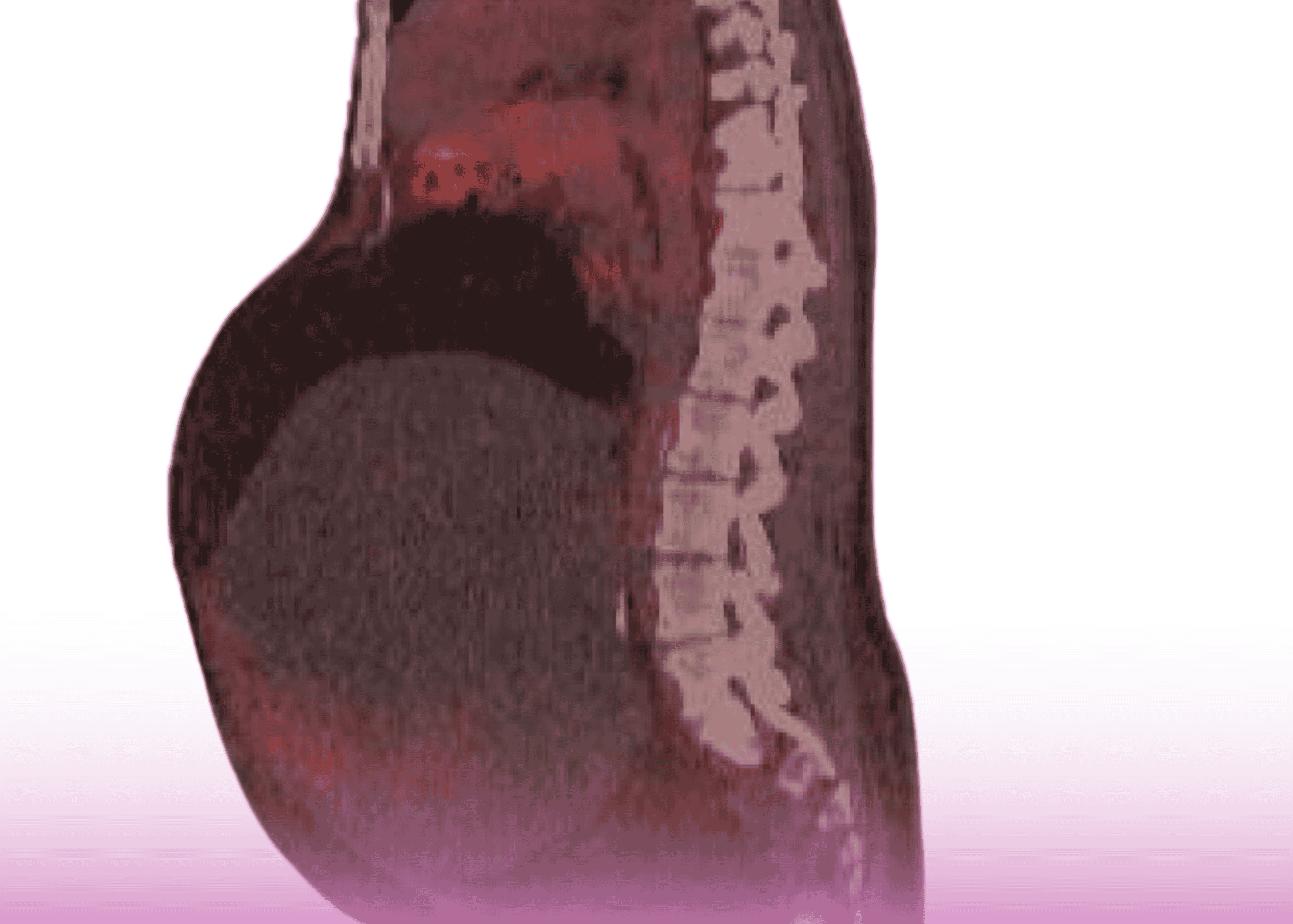
  • CT scan of the abdomen revealing a 13mm pancreatic head mass with significant dilation of both the pancreatic duct (10mm) and bile duct (22mm)
  • CT chest confirming no metastatic disease
  • Elevated tumor marker CA19-9 at 76 (normal range <37)
  • Endoscopic ultrasound showing a 2.5cm pancreatic head mass with biopsy indicating potential malignancy

The case was presented at the hospital’s Oncology Multidisciplinary Team meeting, where specialists collectively recommended proceeding with Whipple’s resection based on the highly suspicious imaging appearances indicative of pancreatic head cancer.

Pioneering Surgical Approach

Prof. Dr. Basil Ammori, Consultant Laparoscopic Bariatric, Gastrointestinal, Hepatobiliary and Pancreatic Surgeon at Burjeel Hospital, performed a complete laparoscopic Whipple procedure—a technically demanding operation rarely performed using minimally invasive techniques.

“The laparoscopic approach to Whipple’s resection is challenging and requires specialized expertise in both laparoscopic and pancreatic surgery,” notes Prof. Ammori. “While the procedure takes longer—approximately 10 hours in this case—the benefits for patient recovery are significant.”

Key advantages of the laparoscopic approach include:

  • Minimal blood loss during surgery
  • No need for intraoperative or postoperative blood transfusions
  • Dramatically shortened hospital stay
  • Accelerated return to normal activities
  • Earlier initiation of adjuvant chemotherapy when needed

Remarkable Recovery

The patient experienced an uneventful recovery and was discharged just four days after surgery—a dramatic improvement over the typical 10-14 day hospitalization period following traditional open Whipple procedures.

Histopathology revealed an undifferentiated carcinosarcoma of the pancreatic head (pT1CN0) with no involvement of the 16 regional lymph nodes examined (0/16) and clear surgical margins, indicating a potentially curative resection.

At the three-week follow-up appointment, the patient was doing well and was scheduled to begin adjuvant chemotherapy based on the Oncology MDT recommendation.

Advancing Surgical Standards

This case highlights the evolution of complex oncological surgery toward minimally invasive approaches. While traditional open Whipple procedures remain standard at many institutions, the laparoscopic approach offers significant benefits for carefully selected patients.

“The rapid recovery associated with laparoscopic surgery means patients can begin adjuvant chemotherapy sooner, potentially improving long-term prognosis,” explains Prof. Ammori. “This integrated approach to cancer care exemplifies modern multidisciplinary treatment.”

The successful implementation of this advanced procedure at Burjeel Hospital Abu Dhabi demonstrates the institution’s commitment to offering state-of-the-art surgical options for patients with complex conditions.

Experts

Minimally Invasive Surgery Advances: Laparoscopic Hernia Repair in Neonates

In pediatric surgery, minimally invasive techniques are revolutionizing treatment options for even the youngest patients. A recent case at Burjeel Hospital Abu Dhabi highlights how laparoscopic approaches are providing excellent outcomes for neonatal patients with inguinal hernias.

The Case

A four-week-old baby girl presented with large bilateral inguinal swellings that had been increasing in size over the previous week. Upon physical examination, the diagnosis was confirmed as reducible large bilateral inguinal hernias, a condition requiring surgical intervention.

Advanced Surgical Approach

The surgical team, led by Prof. Dr. Amulya Saxena, Consultant Pediatric Surgeon at Burjeel Hospital Abu Dhabi, opted for a laparoscopic approach rather than traditional open surgery.

“Laparoscopic procedures offer significant advantages for neonatal patients, including smaller incisions, reduced pain, and faster recovery times,” explains Prof. Saxena. “However, they present unique challenges in such young patients.”

The surgery utilized specialized equipment tailored for neonatal patients:

  • A 5mm 30-degree scope for optimal visualization
  • 3mm instruments for precise manipulation in small spaces
  • Non-resorbable sutures for secure closure of the internal inguinal rings

Technical Challenges Overcome

The primary challenge in this case was navigating the extremely limited abdominal working space typical in neonatal patients. This required exceptional precision with intracorporeal suturing techniques.

The laparoscopic views captured during the procedure show the meticulous process of:

  1. Identifying the open right internal ring
  2. Performing purse-string sutures around the inguinal rings
  3. Precise intracorporeal knot tying to ensure secure closure

Post-Operative Care

Following the successful procedure, the infant remained in the hospital for a single night for apnea monitoring—a standard precaution for neonatal patients. The follow-up appointment one week later confirmed an uneventful recovery with no complications.

Why This Matters

This case exemplifies the advantages of minimally invasive surgical techniques even for the most vulnerable patients. By utilizing laparoscopic approaches, surgeons can achieve excellent outcomes while minimizing trauma, reducing scarring, and promoting faster recovery—critical considerations for neonatal patients.

The successful implementation of these advanced techniques at Burjeel Hospital demonstrates the institution’s commitment to providing cutting-edge surgical care across all age groups, from newborns to adults.

The surgical team included Dr. Samer Shouman Alkahwaty, Consultant Anesthesiologist, whose expertise was essential in managing the anesthetic challenges specific to neonatal patients.

Experts

LINX Device Successfully Treats Severe GERD in Post-Bariatric Surgery Patient: A Day-Case Procedure

Introduction

Burjeel Hospital Abu Dhabi has successfully performed a LINX® device implantation to treat severe gastroesophageal reflux disease (GERD) in a patient with previous bariatric surgery. This case highlights the challenges of managing GERD in post-bariatric surgery patients and demonstrates how innovative medical devices can provide effective symptom relief when traditional surgical approaches are not feasible.

Patient History

A 34-year-old male patient presented with a 4-5 year history of debilitating GERD symptoms, including longstanding heartburn, excessive burping, and dysphagia (difficulty swallowing). Despite previous treatment with proton pump inhibitors (PPIs), his symptoms remained uncontrolled. The patient had a significant surgical history, having undergone laparoscopic butterfly gastroplasty for obesity in 2017.

Diagnostic Workup

A comprehensive evaluation was conducted to assess the severity and physiological basis of the patient’s symptoms:

  1. Gastroscopy: The endoscopic examination revealed grade A esophagitis (mild inflammation of the esophageal lining) and a 3cm sliding hiatal hernia.
  2. pH Study: A 24-hour pH monitoring confirmed severe gastroesophageal reflux with a DeMeester score of 35 to 306 (normal value <14.72), indicating significant acid exposure in the esophagus. The study also demonstrated a strong correlation (93.9%) between reflux episodes and symptom occurrence.

These findings confirmed the diagnosis of severe GERD that was inadequately controlled with medical therapy, warranting surgical intervention.

Surgical Approach

Prof. Dr. Basil Ammori, Consultant in Laparoscopic, Bariatric, General, Gastrointestinal & Hepatobiliary Surgery at Burjeel Hospital Abu Dhabi, performed a laparoscopic hiatal hernia repair and LINX® sphincter-augmentation device placement. The procedure was performed as a day-case surgery with anesthesia provided by Dr. Samer Shouman Alkahwaty.

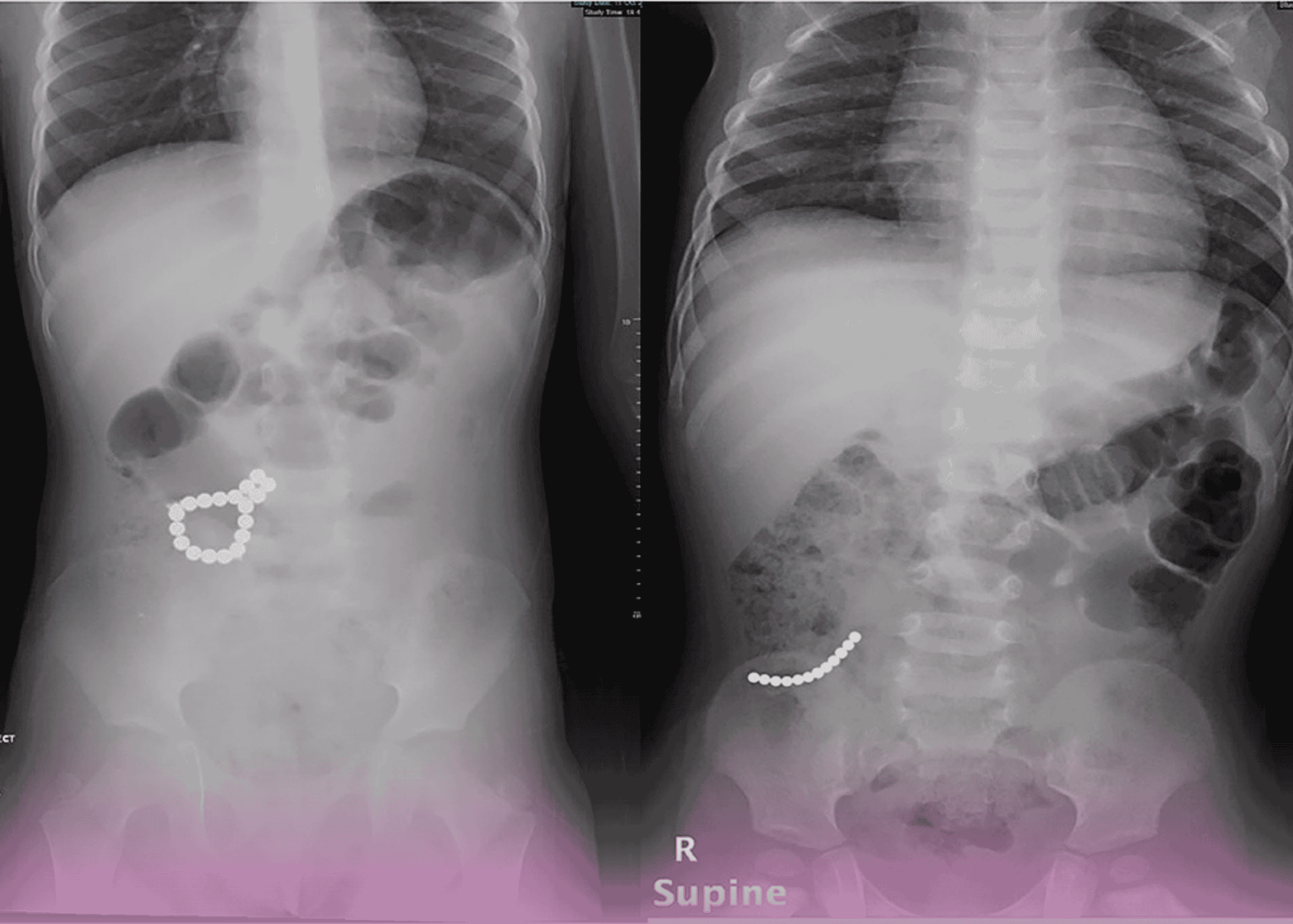
The LINX® device consists of a small, flexible band of interlinked titanium beads with magnetic cores. When placed around the lower esophageal sphincter (LES), it provides additional support to prevent reflux while still allowing food and liquid to pass into the stomach. This approach is particularly valuable in post-bariatric surgery patients where traditional anti-reflux procedures like fundoplication may not be anatomically possible.

Surgical Outcome

The procedure was completed successfully in just 51 minutes, with the patient recovering uneventfully and being discharged the same day. Follow-up evaluations at both 1 and 6 weeks post-surgery confirmed that the patient remained entirely symptom-free, indicating an excellent immediate clinical outcome.

Unique Challenges in Post-Bariatric GERD

This case highlights several important considerations in managing GERD after bariatric surgery:

  1. Limited Surgical Options: Traditional anti-reflux procedures such as fundoplication are often not feasible in post-bariatric patients due to the altered gastric anatomy.
  2. Higher Technical Complexity: Previous abdominal surgery can result in adhesions that increase the technical difficulty of subsequent procedures.
  3. Specialized Device Solution: The LINX® device provides an effective mechanical barrier to reflux while accommodating the modified gastrointestinal anatomy following bariatric surgery.

Risk Considerations

While generally safe and effective, LINX® device implantation carries specific considerations that patients should be aware of:

  1. Esophageal Erosion: Although rare (1-2%), the device can potentially erode into the esophagus over time.
  2. MRI Limitations: Patients with the LINX® device have restrictions regarding MRI procedures due to the magnetic components.
  3. Technical Expertise: The procedure requires specialized surgical skills and experience with the device, which is available at specialized centers like Burjeel Hospital.

Conclusion

This case demonstrates Burjeel Hospital Abu Dhabi’s capability to offer innovative solutions for complex gastroesophageal disorders, particularly in patients with previous bariatric surgery. The successful LINX® device implantation as a day-case procedure with excellent symptom resolution highlights the value of this approach for carefully selected patients with severe GERD who cannot undergo traditional anti-reflux procedures. Prof. Dr. Basil Ammori’s expertise in both bariatric and anti-reflux surgery provided the necessary skill set to achieve this excellent outcome in a challenging clinical scenario.

Experts

Innovative Coil-and-Glue Technique for Hepatic Artery Aneurysm: A Minimally Invasive Life-Saving Solution

Introduction

Burjeel Hospital Abu Dhabi has successfully pioneered an innovative interventional radiology approach for treating a complex hepatic artery aneurysm. This case study highlights how creative technical solutions and advanced interventional expertise can provide life-saving treatment for potentially catastrophic vascular conditions through minimally invasive techniques.

Patient Presentation

A 32-year-old male presented to the emergency department with severe abdominal pain. Computed tomography (CT) revealed a concerning picture: hemoperitoneum (blood in the abdominal cavity) and a large aneurysmal dilation of the left hepatic artery with irregular borders and extra-luminal contrast material. While no active extravasation was visible at the time of imaging, the condition presented an immediate life-threatening risk. Notably, the scan also revealed bilateral renal artery dissections, suggesting a possible systemic vascular pathology.

Technical Challenge

The location and morphology of the hepatic artery aneurysm presented significant challenges for conventional endovascular treatment approaches. Traditional coil embolization alone risked incomplete exclusion of the aneurysm or coil migration, while standard liquid embolic agents could potentially migrate to non-target vessels, causing serious complications.

Innovative Solution

Dr. Mohamed Almarzooqi, Medical Director and Consultant Interventional Radiologist at Burjeel Hospital Abu Dhabi, devised a creative two-stage approach that combined established techniques in a novel way:

  1. Coil Cage Creation: Initially, a framework of embolization coils was carefully deployed to form a stable cage within the aneurysm. This framework served as both structural support and a containment system for the next phase of treatment.
  2. Magic Glue as Filler: Following the establishment of the coil framework, the internal volume was methodically filled with a specially prepared mixture of Magic Glue (n-butyl cyanoacrylate) and non-ionic contrast. This innovative approach allowed the glue to fill the aneurysmal volume without significant risk of migration to non-target vessels. The precise formulation provided enough working time for the controlled and accurate filling of the aneurysm, avoiding premature adhesion to the microcatheter.

Procedural Images

The interventional series clearly demonstrates the progression of this novel technique:

  • Initial angiogram showing the left hepatic artery aneurysm
  • Post-coiling control angiogram with the established coil framework
  • Final angiogram after glue injection showing the dense embolization material contained within the coil cage and complete exclusion of the aneurysm from circulation

Clinical Significance

This case demonstrates several important innovations in interventional radiology:

  1. Adaptable Technical Solutions: By combining existing tools (coils and glue) in a novel approach, complex vascular abnormalities can be treated without requiring specialized devices that may not be immediately available.
  2. Tailored Treatment Approach: The coil-cage technique provided structural integrity while the liquid embolic agent ensured complete filling of the irregular aneurysm, addressing the unique challenges of this specific case.
  3. Minimally Invasive Management: The endovascular approach avoided the need for open surgery in a patient with multiple vascular abnormalities, potentially reducing recovery time and complications.
  4. Comprehensive Vascular Assessment: The identification of bilateral renal artery dissections highlights the importance of complete vascular evaluation in patients with any arterial abnormality.

Conclusion

The successful treatment of this complex hepatic artery aneurysm through an innovative combined coil-and-glue technique showcases Burjeel Hospital Abu Dhabi’s commitment to advancing interventional radiology solutions for life-threatening conditions. Dr. Almarzooqi’s approach demonstrates how technical creativity and expertise can produce excellent outcomes in challenging cases, furthering the field of minimally invasive vascular interventions.

Experts

Smile Makeovers: Transforming Your Confidence with Cosmetic Dentistry 

A smile is often one of the first things people notice about you, and it can leave a lasting impression. Whether it’s a result of genetics, aging, or oral habits, many people feel self-conscious about their smile. Fortunately, modern cosmetic dentistry offers a variety of treatments that can enhance your smile and, more importantly, boost your confidence. 

In this blog, we’ll explore the different options available for smile makeovers, how they work, and how they can help you achieve the smile you’ve always dreamed of. From teeth whitening to dental veneers, the possibilities are endless for creating a healthier, more radiant smile. 

What is a Smile Makeover? 

A smile makeover is a personalized treatment plan designed to address a variety of dental issues, such as crooked teeth, discolored enamel, chipped teeth, or missing teeth. By combining several cosmetic dental procedures, a smile makeover can completely transform the appearance of your smile, leaving you with improved aesthetics and enhanced self-confidence. 

There are several cosmetic dental procedures that can contribute to a smile makeover. Depending on your needs and goals, your dentist may recommend one or more of the following treatments: 

1. Dental Veneers: Achieve a Perfect, Natural-Looking Smile 

Dental veneers are thin shells of porcelain or composite resin that are custom-made to cover the front surface of your teeth. They are ideal for correcting issues such as chipped, stained, or slightly misaligned teeth. Veneers can provide a flawless, natural-looking result that can enhance the overall symmetry and shape of your smile. 

2. Teeth Whitening: A Brighter, Whiter Smile 

Teeth whitening is one of the most popular and least invasive cosmetic treatments. Over time, teeth can become stained or yellow due to various factors such as food, drinks, and tobacco use. Professional teeth whitening treatments can significantly lighten the shade of your teeth, making your smile brighter and more youthful in the most natural and drill-free way.  

3. Dental Implants: Restore Your Smile with Permanent Solutions 

If you’re missing one or more teeth, dental implants can provide a permanent solution that looks and functions like natural teeth. Implants are surgically placed into the jawbone and act as a base for crowns or bridges, restoring both the function and appearance of your smile. 

4. Bonding: A Quick Fix for Small Imperfections 

Dental bonding or tooth-colored restorations involve applying a tooth-colored resin to repair minor imperfections, such as chips, gaps, or discoloration. It’s a cost-effective and quick way to improve your smile without the need for extensive procedures. 

5. Invisalign: Straighten Your Smile with Clear Aligners 

For those with crooked or misaligned teeth, Invisalign offers a discreet alternative to traditional braces. These clear, removable aligners gradually shift your teeth into their ideal position, improving both the appearance and functionality of your smile. 

The Benefits of a Smile Makeover 

Undergoing a smile makeover can provide a wide range of benefits, from aesthetic improvements to psychological boosts. Here are just a few ways a smile makeover can transform your life: 

  • Enhanced Confidence: A beautiful smile can make you feel more confident in social situations, whether at work, with friends, or on a date. 
  • Youthful Appearance: Dental procedures like whitening and veneers can create a youthful, vibrant smile that rejuvenates your overall appearance. 
  • Improved Oral Health: Many smile makeover treatments, such as dental implants or teeth straightening, not only enhance your smile but also improve the health and function of your teeth and gums. 
  • Lasting Results: With proper care, the results of your smile makeover can last for years, providing long-term satisfaction. 

Before and After: What to Expect from a Smile Makeover 

The transformation provided by a smile makeover can be dramatic. Before undergoing the procedures, your dentist will conduct a thorough evaluation of your teeth and gums. They will discuss your goals and create a personalized treatment plan that meets your needs. The timeline for a smile makeover varies depending on the complexity of the procedures, but many treatments can be completed within a few months. 

After your treatments, you’ll be able to enjoy a radiant smile that reflects your personality and boosts your self-esteem. Whether you’re going for subtle improvements or a complete transformation, the results can be life-changing. 

Choosing the Best Dentist for Your Smile Makeover 

The key to a successful smile makeover is finding a skilled and experienced cosmetic dentist who understands your needs and goals. Look for a dentist who has expertise in various cosmetic procedures and can offer a personalized treatment plan. Patient reviews and before-and-after photos of previous patients can also give you a better sense of the dentist’s capabilities. 

Affordable Smile Makeovers: Is It Possible? 

While cosmetic dentistry can sometimes come with a significant price tag, there are options for those seeking a more affordable smile makeover. Many dental offices offer financing plans or payment options that make treatments more accessible. Additionally, some procedures, like bonding or teeth whitening, may be more budget-friendly than others. Consult with your dentist to explore all available options that fit your budget. 

Cosmetic dentistry is constantly evolving, with new technologies and materials making treatments more effective and natural-looking than ever before. From 3D smile design software that allows patients to visualize their results before treatment to advancements in ceramic materials for veneers, the trends in cosmetic dentistry are all about precision, comfort, and aesthetic beauty. 

Self-Care After a Smile Makeover 

To ensure the long-lasting success of your smile makeover, it’s important to maintain good oral hygiene. Regular brushing and flossing, along with routine dental check-ups, are essential to keep your smile looking its best. Additionally, if you’ve had treatments like teeth whitening or veneers, be mindful of your diet and avoid habits that may stain your teeth, such as smoking or excessive coffee consumption. 

Final Thoughts 

A smile makeover can do more than just improve the appearance of your teeth—it can elevate your self-esteem, boost your confidence, and change the way you feel about yourself. Whether you’re looking to address small imperfections or undergo a complete transformation, cosmetic dentistry offers a wide range of solutions that can help you achieve the smile of your dreams. 

Investing in your smile is an investment in yourself, and the right cosmetic dental treatments can provide you with results that last a lifetime. So, why wait? Start your smile transformation today and experience the confidence boost that comes with a radiant, beautiful smile. 

Life After Joint Replacement: What to Expect and How to Thrive 

Joint replacement surgery can be life-changing, offering significant relief from pain and improved mobility. Whether you’ve had a hip, knee, or shoulder replacement, the road to recovery and long-term health involves careful attention to rehabilitation, physical therapy, and lifestyle adjustments. This guide will help you understand what to expect after surgery, how to optimize recovery, and how to thrive post-joint replacement. 

1. Immediate Recovery: What to Expect Right After Surgery 

The first phase of recovery after joint replacement is the immediate post-surgical period. Right after the procedure, you’ll spend time in a recovery room where healthcare professionals monitor your vital signs and pain levels. Pain management is a key focus during this period to ensure that you remain as comfortable as possible. You’ll likely be prescribed pain medications, and your healthcare team may use ice packs or other techniques to reduce swelling. 

What to Expect: 

  • You may stay in the hospital for a few days, depending on your recovery progress and the type of surgery. 
  • Swelling, bruising, and discomfort are common in the initial recovery phase. 
  • You will begin using a walker or crutches as you start to mobilize, even while still in the hospital. 

2. Post-Surgery Rehabilitation: Key to Regaining Mobility 

One of the most important aspects of life after joint replacement is rehabilitation. Physical therapy begins soon after surgery and plays a crucial role in your recovery. Therapy helps to rebuild strength, increase joint flexibility, and improve mobility. You will work closely with a physical therapist to learn exercises designed to help you regain normal function in the joint. 

What to Expect: 

  • You’ll start with gentle, range-of-motion exercises to help the joint heal and prevent stiffness. 
  • Over time, you’ll move on to strengthening exercises to build the muscles around the new joint. 
  • Your physical therapist will monitor your progress and may adjust your exercises to ensure you’re recovering at a healthy pace. 

3. Pain Management: Finding the Right Approach 

While joint replacement surgery offers long-term pain relief, the initial stages of recovery may involve some discomfort. Pain management is essential during this time to help with mobility and daily activities. Your doctor will prescribe pain relief options tailored to your needs, which may include oral medications, topical treatments, or even nerve blocks for more significant pain. 

What to Expect: 

  • Pain levels typically decrease significantly after the first few weeks. 
  • It’s essential to follow your doctor’s instructions on how and when to take medications to manage pain effectively. 
  • Non-medication methods such as ice, heat, and elevation can also help manage swelling and discomfort. 

4. Improving Mobility: Slowly Getting Back to Daily Activities 

One of the most exciting outcomes of joint replacement is the improvement in mobility. As the months progress, you’ll find that your ability to move and participate in daily activities becomes easier. Simple tasks like walking, climbing stairs, and sitting down and standing up should become more manageable. 

What to Expect: 

  • With consistent physical therapy, you’ll notice gradual improvements in your joint’s range of motion and overall function. 
  • It’s normal to experience some stiffness, especially in the mornings or after periods of inactivity, but regular movement will help prevent this. 
  • Your physical therapist will encourage you to practice walking, using stairs, and engaging in low-impact activities as you progress. 

5. Lifestyle Changes: Supporting Long-Term Health 

After joint replacement surgery, some lifestyle changes may be necessary to ensure the long-term success of the prosthetic joint and maintain overall health. Maintaining a healthy weight is crucial to reduce excess stress on the joint and preserve its functionality. Additionally, adopting a more active lifestyle with joint-friendly exercises can improve your overall well-being. 

What to Expect: 

  • Healthy eating and maintaining an appropriate weight can significantly improve your recovery and prevent undue stress on your new joint. 
  • Low-impact exercises such as swimming, cycling, and walking are excellent choices for maintaining joint health. 
  • Be mindful of any activities that may put too much strain on your joint, such as running or high-impact sports. 

6. Prosthetic Care: Keeping Your New Joint in Top Condition 

Your new prosthetic joint requires care and maintenance to ensure its longevity. While joint replacement surgery typically lasts many years, avoiding unnecessary wear and tear is key to maximizing the lifespan of your prosthetic. Your doctor or orthopedic surgeon will provide guidance on how to care for your joint and what to avoid. 

What to Expect: 

  • Regular follow-up appointments with your surgeon to monitor the condition of your prosthetic joint. 
  • You may need X-rays or other imaging tests periodically to ensure the joint is in proper alignment. 
  • It’s crucial to avoid high-impact activities that could stress the new joint prematurely. 

7. Long-Term Outcomes: Expecting a Better Quality of Life 

The majority of joint replacement patients experience significant improvement in their quality of life. Most people report reduced pain and increased mobility, making it easier to return to activities they enjoy. While there are risks associated with any surgery, the benefits of joint replacement often far outweigh the drawbacks, especially in older adults or those with severe joint deterioration. 

What to Expect: 

  • Most patients see a noticeable reduction in joint pain and increased ability to engage in everyday activities. 
  • The prosthetic joint may wear out over time, but modern materials and surgical techniques allow most replacements to last 15-20 years or more. 
  • You’ll likely have a better range of motion and greater independence in daily life. 

8. Patient Education: Being Your Own Advocate 

Patient education is an essential part of life after joint replacement. Understanding your recovery process, how to care for your new joint, and what to do if complications arise will help you make informed decisions and improve outcomes. Regular communication with your healthcare team and a commitment to learning about the recovery process are vital for successful rehabilitation. 

What to Expect: 

  • You’ll receive detailed instructions about exercises, pain management, and activity modifications. 
  • Be proactive in asking questions about what you can do to support your recovery, including potential lifestyle changes. 
  • Learning about signs of complications (such as infection or joint loosening) is crucial for timely intervention. 

Final Thoughts: Thriving After Joint Replacement 

Joint replacement surgery offers a promising path to pain relief and improved mobility. With the right post-surgery rehabilitation, pain management, and lifestyle adjustments, you can regain your independence and enjoy a higher quality of life. By following your healthcare team’s guidance, staying consistent with physical therapy, and taking steps to protect your joint, you can expect long-term benefits and a vibrant, active future. 

Our Experts

See more

Advanced Neuro-Navigation Enables Successful Resection of Complex Brain Tumor

Introduction

Burjeel Royal Hospital Al Ain has demonstrated exceptional neurosurgical capabilities with the successful resection of a large and complex supratentorial tumor. This case highlights the critical role of advanced neuro-navigation technology, multidisciplinary collaboration, and precise surgical technique in managing challenging brain lesions while preserving neurological function.

Patient Presentation

The patient presented with multiple seizure episodes associated with a large supratentorial tumor. Imaging studies revealed an extensive intra-axial mass lesion in the left frontal-parietal region measuring approximately 7.8 × 6 × 6.4 cm with surrounding edema and significant mass effect. The tumor was causing partial effacement of the left lateral ventricle and extending into critical neurological structures, including:

  • Left basal ganglia
  • Body of corpus callosum
  • Motor cortex regions
  • Left insular cortex
  • Primary auditory cortex regions
  • Wernicke’s area (language processing center)

This extensive involvement of eloquent brain regions posed significant challenges, as damage to these areas could result in permanent motor, speech, and language deficits.

Surgical Planning and Approach

The neurosurgical team, led by Prof. Dr. Tamer Ibrahim Metwaly and Dr. Mutaz Jamal Abu Nassar, Consultants in Neurosurgery at Burjeel Royal Hospital Al Ain, employed advanced surgical planning and intraoperative technology:

  1. Preoperative Imaging: Detailed MRI sequences were used to map the tumor’s boundaries and its relationship to critical brain structures.
  2. Advanced Neuro-Navigation: This GPS-like technology provided real-time guidance during the procedure, allowing precise localization of the tumor and surrounding critical structures.
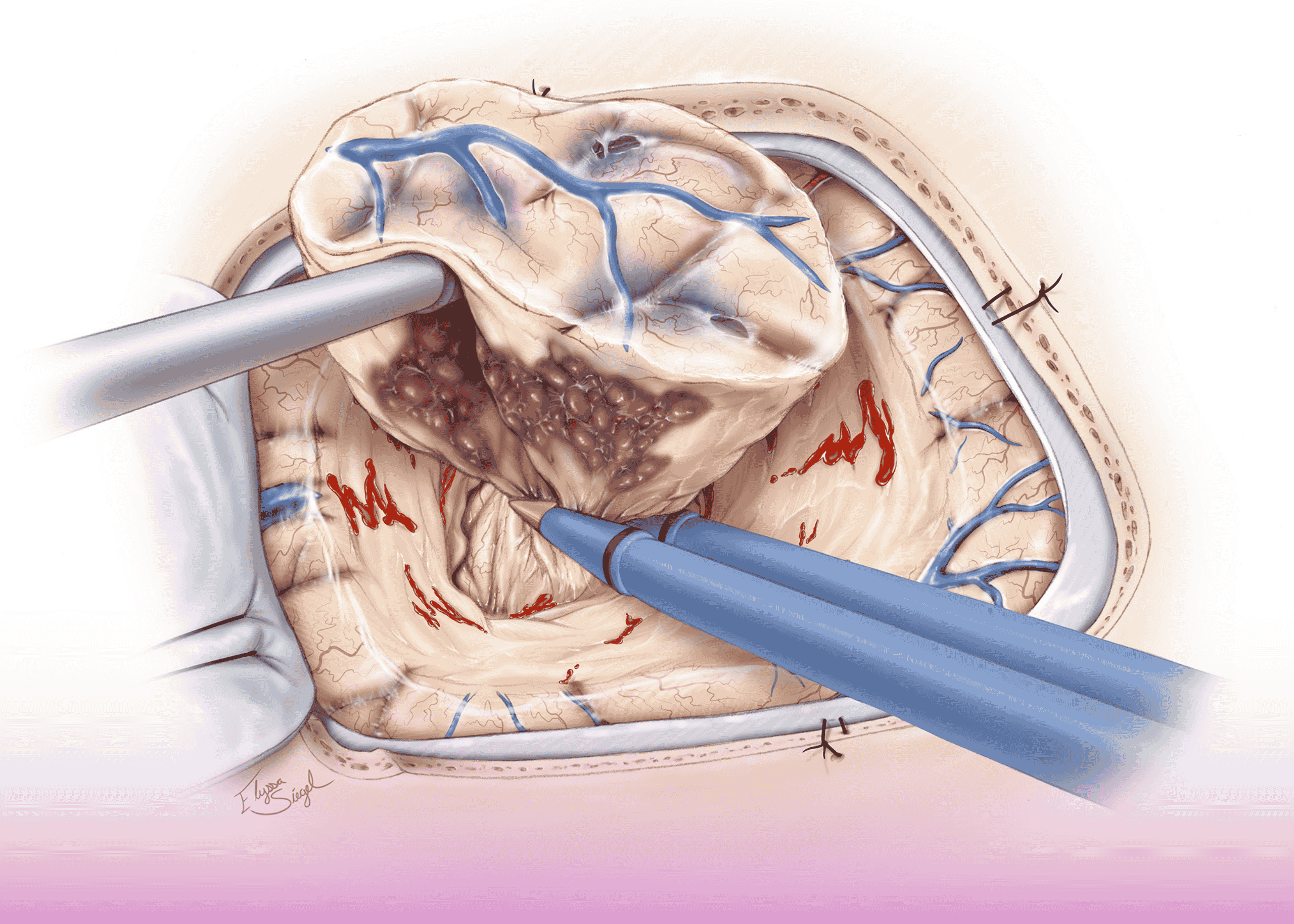
  3. Surgical Strategy: The team performed a frontal craniotomy to access the tumor, meticulously excising the glioma while continuously monitoring to avoid damage to functional brain tissue.
  4. Decompression: Significant effort was directed at relieving intracranial pressure caused by the tumor and surrounding edema.
  5. Reconstruction: Following tumor removal, careful cranio- and duroplasty procedures restored the integrity of the brain’s protective layers.

Perioperative Management

The complex nature of the case required meticulous perioperative care:

  1. Seizure Management: Both pre- and postoperatively, the patient experienced focal seizures, which were promptly addressed with dual-dose antiepileptic treatment based on neurologist recommendations.
  2. Neurological Monitoring: Continuous assessment of neurological status guided postoperative management and rehabilitation.
  3. Multidisciplinary Approach: Collaboration between neurosurgeons, neurologists, intensivists, and rehabilitation specialists ensured comprehensive care.

Recovery and Outcomes

Despite the formidable challenges posed by this case, the patient demonstrated a remarkable recovery trajectory:

  • Stable hemodynamic parameters throughout the perioperative period
  • Progressive improvement in consciousness level
  • Recovery of verbal communication abilities
  • No further seizure episodes following optimization of antiepileptic therapy
  • Favorable prognosis confirmed by the intensivist team

Technological Advantages

The successful management of this case was significantly enhanced by Burjeel Royal Hospital Al Ain’s advanced neurosurgical infrastructure:

  1. Intraoperative Imaging: The hospital’s integrated MRI operating room provided precise guidance for tumor removal.
  2. KINEVO 900 Microscope: This state-of-the-art operative microscope features robotic visualization, 4K imaging technology, and advanced fluorescence capabilities, allowing for optimal visualization throughout the procedure.
  3. Neuro-Navigation System: Advanced computer-assisted navigation allowed real-time tracking of surgical instruments in relation to the patient’s anatomy.

Conclusion

This case exemplifies the exceptional level of neurosurgical care available at Burjeel Royal Hospital Al Ain. By combining advanced technology, surgical expertise, and multidisciplinary collaboration, the team successfully managed a challenging brain tumor that involved multiple critical neurological structures. The patient’s favorable recovery highlights the importance of specialized neurosurgical capabilities in achieving optimal outcomes in complex intracranial cases.

Experts

Custom 3D-Printed Implants Revolutionize Complex Hip Revision Surgery

Introduction

Burjeel Hospital Abu Dhabi has successfully performed a groundbreaking hip revision surgery utilizing custom-designed 3D-printed implants, offering new hope for patients with severe pelvic bone loss. This case demonstrates how advanced imaging, collaborative biomedical engineering, and surgical expertise can transform seemingly impossible cases into successful outcomes, even after previous implant failures.

Patient History

The patient had undergone a fully cemented left hip replacement more than a decade ago outside the UAE. Six months prior to the current intervention, the patient was diagnosed with a left periprosthetic infection in the UAE. In July 2024, the infected total hip replacement was explanted, and the patient was left with static antibiotic-loaded spacers in a disarticulated hanging hip configuration due to the severity of bone stock loss, which made implantation of a traditional hip spacer impossible. An X-ray guided hip needle aspiration for culture in October 2024 confirmed that the infection had been successfully eliminated.

Complex Challenges

The surgical team, led by Dr. Gianluca Cusma, Consultant Trauma & Orthopedic Surgeon at Burjeel Hospital Abu Dhabi, faced an exceptionally challenging case:

  1. Severe Bone Loss: Imaging studies revealed catastrophic bone deficiencies:
    • Complete deficiency of the medial wall
    • Partial deficiency of the posterior wall
    • Complete deficiency of the superior wall
    • Complete resorption of the greater trochanter
  2. Classification: The condition was classified as Paprosky 3B pelvic bone loss, representing one of the most severe scenarios in revision hip arthroplasty.
  3. Functional Limitations: The patient had been in a disarticulated state for five months, resulting in significant soft tissue adaptations, muscle damage, and limb shortening.

Innovative Solution

The management approach required innovative solutions customized specifically to this patient’s unique anatomy:

  1. Custom Implant Design: In collaboration with specialized biomedical engineers, the team designed a CT-based custom-made acetabular component. The process involved:
    • Detailed 3D reconstruction of the remaining bone anatomy
    • Digital design of a tri-flanged component
    • Selection of 3D-printed trabecular titanium material to promote bone ingrowth
    • Engineering of flanges positioned to secure to the remaining viable bone
    • Integration of features to reconstruct the medial wall separating the acetabular cavity from the abdominal cavity
  2. Manufacturing Process: The design-to-delivery process took approximately four weeks, culminating in the receipt of a sterile, patient-specific implant.

Surgical Procedure

Under spinal anesthesia provided by Dr. Srinivas Boddupally, Dr. Cusma performed the complex revision hip replacement:

  1. Acetabular Reconstruction: The custom-made cementless acetabular component was precisely positioned to engage with the remaining bone structure.
  2. Femoral Reconstruction: A cementless off-the-shelf conical stem was implanted to provide stable femoral fixation.
  3. Articulation System: The surgeon selected a dual mobility ceramic-on-polyethylene tribology system to enhance stability while minimizing wear.

Postoperative Course and Recovery

The patient experienced an excellent immediate postoperative recovery:

  • Neurovascular status remained intact
  • No blood transfusions were required
  • The surgical wound healed well
  • Daily physiotherapy focused on regaining active hip flexion/extension control
  • The patient achieved comfortable positional transfers and in-room ambulation
  • Discharge occurred on the fifth postoperative day

Technical and Emotional Challenges

This case highlighted several critical challenges in complex revision hip surgery:

  1. Technical Challenges:
    • Designing a custom implant that would integrate with severely compromised bone
    • Executing precise surgical exposure and preparation for the implant
    • Managing damaged soft tissues and muscles
    • Establishing appropriate limb length and tension to balance hip stability and gait after prolonged disarticulation
  2. Psychological Aspects:
    • Managing the patient’s emotions and expectations throughout the process
    • Providing confidence in a positive outcome despite the extreme complexity

Conclusion

This case exemplifies Burjeel Hospital Abu Dhabi’s capability to tackle exceptionally complex orthopedic challenges through innovative approaches and multidisciplinary collaboration. By combining advanced imaging, custom 3D-printed implant technology, and skilled surgical technique, the team successfully restored hip function in a case that would have been deemed inoperable using conventional methods. This approach represents the cutting edge of orthopedic reconstruction and offers new possibilities for patients with severe bone loss following failed arthroplasty.

Experts

Open Transvesical Prostatectomy for Massive Prostatic Hyperplasia

Patient Presentation

A 66-year-old male presented with symptoms of benign prostatic hyperplasia (BPH) that had become increasingly refractory to medical management. His symptoms included weak urinary stream, increased frequency of micturition, sensation of incomplete bladder emptying, nocturia (4-5 times nightly), and urgency.

Diagnostic Findings

Ultrasound examination revealed a significantly enlarged prostate measuring approximately 180 grams with post-void residual urine exceeding 200 ml.

Treatment Considerations

Dr. Qaraschouli thoroughly discussed all available surgical options with the patient and his family, including:

  • Transurethral resection of the prostate (TURP)
  • Holmium laser enucleation
  • Other laser therapy options
  • Steam therapy (Resume)
  • Open transvesical prostatectomy

After careful consideration of the massive prostate size and the patient’s clinical presentation, the decision was made to proceed with open transvesical prostatectomy.

Surgical Outcome

The surgical and postoperative courses were uneventful. Following removal of the indwelling catheter, the patient experienced satisfactory micturition with only mild stress incontinence (Grade 1) during the first postoperative week, which subsequently resolved completely. Follow-up ultrasound confirmed the absence of residual urine, in stark contrast to the preoperative findings.

Histopathology confirmed prostatic adenoma with chronic prostatitis, with no evidence of malignancy.

The Continued Relevance of Open Surgery

These cases demonstrate that open surgery remains the optimal choice in specific urological scenarios. Key advantages in these cases included:

  1. For Complex Renal Tumors:
    • Better control of bleeding in high-risk patients with coagulopathy
    • Reduced operative time in patients with multiple comorbidities
    • Enhanced ability to manage unexpected intraoperative findings
    • Lower risk of major vascular complications
  2. For Massive Prostatic Hyperplasia:
    • Complete removal of prostatic adenoma regardless of size
    • Definitive solution with lower recurrence rates compared to some minimally invasive approaches
    • Shorter operative duration in cases of extremely enlarged prostates
    • Excellent functional outcomes with rapid recovery

Conclusion

While minimally invasive and robotic approaches continue to advance, these cases from Burjeel Royal Hospital Asharej highlight that open surgical techniques remain essential tools in the modern urologist’s armamentarium. The surgical team’s ability to select the most appropriate surgical approach based on individual patient factors—rather than simply following technological trends—represents the true art of surgical practice. This patient-centered approach ensures optimal outcomes in complex urological cases where traditional open techniques may still offer significant advantages.

Experts

Open Radical Nephrectomy for Large Renal Cell Carcinoma

Patient Presentation

A 62-year-old female presented with irregular left flank pain persisting over several months. The pain pattern was intermittent and occasionally relieved with analgesics. The patient also reported occasional dysuria but no visible hematuria. Despite these concerning symptoms, she had not sought earlier medical attention.

Diagnostic Findings

Initial ultrasound examination revealed a large 10 cm diameter tumor in the left kidney. Subsequent CT scanning confirmed a 10 x 10 cm mass with features suspicious for malignancy, accompanied by multiple prominent collateral vessels in the perinephric region.

Preoperative assessment identified the patient as having high surgical risk, supported by abnormal laboratory parameters including:

  • Significantly elevated APTT
  • Increased INR and prothrombin levels
  • Elevated creatinine indicating impaired renal function

Surgical Approach and Outcome

After thorough discussion with the patient and her family regarding available options, Dr. Humam Qaraschouli, Consultant Urologist, proceeded with an open radical tumor nephrectomy. The surgery was completed without complications, and the patient experienced an uneventful postoperative course, allowing for discharge as scheduled.

Histopathological examination confirmed renal cell carcinoma (pT2a, size 9.6 cm, G4, R0). Postoperative CT scanning of the thorax and abdomen showed no evidence of metastatic disease and demonstrated a satisfactory appearance of the left renal fossa. The oncology team subsequently established a regular follow-up protocol.

Experts