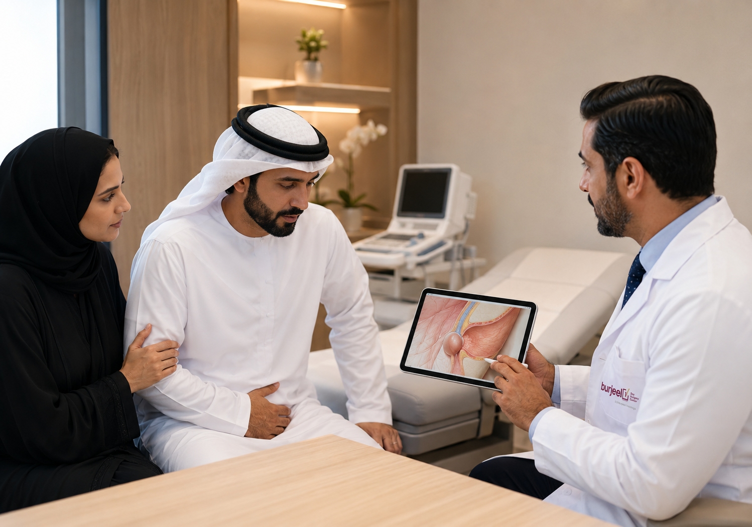
400-Gram Goiter Removed Through 4cm Incision: Revolutionary Thyroid Surgery Transforms Patient’s Life

For years, he lived with a massive swelling in his neck. It made breathing difficult, caused loud snoring at night, and drew unwanted attention from strangers. Multiple surgeons had refused to operate, citing high risks of nerve damage, voice loss, and permanent hormonal complications. But at Burjeel Hospital’s Thyroid Surgery Center of Excellence, advanced technology and surgical expertise offered hope—and a life-changing result. 

4 – 40 – 400: The Numbers That Changed His Life 

  • 4 centimeters: The tiny incision size 
  • 40 minutes: Total surgery duration 
  • 400 grams: Weight of the massive thyroid removed (normal thyroid weighs only 20 grams) 

These numbers tell the story of surgical innovation transforming what seemed impossible into routine excellence. 

Years of Suffering 

The middle-aged patient had endured his condition for years: 

  • Massive bilateral goiter visible to everyone 
  • Difficulty breathing, especially at night 
  • Loud snoring affecting sleep quality 
  • Self-consciousness and social anxiety 
  • Strangers constantly asking: “What is that big thing in your neck?” 
  • Declining confidence and quality of life 

Multiple Refusals 

Several surgeons in Abu Dhabi had previously declined to operate due to serious risks: 

1. Recurrent Laryngeal Nerve Injury 

  • Controls vocal cords 
  • Damage causes permanent hoarseness 
  • Bilateral injury can cause breathing difficulty 
  • High risk with large goiters 

2. Permanent Hypocalcemia 

  • Parathyroid glands control calcium 
  • Located near thyroid 
  • Can be damaged during surgery 
  • Results in lifelong calcium supplementation 

3. Bleeding Complications 

  • Large goiters are highly vascular 
  • Major blood vessels in the neck 
  • Risk of significant hemorrhage 

4. Substernal Extension 

  • Goiter extended down into chest 
  • Complicates surgical access 
  • Increases technical difficulty 

The Burjeel Solution: Advanced Technology 

At Burjeel Hospital’s Thyroid Surgery Center of Excellence (accredited by SCR and Eurocrine), two revolutionary technologies made the impossible possible: 

Technology #1: ICG Near-Infrared Fluorescence (Burjeel Protocol) 

What it does: 

  • Special imaging makes parathyroid glands “light up” 
  • Surgeon can see them clearly during surgery 
  • Preserves their blood supply 
  • Prevents post-operative low calcium 

Why it matters: 

  • Traditional surgery relies on visual identification 
  • Small glands (size of rice grain) easily missed 
  • Accidental removal or damage common 
  • ICG fluorescence: 90%+ preservation rate 

Burjeel’s Published Protocol: 

  • Unique methodology developed at Burjeel 
  • Published in medical literature 
  • Used as reference by surgeons worldwide 
  • Proves center’s expertise and innovation 

Technology #2: Neural Mapping (Abu Dhabi Protocol) 

What it does: 

  • Real-time monitoring of voice nerves 
  • Alerts surgeon if nerves at risk 
  • Allows precise dissection near nerves 
  • Prevents vocal cord damage 

Why it matters: 

  • Voice nerves run through thyroid gland 
  • Large goiters distort anatomy 
  • Traditional surgery: 5-10% nerve injury risk 
  • Neural mapping: <1% injury rate 

Abu Dhabi Protocol: 

  • Specialized technique developed locally 
  • Continuous nerve monitoring throughout surgery 
  • Functional testing before completing operation 
  • Ensures full vocal cord function 

The 40-Minute Miracle 

Despite the massive size (400 grams—20 times normal), the entire thyroid was removed in just 40 minutes through a 4cm cosmetic incision. 

Surgical Approach: 

  1. Small 4cm horizontal neck incision 
  1. ICG fluorescence activated 
  1. All four parathyroid glands identified and preserved 
  1. Neural monitoring electrodes placed 
  1. Systematic dissection with real-time nerve monitoring 
  1. Substernal component mobilized from chest 
  1. Complete removal with clear margins 
  1. No wound drain required 
  1. Cosmetic closure 

Intraoperative Results: 

  • Zero blood transfusions 
  • Minimal blood loss 
  • All parathyroid glands preserved 
  • Both voice nerves intact 
  • No complications 

Immediate Post-Operative Benefits 

Day 1: 

  • Normal voice 
  • Normal calcium levels 
  • No breathing difficulty 
  • Minimal pain 
  • Mobilized immediately 

Hospital Stay: 

  • Discharged next day 
  • No medications required 
  • Normal diet 
  • Returned to work within days 

Long-Term: 

  • No hoarseness 
  • No calcium supplementation needed 
  • Normal thyroid hormone replacement only 
  • Minimal scar (4cm line) 
  • Restored confidence 
  • Normal quality of life 

Understanding Multinodular Goiter 

What is a goiter? 

  • Enlarged thyroid gland 
  • Can be diffuse or nodular 
  • May produce normal, high, or low hormone levels 
  • Grows slowly over years 

Symptoms include: 

  • Visible neck swelling 
  • Difficulty swallowing 
  • Breathing problems 
  • Voice changes 
  • Snoring and Coughing 
  • Feeling of pressure 

When surgery is needed: 

  • Large size causing symptoms 
  • Substernal extension (into chest) 
  • Suspicious nodules 
  • Cosmetic concerns 
  • Failed medical management 

Why Traditional Surgery Was Refused 

In this case, previous surgeons declined because: 

  • Massive size: 400 grams (20x normal) 
  • Bilateral involvement: Both lobes affected 
  • Substernal extension: Extended into chest 
  • Vascular distortion: Blood vessels displaced 
  • Nerve risk: Large goiter distorts nerve anatomy 
  • Parathyroid risk: Difficult to identify and preserve 

The Center of Excellence Difference 

Burjeel’s Thyroid Surgery Center offers: 

  • SCR and Eurocrine accreditation 
  • Published protocols (ICG, Neural Mapping) 
  • Subspecialized thyroid surgeons 
  • Advanced technology platforms 
  • Minimally invasive approaches 
  • Superior outcomes 
  • International reputation 

Volume and Experience: 

  • High-volume center 
  • Complex cases from across region 
  • Routine use of advanced technology 
  • Consistent excellent outcomes 
  • Continuous innovation 

Patient Testimonial Impact 

While we respect patient privacy and don’t include direct testimonials here, the transformation was profound: 

  • Breathing normalized 
  • Sleep quality improved 
  • Confidence restored 
  • Social anxiety eliminated 
  • Professional life enhanced 
  • Family life improved 
  • Gratitude expressed for the skilled care 

The Burjeel Thyroid Protocol 

Pre-Operative: 

  • Comprehensive evaluation 
  • Ultrasound with elastography 
  • Fine needle aspiration if needed 
  • Vocal cord assessment 
  • Calcium and parathyroid hormone baseline 
  • Detailed counseling 

Intraoperative: 

  • ICG near-infrared fluorescence 
  • Continuous neural monitoring 
  • Minimally invasive approach 
  • Frozen section if indicated 
  • Meticulous hemostasis 

Post-Operative: 

  • Early calcium monitoring 
  • Voice assessment 
  • Same-day or next-day discharge 
  • Minimal restrictions 
  • Rapid return to normal activities 

Our Experts 

Prof. Dr. Iyad Hassan 

HOD & Consultant – General, Endocrine & Cancer Surgeon 
Burjeel Hospital, Abu Dhabi 

Expert Thyroid Surgery 

Complex thyroid conditions require specialized expertise and advanced technology. Our Center of Excellence provides comprehensive evaluation and superior surgical outcomes. 

For Appointments & Consultations: 

Saving the Smallest Patients: Premature Baby’s Life Transformed by Advanced Neonatal Surgery

Neonatal surgery represents one of medicine’s greatest challenges—operating on the tiniest, most vulnerable patients whose bodies are still developing. When a premature baby born at just 35 weeks and 4 days developed a rare right-sided congenital diaphragmatic hernia, the multidisciplinary team at Burjeel Specialty Hospital, Sharjah, performed a complex thoracoscopic and thoracotomy repair, showcasing the advanced neonatal surgical capabilities now available in the region. 

A Complicated Beginning 

Birth Details: 

  • Gestational age: 35 weeks + 4 days (premature) 
  • Birth weight: 2.6 kg 
  • Maternal history: G5P1, no comorbidities 

Immediate Post-Birth Complications: 

  • Respiratory distress requiring surfactant therapy 
  • Early-onset sepsis (blood culture positive for Group B Streptococcus) 
  • Suspected meningitis (treated empirically) 

Initial Imaging: 

  • Day 1: Clear chest X-ray 
  • Subsequent imaging: Right-sided pneumonia with pleural effusion 
  • Day 9: Echocardiography revealed mediastinal mass with leftward shift of mediastinal structures 

Clinical Progression: 

  • Developed tachypnea (rapid breathing) 
  • Required High-Flow Nasal Cannula (HFNC) support (FiO₂ 25%) 
  • Continued respiratory compromise 

The Diagnostic Discovery 

Chest X-ray and CT Scan revealed: 

  • Defect in the right hemidiaphragm 
  • Herniation of right lobe of liver into thoracic cavity 
  • Bowel loops displaced into chest 
  • Compression of lung tissue 

Final Diagnosis: Right Congenital Diaphragmatic Hernia (CDH) 

Understanding Congenital Diaphragmatic Hernia 

CDH is a rare birth defect where: 

  • The diaphragm (muscle separating chest and abdomen) doesn’t form completely 
  • Abdominal organs herniate into the chest cavity 
  • Lung development is compromised 
  • Can cause severe respiratory problems 
  • Requires surgical repair 

Incidence: Approximately 1 in 2,500 to 1 in 3,000 births 

Right-sided CDH is particularly rare: 

  • Only 10-15% of CDH cases 
  • Often more challenging surgically 
  • Involves liver herniation (heavy organ) 

The Dual Surgical Approach 

After optimization and control of sepsis, surgery was planned on Day 16 of life. 

Initial Approach: Thoracoscopic Repair 

  • Minimally invasive using 5mm neonatal-specific instruments 
  • CO₂ insufflation with careful ETCO₂ monitoring 
  • Identified hernia contents: right liver lobe, large intestine, small bowel 
  • Gentle manipulation to reduce organs back to abdomen 

Intraoperative Finding: 

  • Defect occupied nearly 50% of the diaphragm 
  • Well-developed lung (positive sign) 
  • Size and complexity necessitated conversion 

Conversion to Open Thoracotomy: 

  • Ensured adequate exposure 
  • Allowed precise repair 
  • Used non-absorbable Ethibond sutures 
  • Intercostal drainage tube placed 

Total operative time: Approximately one and a half hours 

Post-Operative Excellence 

PICU Care (48 hours): 

  • Smooth post-operative recovery 
  • Monitoring of respiratory function 
  • Pain management 
  • Early feeding initiation 

Hospital Course: 

  • Intercostal drainage tube removed on Day 1 post-op 
  • Progressive oral feeding 
  • Stable respiratory status 
  • Discharged in stable general condition 

Follow-Up: Well-developed lung with satisfactory function 

The Complexity of Neonatal Surgery 

Operating on a premature neonate presents unique challenges: 

1. Size Constraints 

  • Limited anatomical space 
  • Tiny organs and structures 
  • Requires specialized miniature instruments 
  • Magnification often necessary 

2. Physiological Vulnerabilities 

  • Immature organ systems 
  • Temperature regulation challenges 
  • Fluid and electrolyte balance critical 
  • Respiratory system not fully developed 

3. Anesthetic Challenges 

  • Precise medication dosing 
  • Single-lung ventilation in tiny patient 
  • Maintaining oxygenation 
  • Hemodynamic monitoring 

4. Surgical Precision 

  • Delicate tissue handling 
  • Minimal bleeding tolerance 
  • Suturing tiny structures 
  • Preventing complications 

5. Post-Operative Care 

  • Specialized NICU/PICU monitoring 
  • Respiratory support management 
  • Infection prevention 
  • Nutritional support 

Why Thoracoscopy First? 

The initial thoracoscopic approach offered several advantages: 

  • Minimal incision 
  • Better visualization with camera magnification 
  • Less post-operative pain 
  • Faster recovery if successful 
  • Option to convert if needed 

Conversion to open procedure was appropriate when: 

  • Defect size was larger than anticipated (50% of diaphragm) 
  • Ensured complete, secure repair 
  • Patient safety prioritized over minimally invasive approach 

The Multidisciplinary Excellence 

Success required seamless collaboration: 

  • Pediatric Surgery: Dr. Bhushanrao Bhagawan Jadhav (surgical expertise) 
  • Neonatology: Dr. Yamen Fayez Elmughanni (NICU support and optimization) 
  • Anesthesia: Dr. Mohamed Eid Ali (expert neonatal anesthetic care) 
  • NICU Nursing: Ms. Julie and team (specialized post-operative care) 
  • OT Nursing: Mr. Rida, Ms. Bindu (ensuring specialized neonatal instruments available) 

A Milestone Achievement 

This case represents: 

  • First neonatal thoracoscopic-thoracotomy surgery at Burjeel Specialty Hospital, Sharjah 
  • Advancement in regional pediatric surgical capabilities 
  • Establishment of complex neonatal surgery program 
  • Foundation for future advanced pediatric procedures 
  • Demonstration of multidisciplinary excellence 

Why This Matters for the Region 

Previously, such complex neonatal cases often required: 

  • Transfer to specialized centers abroad 
  • Family separation during critical time 
  • Higher costs 
  • Travel risks for unstable neonates 

Now available locally: 

  • Expert neonatal surgical care 
  • Family-centered environment 
  • Immediate access to specialized services 
  • Comprehensive follow-up care 
  • Cost-effective quality healthcare 

Long-Term Outlook 

With successful CDH repair, children typically: 

  • Develop normal respiratory function 
  • Reach normal developmental milestones 
  • Participate in regular activities 
  • Require periodic follow-up monitoring 
  • Have excellent quality of life 

Our Experts 

Dr. Bhushanrao Bhagawan Jadhav 

Consultant – Pediatric Surgery 
Burjeel Specialty Hospital, Sharjah

Dr. Mohamed Eid Ali 

Consultant – Anesthesia 
Burjeel Specialty Hospital, Sharjah

Dr. Yamen Fayez Elmughanni 

Consultant – Pediatric & Neonatology 
Burjeel Specialty Hospital, Sharjah 

Advanced Neonatal & Pediatric Surgery 

Complex neonatal conditions require specialized expertise and family-centered care. Our multidisciplinary team provides comprehensive evaluation and advanced surgical solutions for the smallest patients. 

For Appointments & Consultations: 

14 Years of Breathing Difficulties End: Teen’s Life Transformed by Advanced Lung Surgery

Imagine struggling to breathe for 14 years. Every day marked by chronic cough, breathing difficulties, and fatigue. Missing school, unable to participate in activities other teenagers enjoy, and watching friends run and play while you struggle to catch your breath. This was the reality for a 17-year-old girl whose life was transformed by advanced thoracic surgery. 

A Childhood Defined by Illness 

The patient first experienced symptoms at age three. What began as severe pneumonia in early childhood progressed into a chronic lung infection that persisted for 14 years. Despite consultations at multiple healthcare facilities, her symptoms continued and worsened, significantly affecting: 

  • Daily activities 
  • Academic performance 
  • Physical abilities 
  • Quality of life 
  • Mental well-being 

Finally, A Diagnosis 

Clinical evaluation revealed the extent of the damage: 

  • Chronic lung infection with significant damage to the right middle lobe 
  • Pulmonary function tests showing reduced ventilatory function (FEV1 – 77%, FVC – 88%) 
  • Imaging showed a destroyed right middle lobe with possible spread of infection to adjacent lung tissue 

Final Diagnosis: 

  • Chronic lung infection with destruction of the right middle lobe (post-pneumonia sequelae) 
  • Secondary restrictive lung disease due to chronic parenchymal damage 

The Surgical Solution: Uni-Portal VATS 

After careful patient stabilization, the decision was made: minimally invasive uni-portal Video-Assisted Thoracoscopic Surgery (VATS) for right middle lobectomy

Why VATS? 

  • Single small incision instead of large thoracotomy 
  • Less post-operative pain 
  • Faster recovery 
  • Better cosmetic result 
  • Preserved chest wall function 
  • Earlier return to normal activities 

Anesthetic Challenge: Single-lung ventilation (left lung) during surgery—allowing the surgical team to work on the right lung while maintaining adequate oxygenation. 

The Complex Procedure 

Operative Approach: 

  • Minimally invasive uni-portal VATS technique 
  • Right middle lobectomy (removal of infected, destroyed lung lobe) 
  • Preservation of healthy lung tissue 
  • Minimal blood loss 
  • Precise dissection and removal 

Post-Operative Care: 

  • Chest tube removed on postoperative day 1 
  • Early mobilization and breathing exercises 
  • Monitored recovery 

Life-Changing Results 

The transformation was remarkable: 

Immediate Improvements: 

  • Reduced cough 
  • Improved breathing capacity 
  • Increased energy levels 

Return to Normal Life: 

  • Resumed academics 
  • Started running again 
  • Began drawing and writing (hobbies previously too exhausting) 
  • Participated in activities with peers 

Follow-up Pulmonary Function Tests: FEV1 – 77%, FVC – 88% (satisfactory recovery despite removal of lung lobe) 

Understanding Lung Lobectomy 

The lungs are divided into lobes: 

  • Right lung: 3 lobes (upper, middle, lower) 
  • Left lung: 2 lobes (upper, lower) 

When one lobe is severely damaged by infection or disease, removing it allows: 

  • Remaining healthy lung tissue to expand 
  • Elimination of chronic infection source 
  • Improved overall lung function 
  • Better quality of life 

The body compensates remarkably well: 

  • Remaining lobes expand 
  • Breathing capacity improves 
  • Normal activities resume 
  • Quality of life significantly enhanced 

The Challenges of This Case 

1. Long-Standing Misdiagnosis (14 years) 

  • Patient endured symptoms without definitive diagnosis 
  • Prolonged morbidity affecting development 
  • Multiple failed treatment attempts 

2. Psychological Impact 

  • Chronic illness affected mental well-being 
  • Social isolation from peers 
  • Anxiety about the future 
  • Required emotional support before surgery 

3. Surgical Complexity 

  • VATS lobectomy in chronically infected, scarred lung 
  • Required precise technique 
  • Risk of complications in damaged tissue 
  • Need for specialized thoracic surgery expertise 

4. Physiological Adaptation 

  • Post-lobectomy, patient lost approximately 10% lung function 
  • Remaining lung capacity needed to compensate 
  • Successful adaptation allowed full recovery 

5. Multidisciplinary Approach 

  • Collaboration between pulmonology, thoracic surgery, and anesthesia 
  • Coordinated pre-operative optimization 
  • Specialized post-operative care 
  • Essential for successful outcome 

Why Minimally Invasive Thoracic Surgery Matters 

Traditional Open Thoracotomy: 

  • Large incision between ribs 
  • Significant post-operative pain 
  • Longer hospital stay (7-10 days) 
  • Extended recovery (weeks to months) 
  • Larger scar 
  • Reduced chest wall function 

Uni-Portal VATS Approach: 

  • Single small incision 
  • Minimal pain 
  • Shorter hospital stay (3-5 days) 
  • Faster recovery (days to weeks) 
  • Better cosmetic result 
  • Preserved chest wall mechanics 
  • Earlier return to normal activities 

The Surgical Precision Required 

Uni-portal VATS is technically demanding: 

  • All instruments through one small incision 
  • Requires advanced thoracoscopic skills 
  • Limited centers worldwide offer this expertise 
  • Especially challenging in chronically infected, scarred tissue 
  • Demands precise visualization and instrument control 

Burjeel’s Advanced Thoracic Surgery Capabilities 

  • Specialized thoracic surgeons 
  • Minimally invasive VATS expertise 
  • Advanced thoracoscopic equipment 
  • Comprehensive pulmonology services 
  • Multidisciplinary thoracic disease management 
  • Pediatric and adult thoracic surgery 
  • Post-operative rehabilitation programs 

Our Experts 

Dr. Taj Mohammed Fiyaz Chowdhry 

Consultant – Thoracic Surgery 
Burjeel Royal Hospital, Al Ain 

Advanced Thoracic Surgery 

Complex lung conditions require specialized expertise. Our thoracic surgery team provides comprehensive evaluation and advanced minimally invasive surgical solutions. 

For Appointments & Consultations: 

Revolutionary Cancer Surgery: 73-Year-Old Recovers in Days After Complex Esophageal Operation

Esophageal cancer surgery is among the most complex operations in gastrointestinal surgery. Traditionally requiring large incisions, prolonged hospital stays, and lengthy recovery periods, it’s a procedure many patients fear. But advanced minimally invasive techniques are changing that narrative. A 73-year-old patient recently underwent a groundbreaking laparoscopic-thoracoscopic Ivor Lewis esophagectomy at Burjeel Hospital, Abu Dhabi, and was discharged just four days later—a testament to surgical innovation and expertise. 

The Patient’s Journey 

For three years, the patient suffered from gastroesophageal reflux symptoms that responded to PPI therapy. However, he recently developed dysphagia (difficulty swallowing solids), prompting further investigation. 

Medical history included: 

  • Long-segment Barrett’s esophagus under surveillance 
  • Hyperlipidemia 
  • Hypertension 
  • Coronary artery disease (3 coronary stents, dual antiplatelet therapy) 
  • Non-smoker 

Endoscopy revealed what surveillance was designed to detect: a biopsy-proven moderately differentiated adenocarcinoma of the distal esophagus. 

Comprehensive Staging 

CT Scan findings: 

  • Long-segment asymmetric circumferential wall thickening (9mm) 
  • Involving distal 6.7cm of esophagus 
  • Mild luminal narrowing with proximal dilatation 
  • No evidence of metastatic disease 

PET Scan results: 

  • FDG-avid (SUV 4.36) hypermetabolic wall thickening 
  • Few hypermetabolic hilar and paratracheal lymph nodes (SUV 2.5-3.0) 
  • No distant organ deposits 

Endoscopic Ultrasound (EUS): Dr. Khalid Elsayed, Consultant Gastroenterologist, performed EUS showing a superficial esophageal malignant nodule at 35cm, which he removed using endoscopic submucosal dissection (ESD) technique. 

Histopathology revealed: Moderately differentiated adenocarcinoma, at least stage pT1b with positive deep resection margin. 

The Oncology MDT Decision 

Given the incomplete ESD and cancer stage of at least pT1b (carrying up to 20% risk of lymph node metastases), the Multidisciplinary Team decided to offer esophagectomy for complete cancer removal. 

The Minimally Invasive Approach 

The patient underwent laparoscopic-thoracoscopic Ivor Lewis cardio-esophagectomy with D2 lymphadenectomy and pyloroplasty

Why this matters: Most centers perform this operation through large open incisions. The minimally invasive approach offers: 

  • Smaller incisions 
  • Less post-operative pain 
  • Faster recovery 
  • Shorter hospital stay 
  • Better cosmetic outcome 
  • Reduced complications 

The Five-Hour Surgical Journey 

Stage 1: Abdominal Phase (Lloyd Davis Position) 

  • Proximal gastrectomy en bloc with regional lymphadenectomy 
  • Construction of gastric tube 
  • Pyloroplasty 
  • Laparoscopic approach throughout 

Stage 2: Thoracoscopic Phase (Prone Position, Single Lung Ventilation) 

  • Esophagectomy completed by dividing mid-esophagus with stapler 
  • Regional lymphadenectomy performed 
  • Gastric tube pulled into chest (avoiding twisting) 
  • Thoracoscopically-sutured end-to-side esophago-gastric anastomosis 
  • Methylene blue leak test: negative 
  • Specimen placed in water-impervious bag and pushed through hiatus into abdomen 

Stage 3: Final Abdominal Phase 

  • Hiatus closed (preventing visceral herniation into chest) 
  • Specimen retrieved through Pfannensteil incision (avoiding painful thoracotomy) 
  • Thoraco-abdominal drain placed 

Blood loss: 50ml 
Blood transfusions: None (intraoperative or postoperative) 

Frozen Section Verification 

During surgery, frozen sections of proximal and distal resection margins returned negative—confirming complete tumor removal with clear margins. 

Remarkable Recovery 

Postoperative Course: 

  • No complications 
  • Discharged on postoperative day 4 
  • On full liquid diet 
  • Remained well at 6-week follow-up 

Traditional approach comparison: 

  • Open surgery typically requires 10-14 day hospital stay 
  • More pain and slower mobilization 
  • Larger incisions with extended healing time 

Final Pathology Results 

  • Moderately differentiated esophageal adenocarcinoma 
  • Stage: pT1bN0 
  • All 32 lymph nodes: negative for metastases 
  • Resection margins: negative (R0 resection—complete removal) 

Oncology Follow-up Management 

The Oncology MDT reviewed the final pathology and determined: Follow-up only (no adjuvant chemoradiotherapy required). 

This excellent outcome reflects: 

  • Early detection through surveillance 
  • Complete surgical removal 
  • No lymph node involvement 
  • Clear margins 

Understanding Barrett’s Esophagus and Cancer Risk 

Barrett’s esophagus is a condition where chronic acid reflux changes the lining of the esophagus. While most people with Barrett’s never develop cancer, regular surveillance is crucial because: 

  • Small percentage progress to cancer 
  • Early detection significantly improves outcomes 
  • Minimally invasive treatment options available for early-stage disease 
  • Cure rates are high when caught early 

The Surgical Excellence Behind This Success 

This case demonstrates several aspects of advanced surgical care: 

  1. Minimally Invasive Expertise: Few surgeons worldwide perform this complex operation laparoscopically 
  1. Multidisciplinary Collaboration: Gastroenterology, surgery, oncology, pathology, and anesthesia working seamlessly 
  1. Advanced Techniques: Thoracoscopic anastomosis construction, Pfannensteil retrieval avoiding thoracotomy pain 
  1. Patient-Centered Care: Balancing cancer cure with quality of life and recovery 

Why Choose Burjeel for Esophageal Cancer 

  • Minimally invasive esophageal surgery expertise 
  • Multidisciplinary tumor board 
  • Advanced endoscopic techniques (ESD, EUS) 
  • Intraoperative frozen section capability 
  • Comprehensive oncology services 
  • Enhanced recovery protocols (ERAS) 
  • Shorter hospital stays 
  • Better outcomes 

Our Experts 

Prof. Dr. Basil Ammori 

Consultant – Laparoscopic Bariatric, Gastrointestinal, Hepatobiliary and Pancreatic Surgeon 
Burjeel Hospital, Abu Dhabi 

Advanced Gastrointestinal Cancer Care 

Esophageal cancer requires specialized expertise and a multidisciplinary approach. Our team provides comprehensive evaluation, advanced surgical options, and ongoing oncology support. 

For Appointments & Consultations: 


Seven Months of Struggle, One Surgery for Hope: Treating Rare Esophageal Achalasia in an Infant

For seven months, a baby suffered with a condition so rare it affects only 1 in 100,000 newborns. Persistent vomiting since birth, recurring chest infections, and the inability to feed normally left the child and family frustrated and exhausted. The diagnosis: esophageal achalasia, a condition where the lower esophageal sphincter fails to relax, preventing food from passing into the stomach. 

The Long Road to Diagnosis 

The 7-month-old child presented to the Emergency Room with a troubling medical history: 

  • Persistent vomiting since birth 
  • Fed exclusively through nasogastric tube 
  • Recurrent episodes of aspiration 
  • Multiple chest infections 
  • Frequent nasogastric tube replacements 

The child and family were understandably distressed by the ongoing situation. Previous investigations at another facility finally revealed the cause: a contrast study showing esophageal achalasia—distal esophageal obstruction present from birth. 

Understanding Esophageal Achalasia 

Esophageal achalasia is an extremely rare condition in pediatric patients, characterized by: 

  • Failure of the lower esophageal sphincter to relax 
  • Inability to swallow food normally 
  • Accumulation of food in the esophagus 
  • Risk of aspiration into the lungs 
  • Chronic malnutrition and failure to thrive 

In children, symptoms include: 

  • Persistent vomiting 
  • Feed intolerance 
  • Underweight/failure to thrive 
  • Recurrent aspiration pneumonia 
  • Respiratory failure in severe cases 

The Surgical Solution: Laparoscopic Heller Myotomy 

After careful stabilization, the child underwent minimally invasive surgery under general anesthesia: Laparoscopic Heller myotomy with 270-degree fundoplication

Why this approach? 

  • Minimally invasive (small incisions) 
  • Precise relief of esophageal obstruction 
  • Fundoplication prevents reflux after opening the sphincter 
  • Faster recovery than open surgery 
  • Less post-operative pain 

Operative time: One and a half hours 

Remarkable Recovery 

The post-operative course was exceptional: 

Post-Operative Day 1: 

  • Chest tube removed 
  • Started on oral feeds 

Post-Operative Day 4: 

  • Reached full oral feeds 
  • No feeding difficulties 

Post-Operative Day 7: 

  • Discharged home in stable condition 
  • Normal feeding pattern established 

Follow-up pulmonary function tests: FEV1 – 77%, FVC – 88% (satisfactory recovery) 

The Complexity of Pediatric Minimally Invasive Surgery 

Performing laparoscopic surgery on a 7-month-old infant presents unique challenges: 

  • Limited anatomical space: Small body size requires precision 
  • Delicate tissue handling: Infant tissues are fragile 
  • Anesthesia considerations: Careful management required 
  • Post-operative care: Specialized PICU monitoring needed 

Success requires: 

  • High-level surgical skill 
  • Specialized pediatric anesthesia expertise 
  • Advanced laparoscopic equipment designed for infants 
  • Experienced PICU team 
  • Coordinated multidisciplinary approach 

Long-term Outlook 

With successful surgery, children with esophageal achalasia can: 

  • Feed normally by mouth 
  • Gain weight appropriately 
  • Avoid recurrent respiratory infections 
  • Develop normally 
  • Enjoy good quality of life 

Regular follow-up ensures the surgical repair remains effective and the child continues to thrive. 

Why Minimally Invasive Matters for Children 

Traditional open surgery would have required: 

  • Large abdominal incision 
  • Longer hospital stay 
  • More post-operative pain 
  • Larger scar 
  • Extended recovery time 

Laparoscopic approach offers: 

  • Small keyhole incisions 
  • Minimal scarring 
  • Reduced pain 
  • Faster recovery 
  • Earlier return to normal feeding 
  • Better cosmetic outcome as the child grows 

Burjeel’s Pediatric Surgery Excellence 

Our pediatric surgical capabilities include: 

  • Specialized pediatric surgeons 
  • Advanced laparoscopic equipment for infants 
  • Pediatric anesthesia team 
  • PICU with neonatal/infant expertise 
  • Multidisciplinary pediatric care 
  • Family-centered approach 

Our Experts

Dr. Mohsen Mostafa 
Consultant & HOD – Anesthesia 
Burjeel Hospital, Abu Dhabi 

Expert Pediatric Surgical Care 

Complex pediatric conditions require specialized expertise and child-focused care. Our team provides comprehensive evaluation and advanced surgical solutions for infants and children. 

For Appointments & Consultations: 

30 Minutes That Saved a Life: How Rapid Trauma Response Rescued a 25-Year-Old After Devastating Road Accident

Every second counts in trauma care. For a 25-year-old man rushed to our Emergency Department following a high-impact road traffic accident, those seconds made the difference between life and death. 

The Critical Moment 

When the ambulance doors opened, our trauma team knew they were racing against time. The patient was unresponsive, in severe hypovolemic shock, with blood pressure dangerously low at 70/40 mmHg. His rapid pulse and pallor told the story—massive internal bleeding was stealing his life away. 

The Golden Hour Response 

Under our hospital’s major trauma protocol, a specialized team assembled within minutes. The primary survey revealed the severity: 

  • Airway: Patent but weak response 
  • Breathing: Rapid, shallow, oxygen saturation at 85% 
  • Circulation: Weak thready pulse, hypotensive 
  • Disability: GCS 10/15 
  • Exposure: Abdominal distension and tenderness 

Diagnosis at the Speed of Life 

A bedside FAST scan (Focused Assessment with Sonography for Trauma) immediately showed free fluid in the abdominal cavity—the telltale sign of internal hemorrhage. Within minutes, an urgent CT scan confirmed the diagnosis: massive hemoperitoneum with complete splenic rupture. 

From Emergency Room to Operating Theatre in 30 Minutes 

The decision was immediate—emergency laparotomy. In what exemplifies exceptional coordination and operational efficiency, only 30 minutes elapsed from arrival to the first surgical incision. 

Intraoperative findings revealed: 

  • Approximately 2 liters of blood and clots in the peritoneal cavity 
  • Complete splenic rupture with active bleeding from the hilum 
  • Life-threatening hemorrhage requiring immediate intervention 

A swift splenectomy was performed, achieving hemostasis and thoroughly irrigating the abdominal cavity. The patient received 4 units of packed RBCs and 2 units of plasma during surgery. 

Recovery: A Testament to Teamwork 

Post-surgery, the patient was transferred to the Intensive Care Unit for close monitoring. Within 24 hours, his hemodynamic status stabilized. He progressed to the surgical ward, was gradually mobilized, and received appropriate antibiotic prophylaxis and post-splenectomy vaccination protocols. 

Outcome: Full recovery and discharge in stable condition within days. 

Why the 30-Minute Response Matters 

This case demonstrates several critical factors in trauma survival: 

  1. Immediate Protocol Activation: Our major trauma protocol ensures the right specialists are present within minutes 
  1. Bedside Diagnostics: FAST scan capability at the bedside eliminates delays 
  1. OR Readiness: 24/7 operating theatre availability with trauma-ready teams 
  1. Multidisciplinary Excellence: Seamless coordination between emergency, anesthesia, surgery, and critical care 

Understanding Splenic Rupture 

The spleen, located in the upper left abdomen, is highly vascular and vulnerable to trauma. When ruptured, massive internal bleeding occurs rapidly, blood pressure drops dangerously, and without immediate surgery, survival is unlikely. 

Common causes include: 

  • Motor vehicle accidents 
  • Sports injuries 
  • Falls from height 
  • Direct abdominal trauma 

Post-Splenectomy Life 

Modern medicine has protocols to ensure patients thrive after spleen removal: 

  • Vaccination against encapsulated bacteria (pneumococcus, meningococcus, H. influenzae) 
  • Antibiotic prophylaxis in certain cases 
  • Patient education on infection risks 
  • Regular follow-up care 

Most patients resume completely normal lives with appropriate precautions. 

Burjeel’s Trauma Excellence 

Our trauma capabilities include: 

  • 24/7 specialized trauma teams 
  • Immediate CT and imaging availability 
  • State-of-the-art operating theatres 
  • ICU with advanced monitoring 
  • Blood bank with immediate access 
  • Multidisciplinary approach 

Our Experts 

Dr. Mohamed Elsayed Eraki Ibrahium 
Medical Director & Consultant – General, Laparoscopic Surgery & Oncosurgery  
Burjeel Specialty Hospital, Sharjah 

Dr. Amir Husain 
Specialist – Anesthesia 
Burjeel Specialty Hospital, Sharjah

When Seconds Count, Choose Experience 

If you or a loved one experiences trauma, remember that rapid, expert care makes all the difference. Burjeel Hospital’s trauma teams are ready 24/7 to provide life-saving care. 

For Appointments & Consultations: 

Minimally Invasive Heart Valve Surgery: A Patient Guide

Introduction 

If a damaged heart valve is affecting your quality of life, it’s natural to worry about surgery. Fortunately, minimally invasive heart valve surgery offers a much gentler alternative compared to traditional open-heart surgery. At Burjeel Specialty Hospital, Sharjah, our advanced cardiology and cardiac surgery team combines modern techniques with compassionate care — giving patients safe, effective treatment and quicker recovery.  

Here’s what you need to know if you or a loved one is considering heart valve surgery. 

What Is Minimally Invasive Heart Valve Surgery? 

Unlike traditional open-heart surgery, which requires a large sternotomy (full breastbone opening), minimally invasive techniques use small incisions — often between the ribs or via partial sternotomy — to access the heart. Surgeons use specialized instruments and sometimes robotic or thoracoscopic assistance for precision. 

At Burjeel, this advanced method allows treatment of various valve disorders while reducing trauma, pain, and recovery time.  

Common heart conditions treated include: 

  • Aortic valve stenosis 
  • Mitral valve regurgitation or prolapse 
  • Tricuspid valve disorders 
  • Other congenital or acquired valve defects 

Patients may receive a valve repair (preferred when possible) or a valve replacement (with mechanical or biological valves), depending on individual condition and surgeon recommendation. 

How the Procedure Works at Burjeel 

After initial evaluation — including echocardiograms, ECG, CT scans or cardiac catheterization — the surgical team chooses the best approach. Depending on the valve and condition: 

  • Mini-thoracotomy: A small incision on the side of the chest (common for mitral/tricuspid valves) 
  • Mini-sternotomy: Partial opening of the breastbone (common for aortic valve surgery) 
  • Robotic-assisted / video-assisted techniques: For greater precision and less tissue damage 
  • Transcatheter (catheter-based) procedures: In some cases, for valve replacement without surgery 

Burjeel’s experienced cardiac surgeons and interventional cardiologists work together to plan and perform the most suitable procedure.  

Benefits: Why Minimally Invasive Surgery Is Often Preferable 

  • Smaller incision → less visible scarring 
  • Reduced post-operative pain and faster mobilization 
  • Lower risk of infection 
  • Shorter hospital stay (often 3–5 days) 
  • Faster recovery — many patients return to normal activities within 4–6 weeks 
  • Equivalent long-term outcomes compared to open-heart surgery 

For many eligible patients, minimally invasive valve surgery offers the best balance between effectiveness and comfort. 

Who is a Good Candidate? 

Minimally invasive surgery is not for everyone. Suitability depends on factors such as: 

  • Type and severity of valve disease 
  • Patient’s general health, age, and presence of other heart or medical conditions 
  • Anatomy of the heart and arteries, previous surgeries (if any) 
  • Diagnostic imaging results (ECHO, CT, angiography) 

At Burjeel, the cardiology and cardiac surgery team conducts thorough pre-operative evaluation to recommend the best personalized treatment plan for each patient.  

Symptoms Suggesting You Might Need Valve Treatment 

Valve disease may develop slowly. Watch out for: 

  • Shortness of breath, especially during activity or when lying down 
  • Fatigue or decreased stamina 
  • Chest discomfort 
  • Palpitations or irregular heartbeat 
  • Swelling in legs, ankles, or abdomen 
  • Fainting spells, dizziness, or lightheadedness 

If you notice one or more of these, an assessment by Burjeel’s cardiac team can determine whether valve repair or replacement — possibly via minimally invasive surgery — is needed. 

Recovery and What to Expect 

Most patients at Burjeel recover faster than with traditional surgery. Typical timeline: 

  • Hospital stay: ~3–5 days 
  • Return home: After discharge with follow-up care 
  • Full recovery / normal activity: 4–6 weeks (varies by patient) 

Follow-up includes medications, regular checkups, echocardiograms, and sometimes cardiac rehabilitation. The goal is to save healing and restore quality of life.  

Risks and Safety Considerations 

As with any surgery, there are risks — but minimally invasive approaches tend to reduce them. Potential complications: 

  • Bleeding or infection 
  • Temporary arrhythmias 
  • Reaction to anesthesia 
  • Rarely: blood clots, stroke, or valve-related complications 

At Burjeel, strict safety protocols, experienced surgeons, and advanced technology help minimize these risks.  

Minimally Invasive vs Traditional Open-Heart Surgery 

Feature Minimally Invasive Valve Surgery Traditional Open-Heart Surgery 
Incision size Small (5–7 cm or less) Full sternotomy (large opening) 
Post-operative pain Relatively low Higher 
Hospital stay Short (3–5 days) Longer (7–10 days) 
Recovery time Faster Slower 
Scarring Minimal Noticeable 
Infection risk Lower Higher 
Suitability Selected by careful evaluation Broader, but more traumatic 

For eligible patients, the less-invasive approach tends to deliver the same therapeutic benefit with greater comfort and quicker return to normal life. 

Take the First Step — Consult Burjeel’s Cardiac Team 

If you or someone you know is showing signs of heart valve disease — or you’ve already been diagnosed — don’t wait. Contact Burjeel Specialty Hospital, Sharjah for a detailed evaluation. 

Our combined team of interventional cardiologists and cardiac surgeons will review your condition and recommend whether minimally invasive valve surgery is a safe and effective option for you.  

  • Call +971 800 55 to book a consultation 
  • Or visit our website to book an appointment online — select “Cardiology & Cardiovascular Surgery” for heart valve evaluation.  

Final Thoughts 

At Burjeel Specialty Hospital, Sharjah, we believe every patient deserves compassionate, cutting-edge cardiac care. Whether you’re acting on warning signs or exploring valve treatment options — our cardiology team is ready to support your heart health journey. 

Let your heart lead a healthy life — with expert care, timely intervention, and peace of mind. 

5 Warning Signs You Should See a Cardiologist Right Away

Heart disease remains one of the leading causes of health concerns around the world. While not every symptom means something serious, your heart often sends early warning signs when something isn’t right. Recognizing these signs and consulting a cardiologist early can make a life-saving difference. 

This guide explains the top warning signs you should see a cardiologist right away, how they relate to heart health, and why timely diagnosis is essential for your overall well-being. 

1) Chest Pain or Discomfort That Doesn’t Go Away 

Chest pain is one of the most well-known signs of a heart problem. However, many people misunderstand its seriousness. 

You should not ignore: 

  • Pressure or tightness in the chest 
  • Burning or squeezing sensation 
  • Pain that spreads to arms, neck, jaw, or back 
  • Pain that comes on with activity or stress 
  • Pain lasting more than a few minutes or recurring frequently 

Chest discomfort could indicate coronary artery disease or even an early heart attack. Even if the pain comes and goes, a cardiologist can check for blocked arteries and prevent complications. 

Important: Always seek emergency care for severe or sudden chest pain. 

2) Shortness of Breath Without a Clear Reason 

Feeling breathless after climbing stairs or exercising can be normal. But if you struggle to breathe without major physical effort, this could be a cardiac symptom

Shortness of breath can be related to: 

  • Heart failure 
  • Valve disorders 
  • Weak heart muscle 
  • Low oxygen circulation due to heart issues 

If breathing feels harder while lying down or if you wake up at night gasping for air, these are urgent signs that your heart may not pump blood efficiently. 

A cardiologist can evaluate your heart and lung function to find the cause quickly. 

3) Irregular Heartbeat or Frequent Palpitations 

Your heart may occasionally beat faster due to stress, caffeine, or excitement. But a continuous irregular rhythm may indicate arrhythmia

Symptoms to watch for: 

  • Fluttering or pounding feeling 
  • Skipping beats 
  • Very fast or very slow pulse 
  • Dizziness or fainting along with palpitations 

Some arrhythmias are harmless, but others can increase the risk of: 

  • Stroke 
  • Heart failure 
  • Sudden cardiac events 

Tests like ECGHolter monitoring, and stress testing help cardiologists diagnose heartbeat problems early. 

4) High Blood Pressure or Family History of Heart Disease 

High blood pressure (hypertension) is often called the “silent killer” because it usually shows no symptoms until major damage occurs. 

If you have: 

  • Persistent high readings above recommended levels 
  • Diabetes, high cholesterol, or obesity 
  • Family members with heart disease at a young age 

…you should schedule a preventive cardiology consultation

A cardiologist can provide: 

  • Risk assessment 
  • Medication if required 
  • Lifestyle strategies to protect your heart 

Knowing your heart disease risk early gives you a better chance of preventing future complications. 

5) Swelling in Ankles, Feet, or Abdomen 

Fluid buildup in the body—called edema—can be a sign that the heart is struggling to circulate blood efficiently. When the heart weakens, blood pressure increases in the veins, causing fluid to leak into surrounding tissues. 

Swelling may be accompanied by: 

  • Weight gain 
  • Heavy, tired legs 
  • Frequent urination at night 
  • Shortness of breath 

This could indicate congestive heart failure or other serious cardiac concerns. Seeing a cardiologist early helps improve treatment outcomes and quality of life. 

Additional Warning Symptoms to Take Seriously 

If you notice any of the following, especially combined with the top five signs above, it’s best to consult a cardiologist: 

  • Extreme fatigue that limits daily activities 
  • Fainting or near-fainting episodes 
  • Persistent dizziness or confusion 
  • Bluish lips or fingertips indicating low oxygen 
  • Pain during physical activity 

Any unexplained or recurring symptoms related to breathing, circulation, or heartbeat need professional evaluation. 

Why Early Evaluation Matters 

When detected early, heart disease can often be: 

  • Managed with lifestyle changes 
  • Treated with medications 
  • Corrected using minimally invasive procedures 

A cardiologist will perform diagnostic tests such as: 

  • ECG (electrocardiogram) 
  • Echocardiogram 
  • Stress test 
  • Cardiac blood tests 

Early diagnosis ensures: 

  • Better long-term heart health 
  • Lower hospitalization risks 
  • Improved quality of life 

Remember: It’s always better to get checked and be safe than to ignore potential signs. 

How to Protect Your Heart Health Every Day 

Here are some simple steps to reduce heart risk: 

  • Maintain a healthy weight and regular exercise routine 
  • Quit smoking and limit alcohol 
  • Reduce salt, sugar, and processed foods 
  • Include heart-healthy foods like fruits, vegetables, nuts, and fish 
  • Manage stress through relaxation techniques 
  • Monitor blood pressure, cholesterol, and blood sugar regularly 

Prevention is the strongest heart treatment. 

When in Doubt, See a Cardiologist 

Your heart is your body’s most vital organ — take every sign seriously. If you or a loved one experiences any of the warning signs listed above, scheduling a cardiology assessment can ensure timely care and peace of mind. 

Why Early Evaluation at Burjeel Matters 

  • State-of-the-art diagnostics: ECG, echocardiogram, stress testing, Holter monitoring, and more.  
  • Comprehensive care: From preventive cardiology to advanced interventions, patients get personalized treatment plans.  

Take Charge of Your Heart Health — Book a Consultation 

If you notice any of the warning signs above — or have concerns because of risk factors — don’t wait. Schedule an appointment with Burjeel’s Cardiology Department today. Early intervention can prevent major complications later. 

Minimally Invasive Hysterectomy vs Open Surgery: Which Is Right for You?

A hysterectomy — the surgical removal of the uterus — is one of the most common procedures performed in women’s health. It is recommended for several conditions such as fibroids, endometriosis, abnormal uterine bleeding, adenomyosis, pelvic pain, and certain gynecological cancers. Today, women have more than one surgical option, and the choice can significantly impact recovery time, pain levels, scarring, and overall comfort. 

At Burjeel Specialty Hospital, Sharjah, our gynecology experts provide personalised care to help women choose the most suitable surgical approach based on their medical needs and lifestyle. 

Understanding the Two Main Types of Hysterectomy Procedures 

1. Open Hysterectomy (Traditional Surgery) 

An open hysterectomy involves removing the uterus through a larger incision in the lower abdomen. 

Advantages 

  • Suitable for complex conditions or extensive disease 
  • Allows greater visibility and access for the surgeon 

Considerations 

  • Larger scar 
  • Longer recovery period (typically 6–8 weeks) 
  • Higher postoperative discomfort compared to minimally invasive methods 

2. Minimally Invasive Hysterectomy 

This technique uses small incisions and advanced surgical tools. Forms include laparoscopic hysterectomy, robotic-assisted hysterectomy, and vaginal hysterectomy. 

Advantages 

  • Smaller incisions and minimal scarring 
  • Less postoperative pain 
  • Shorter hospital stay 
  • Faster recovery (usually 2–4 weeks) 
  • Reduced risk of infection and blood loss 

Considerations 

  • Not suitable for every patient 
  • Requires evaluation by an experienced gynecologic surgeon 

Which Hysterectomy Is Right for You? Key Factors 

Choosing between minimally invasive and open surgery depends on: 

  • Underlying medical condition 
  • Size and position of the uterus or fibroids 
  • Prior abdominal or pelvic surgeries 
  • Patient’s age and overall health 
  • Presence of pelvic scar tissue 
  • Preference and recovery goals 

A comprehensive evaluation allows specialists to recommend the safest and most effective method. 

Recovery and Postoperative Care 

No matter the surgical approach, proper recovery care ensures the best outcomes. 

Tips for Recovery 

  • Follow all postoperative instructions 
  • Take prescribed pain medication as directed 
  • Avoid lifting heavy objects 
  • Incorporate light walking to promote circulation 
  • Monitor incision site for signs of infection 
  • Attend scheduled follow-up appointments 

With minimally invasive surgery, most women return to daily activities sooner — often with significantly less physical strain. 

Meet Our Women’s Surgical Care Expert 

At Burjeel Specialty Hospital, Sharjah, hysterectomy procedures are performed with precision and compassion under the expertise of: 

Dr. Rabia Bashir Natash

Specialist Obstetrician & Gynecologist, Laparoscopic & Hysteroscopic Surgeon

She specializes in minimally invasive gynecologic surgery, laparoscopic procedures, management of fibroids and endometriosis, and safe hysterectomy planning tailored to women’s reproductive and long-term health. 

FAQ 

1. What is the difference between minimally invasive hysterectomy and open surgery? 

Minimally invasive hysterectomy uses small incisions and offers faster recovery, while open surgery uses a larger incision and requires a longer healing period. 

2. Is minimally invasive hysterectomy safer than open surgery? 

Both procedures are safe. Minimally invasive surgery generally results in less pain, lower infection risk, and shorter hospital stays, but suitability depends on the patient’s condition. 

3. How long does recovery take for each approach? 

Minimally invasive hysterectomy usually takes 2–4 weeks for recovery, whereas open hysterectomy typically takes 6–8 weeks. 

4. Who is a good candidate for minimally invasive hysterectomy? 

Women with fibroids, abnormal bleeding, or early-stage endometriosis may be good candidates, but evaluation by a gynecologist is required. 

5. Who performs minimally invasive hysterectomy in Sharjah? 

Women can consult Dr. Rabia Bashir Natash at Burjeel Specialty Hospital, Sharjah for specialised evaluation and minimally invasive hysterectomy expertise. 

Making the Right Surgical Decision With Confidence 

There is no one approach that fits every woman — the goal is to choose the procedure that provides the best medical outcome with the safest and most comfortable recovery. 

If you have been advised to undergo a hysterectomy or are seeking a second opinion: 

Book a consultation at Burjeel Specialty Hospital, Sharjah to explore your options and receive a customised treatment plan. 

Essential Prenatal and Postnatal Tips for a Healthy Pregnancy

Pregnancy is one of the most transformative phases in a woman’s life — physically, emotionally, and mentally. From conception to postpartum recovery, the right care and lifestyle decisions can make a significant difference for both mother and baby. Whether you’re preparing to welcome your first child or expand your family, understanding the essentials of prenatal and postnatal care plays a vital role in ensuring a healthy pregnancy and smooth recovery. 

At Burjeel Specialty Hospital, Sharjah, our experienced women’s health specialists offer personalized guidance and medical support to help expectants and new mothers enjoy a safe, comfortable, and joyful journey into motherhood. 

Prenatal Care: Preparing for a Healthy Pregnancy 

Prenatal (before birth) care ensures a healthy pregnancy and reduces the risks of complications for both mother and baby. It includes medical support, lifestyle planning, and emotional well-being. 

1. Schedule Early and Regular Prenatal Check-Ups 

Routine prenatal visits help monitor: 

  • Baby’s growth and development 
  • Mother’s blood pressure, blood sugar, thyroid, and nutrition levels 
  • High-risk pregnancy markers 

Early screening allows timely interventions for the best outcomes. 

2. Eat a Balanced, Nutrient-Rich Diet 

A pregnancy-friendly diet should include: 

  • Iron-rich foods (leafy vegetables, meat, beans) 
  • Calcium (milk, eggs, yogurt, nuts) 
  • Protein (fish, legumes, lean meat) 
  • Folate and Vitamin B-complex 

Hydration is equally essential to support blood volume and amniotic fluid levels. 

3. Adopt Safe Exercise and Movement 

Light to moderate activities — such as walking, yoga, and prenatal exercises — improve flexibility, stamina, and blood circulation. They also help reduce back pain and prepare the body for delivery. 

4. Prioritize Mental and Emotional Well-Being 

Pregnancy hormones may affect mood and energy levels. Practices like meditation, bonding with family, and good sleep hygiene can reduce stress and support emotional balance. 

5. Avoid Smoking, Alcohol, and Unsupervised Medication 

Certain substances and medications can harm fetal development. Always consult a specialist before taking any medicine or supplement during pregnancy. 

Postnatal Care: Supporting Recovery After Childbirth 

Postnatal (after birth) care is equally important as prenatal care — ensuring the mother’s physical and emotional recovery and helping with newborn care. 

1. Allow the Body Time to Heal 

Rest is crucial in the first few weeks. Adequate sleep, hydration, and gradual movement support faster postpartum recovery. 

2. Continue Nutritious Eating 

Postnatal nutrition supports healing and strengthens the body. Breastfeeding mothers also require enhanced nutrients to support milk production. 

3. Monitor Mental Health 

Postpartum blues and postpartum depression are common but often overlooked. If feelings of sadness, anxiety, or guilt persist, prompt medical support is essential. 

4. Practice Safe Postnatal Physical Activity 

Once the doctor approves, gentle exercises can improve muscle tone, pelvic strength, and overall energy. 

5. Stay Up-to-Date with Postnatal Follow-Up Appointments 

Regular check-ups ensure: 

  • Wound or surgical healing (if applicable) 
  • Hormonal and emotional health 
  • Breastfeeding support 
  • Contraception counselling, if required 

Meet Our Women’s Health Experts 

At Burjeel Specialty Hospital, Sharjah, pregnancy care is led by highly skilled and compassionate specialists: 

Dr. Rabia Bashir Natash

Specialist Obstetrician & Gynecologist, Laparoscopic & Hysteroscopic Surgeon

Dr. Zarqa Noreen Shah

Specialist Obstetrics & Gynecology

Both experts provide comprehensive prenatal and postnatal support, high-risk pregnancy management, lactation guidance, and maternal wellness programmes — ensuring safety and comfort for every mother and baby. 

FAQ Section 

1. Why are prenatal check-ups important? 

Regular prenatal visits help track the baby’s development, identify risks early, and ensure the mother’s health is stable throughout pregnancy. 

2. What foods should I eat during pregnancy? 

A diet rich in iron, calcium, protein, folic acid, whole grains, fruits, and vegetables provides essential nutrients for mother and baby. 

3. When can I start exercising after childbirth? 

Light movement is helpful, but structured postnatal exercises should start only after medical clearance to ensure safe recovery. 

4. What is postpartum depression and should I seek help? 

Postpartum depression involves persistent sadness, anxiety, or emotional distress after childbirth. If symptoms persist, medical guidance is important for early recovery. 

5. Who provides prenatal and postnatal care in Sharjah? 

Expectant and new mothers can consult Dr. Rabia Bashir Natash and Dr. Zarqa Noreen Shah at Burjeel Specialty Hospital, Sharjah for specialized care. 

Begin Your Safe and Confident Pregnancy Journey 

A healthy pregnancy doesn’t need to be overwhelming — with the right care, every mother can experience joy, comfort, and reassurance. Whether you are planning a pregnancy, currently expecting, or recovering after childbirth, our experts are here to support you every step of the way. 

Book a consultation at Burjeel Specialty Hospital, Sharjah and enjoy a safe and nourishing pregnancy journey.