Glaucoma Treatment Options: A Complete Guide to Saving Your Sight

Glaucoma is a serious eye condition that can lead to permanent vision loss if left untreated. The good news? With timely diagnosis and proper glaucoma treatment, it can be managed effectively, helping preserve vision and prevent further damage. 

This guide breaks down everything you need to know about glaucoma, its management, and surgical treatment options. 

Understanding Glaucoma 

Glaucoma is a group of eye disorders that damage the optic nerve — the part of your eye responsible for sending visual signals to your brain. This damage is often caused by elevated intraocular pressure (IOP), or pressure inside the eye. 

If left untreated, this pressure can gradually destroy the optic nerve fibers, resulting in irreversible vision loss. 

While anyone can develop glaucoma, it’s more common in people over 60, those with a family history of the disease, and individuals with diabetes or high blood pressure. 

Glaucoma Management 

The goal of glaucoma management is simple — to reduce eye pressure and prevent further optic nerve damage. Depending on the type and severity of the condition, this can be achieved through medication, laser therapy, or surgery

1. Eye Drops and Medications 

Medicated eye drops are typically the first line of treatment. They work by either reducing fluid production in the eye or improving fluid drainage. 

Common types of eye drops include: 

  • Prostaglandin analogs (e.g., latanoprost, bimatoprost) – increase fluid outflow. 
  • Beta blockers (e.g., timolol) – reduce fluid production. 
  • Alpha agonists (e.g., brimonidine) – decrease fluid production and increase drainage. 
  • Carbonic anhydrase inhibitors – lower fluid production. 

Oral medications may also be prescribed if eye drops alone don’t sufficiently lower pressure. 

Note: Always use glaucoma medications exactly as prescribed. Skipping doses or inconsistent use can cause pressure spikes that lead to vision damage. 

2. Laser Treatments 

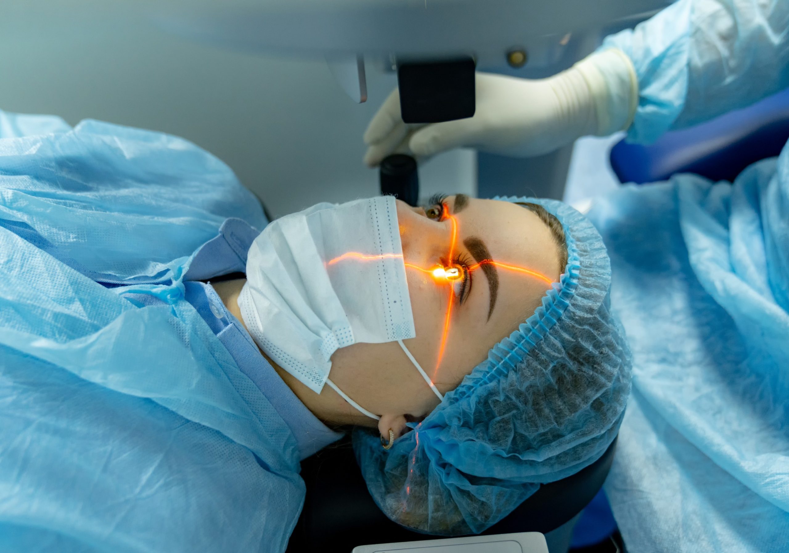
If medications aren’t enough, laser therapy can be an effective next step. Laser treatments are minimally invasive and often performed in a doctor’s office. 

Common types include: 

  • Laser trabeculoplasty: Used for open-angle glaucoma to improve fluid drainage. 
  • Laser iridotomy: Used for angle-closure glaucoma to create a small hole in the iris, allowing fluid to flow freely. 
  • Cyclophotocoagulation: Lowers eye pressure by reducing fluid production in the eye. 

These procedures can be repeated if pressure begins to rise again over time. 

Glaucoma Surgery 

When medications and laser treatments fail to adequately control intraocular pressure, surgery may be recommended. Modern glaucoma surgery is highly effective and often minimally invasive. 

1. Trabeculectomy 

A trabeculectomy creates a new drainage channel for the eye’s fluid to leave, lowering eye pressure. It’s one of the most common types of glaucoma surgery and can help reduce the need for medications afterward. 

2. Glaucoma Drainage Implants 

In this procedure, a tiny drainage device is implanted into the eye to help fluid escape more efficiently. These implants are especially useful in patients with advanced or complex glaucoma cases. 

3. Minimally Invasive Glaucoma Surgery (MIGS) 

MIGS procedures are newer, less invasive options that offer faster recovery times. They’re typically performed during cataract surgery and are designed to reduce IOP with fewer complications. 

Recovery and Aftercare 

After surgery, you may experience mild discomfort, redness, or blurry vision for a few days. It’s essential to follow all post-operative instructions and attend regular check-ups to monitor healing and ensure eye pressure remains stable. 

Frequently Asked Questions 

1. What is the first line of treatment for glaucoma? 

Eye drops are typically the first choice for most patients. They effectively reduce intraocular pressure and help prevent further optic nerve damage. 

2. What are the side effects of glaucoma surgery? 

Possible side effects include temporary blurred vision, infection, or fluctuations in eye pressure. Most patients recover well with proper care and follow-up visits. 

3. Can glaucoma be cured? 

There’s currently no cure for glaucoma, but treatment can slow or stop its progression, preserving vision for many years. 

4. How often should I see my eye doctor if I have glaucoma? 

Regular monitoring is critical. Most patients should see their ophthalmologist every 3–6 months to track eye pressure and optic nerve health. 

Conclusion 

Glaucoma can be managed effectively with early detection, consistent treatment, and proper follow-up care. Whether through medications, laser therapy, or surgery, the goal remains the same — to protect your vision and maintain your quality of life

If you suspect you may have glaucoma or have been diagnosed and need guidance, don’t wait. Early treatment can make all the difference. 

Take action today — Book an Eye Examination or Consult an expert Ophthalmologist to discuss the best glaucoma treatment options for you. 

Cataracts Treatment: Your Complete, Step-by-Step Guide

Cataracts are one of the most common causes of vision impairment across the world. This condition occurs when the lens of the eye becomes cloudy, leading to blurred vision and, if left untreated, potential blindness. 

The good news is that cataracts are highly treatable. With modern surgery and proper management, most people regain clear, sharp vision. This comprehensive guide explains cataract treatment options, management strategies, and preventive remedies to help you make informed decisions about your eye health. 

Understanding Cataracts and Their Impact on Vision 

A cataract forms when proteins in the eye’s natural lens break down and clump together, creating cloudiness that affects how light passes through the eye. Over time, this cloudiness worsens, making it harder to see clearly. 

Causes of Cataracts 

While aging is the most common cause, cataracts can develop due to several factors, including: 

  • Diabetes – high blood sugar can damage eye tissues. 
  • Smoking – increases oxidative stress in the lens. 
  • Prolonged UV exposure – sunlight can accelerate cataract formation. 
  • Eye injury or surgery – trauma may trigger cataract development. 
  • Certain medications – especially long-term steroid use. 

Common Symptoms of Cataracts 

Recognizing the early signs of cataracts is key to effective treatment. Symptoms often include: 

  • Blurred, dim, or cloudy vision 
  • Increased sensitivity to bright lights or glare 
  • Difficulty seeing at night 
  • “Halos” around lights 
  • Frequent changes in eyeglass or contact lens prescriptions 
  • Fading or yellowing of colors 
  • Double vision in one eye 

If you notice these symptoms, it’s time to schedule an eye exam. Early diagnosis can prevent vision loss and make treatment easier. 

Cataracts Treatment: What Are Your Options? 

The treatment approach depends on how much the cataract affects your daily life. In early stages, stronger lighting, updated glasses, or magnifying lenses may help. But when vision loss starts to impact your routine, surgery becomes the most effective solution

Surgical Procedures for Cataract Management 

Cataract surgery is one of the safest and most commonly performed procedures in the world, with a success rate above 95%. It’s typically done on an outpatient basis, meaning you can go home the same day. 

There are two main surgical methods: 

1. Phacoemulsification (Phaco Surgery) 

This modern technique uses ultrasound waves to break up the cloudy lens into tiny pieces, which are then gently removed through a small incision. A clear artificial intraocular lens (IOL) is implanted in its place. 

Benefits: 

  • Small incision 
  • Minimal discomfort 
  • Quick recovery time 

2. Extracapsular Cataract Extraction (ECCE) 

This method involves removing the entire lens in one piece through a slightly larger incision. The back part of the lens capsule remains intact to support the artificial lens. 

When It’s Used: 

  • For advanced cataracts that are too dense for phacoemulsification. 

Both procedures are highly effective in restoring vision. Your ophthalmologist will recommend the best method based on your eye health and specific needs. 

Post-Surgery Care and Recovery 

After cataract surgery, most patients notice clearer vision within a few days. The full healing process usually takes 6–8 weeks

What to Expect After Surgery 

  • Mild discomfort or itching for a few days 
  • Temporary blurry vision, which improves as your eye heals 
  • Use of prescribed eye drops to prevent infection and inflammation 
  • Avoid rubbing your eyes or heavy lifting for at least a week 

Your doctor will schedule follow-up visits to monitor healing and adjust medications if necessary. 

Most people are able to resume normal activities — including reading, watching TV, and light exercise — within a few days. 

Cataract Remedies: Lifestyle Changes and Prevention 

While surgery is the only way to remove cataracts, certain lifestyle habits can help slow their development and maintain healthy vision. 

1. Diet and Nutrition 

Eating foods rich in antioxidants, vitamins C and E, and carotenoids like lutein and zeaxanthin can protect the eyes from oxidative stress. 

Include these in your diet: 

  • Leafy greens (spinach, kale) 
  • Carrots and sweet potatoes 
  • Citrus fruits and berries 
  • Nuts and seeds 
  • Oily fish rich in omega-3s 

2. Protect Your Eyes 

  • Wear UV-protective sunglasses when outdoors. 
  • Quit smoking — it significantly increases cataract risk. 
  • Limit alcohol consumption. 
  • Manage chronic conditions such as diabetes and hypertension. 

3. Regular Eye Exams 

Early detection is key. Adults over 40 should have a comprehensive eye exam every 2 years, while those over 60 should be checked annually. Regular visits help track changes in vision and ensure prompt intervention. 

Frequently Asked Questions About Cataracts Treatment 

1: Is cataract surgery painful? 
No. The procedure is performed under local anesthesia, so you won’t feel pain. Mild discomfort afterward is common but temporary. 

2: How long does cataract surgery take? 
Usually about 15–30 minutes per eye. You’ll be able to go home the same day. 

3: Can cataracts come back after surgery? 
No, cataracts cannot grow back. However, some patients may develop a secondary cataract (posterior capsule opacification), which can be treated easily with a quick laser procedure. 

4: What are the risks of cataract surgery? 
Serious complications are rare but may include infection, inflammation, or retinal detachment. Following your doctor’s post-surgery care instructions minimizes these risks. 

5: How soon can I return to normal activities? 
Most people return to daily activities within a few days, though heavy lifting and strenuous exercise should be avoided for at least a week. 

In Summary 

Cataracts are a natural part of aging, but they don’t have to mean permanent vision loss. With today’s advanced cataract treatment options, patients can expect safe procedures, quick recovery, and excellent visual outcomes. 

If you’re experiencing blurry or cloudy vision, schedule an appointment with an eye specialist to discuss your treatment options. 

Remember: Early detection and timely cataract management can protect your vision and greatly improve your quality of life. 

Vitamin D Deficiency Treatment: Comprehensive Guide to Boosting Your Levels Naturally

Vitamin D plays a powerful role in keeping your body strong and healthy — from supporting bone strength to improving immune function and mood regulation. Yet, vitamin D deficiency affects millions of people globally, often without them even realizing it. 

This guide breaks down everything you need to know about vitamin D deficiency treatment — what causes it, how to identify it, and the best ways to naturally raise your vitamin D levels. 

Understanding Vitamin D Deficiency

Vitamin D is unique — it functions like a hormone and is produced when your skin is exposed to sunlight. You can also get it from certain foods and supplements. 

When your body doesn’t get enough sunlight or dietary vitamin D, levels drop, leading to a deficiency. Over time, this can affect bone density, muscle function, and even your immune system. 

Common causes of vitamin D deficiency include: 

  • Limited sun exposure (especially during winter months) 
  • Darker skin pigmentation, which reduces vitamin D synthesis 
  • Health conditions affecting absorption, such as Crohn’s or celiac disease 
  • Aging, which reduces the skin’s ability to produce vitamin D 
  • A diet lacking in vitamin D–rich foods 

Signs and Symptoms of Vitamin D Deficiency 

Vitamin D deficiency often develops slowly, and symptoms may not be immediately obvious. But over time, you might notice: 

  • Fatigue or low energy that doesn’t improve with rest 
  • Muscle weakness or cramps 
  • Bone or joint pain, particularly in the lower back or legs 
  • Frequent colds or infections due to a weakened immune system 
  • Low mood or depression 
  • Hair thinning or brittle nails 

If these symptoms sound familiar, ask your healthcare provider about checking your vitamin D levels with a simple blood test. 

How to Treat Low Vitamin D Levels 

The good news? Vitamin D deficiency can be corrected. Treatment focuses on raising your vitamin D levels through a combination of sunlight, diet, and supplementation. 

1. Get More Sunlight 

Your body naturally makes vitamin D when exposed to UVB rays. Aim for 10–30 minutes of midday sunlight several times a week, depending on your skin tone and where you live. Always balance sun exposure with skin protection. 

2. Eat Vitamin D–Rich Foods 

Add these foods to your diet for a natural boost: 

  • Fatty fish (salmon, mackerel, tuna, sardines) 
  • Fortified milk or plant-based alternatives 
  • Egg yolks 
  • Beef liver 
  • Fortified cereals 

3. Take Vitamin D Supplements 

If sunlight and diet aren’t enough, supplements can help restore healthy levels. Always consult your doctor before starting supplementation to determine the correct dosage. 

Supplements for Vitamin D Deficiency 

There are two main forms of vitamin D supplements: 

  • Vitamin D2 (ergocalciferol) – plant-derived and often used in fortified foods. 
  • Vitamin D3 (cholecalciferol) – animal-based and more effective at raising and maintaining blood vitamin D levels. 

Your doctor may recommend a specific dosage based on your blood test results. Typical recommendations for adults range between 600–800 IU daily, though higher doses may be prescribed for severe deficiency. 

For best absorption: 

  • Take supplements with meals containing healthy fats. 
  • Follow your doctor’s guidance to avoid taking too much, as excessive vitamin D can cause toxicity. 

FAQs 

1. What causes vitamin D deficiency? 
The most common causes include limited sun exposure, a poor diet, or health conditions that reduce vitamin absorption. 

2. How much vitamin D do I need each day? 
Most adults need around 600–800 IU per day. However, your exact needs depend on your age, health, and where you live. 

3. Can you take too much vitamin D? 
Yes. Excessive vitamin D intake can lead to high calcium levels, nausea, weakness, and kidney problems. Always take supplements as advised by a healthcare provider. 

4. How long does it take to correct a deficiency? 
Mild deficiencies can often be corrected within a few months of consistent supplementation, while more severe cases may take longer. 

Conclusion 

Addressing vitamin D deficiency is one of the simplest yet most impactful steps you can take for your long-term health. Sunlight, diet, and supplements — used wisely — can help restore balance and protect your bones, muscles, and immune system. 

If you suspect a deficiency, don’t self-diagnose. A quick blood test and a personalized treatment plan from your orthopedic expert can help you safely boost your vitamin D levels. 

Take action today: 

  • Consume Vitamin D Supplements to start supporting your body naturally. 

Understanding Vitamin D Deficiency: Causes, Symptoms, and Solutions

Introduction 

Vitamin D — often called the sunshine vitamin — is essential for maintaining good health. Despite its importance, many people don’t get enough of it. Vitamin D deficiency affects millions worldwide and can lead to a range of health problems, from fatigue to bone disorders. 

This guide breaks down what causes low vitamin D levels, how to spot the symptoms, and what you can do to prevent or correct a deficiency. 

What Is Vitamin D Deficiency? 

Vitamin D deficiency happens when your body doesn’t have enough vitamin D to function properly. This nutrient helps your body absorb calcium — a key mineral for bone strength. 

Historically, severe deficiency has been linked to rickets, a childhood bone disorder that leads to soft, weak, or deformed bones. In adults, a long-term deficiency can result in osteomalacia or contribute to osteoporosis. 

Causes of Vitamin D Deficiency 

Several factors can limit your body’s ability to produce or absorb vitamin D. Understanding these helps in both prevention and treatment. 

1. Limited Sunlight Exposure 

Your skin produces vitamin D when it’s exposed to sunlight. People who spend little time outdoors, live in northern regions, or wear clothing that covers most of their skin are at higher risk. 

2. Darker Skin Tone 

Melanin, the pigment that gives skin its color, reduces the skin’s ability to make vitamin D from sunlight. This means individuals with darker skin may need more sun exposure to produce adequate levels. 

3. Kidney and Liver Conditions 

Both organs help convert vitamin D into its active form. Diseases affecting the kidneys or liver can reduce this conversion, leading to lower levels of usable vitamin D. 

4. Digestive Disorders 

Conditions such as Crohn’s disease, celiac disease, or cystic fibrosis can interfere with fat absorption — and since vitamin D is fat-soluble, this limits how much your body can absorb from food. 

Symptoms of Low Vitamin D Levels 

The symptoms of vitamin D deficiency can vary and are often subtle at first. Some of the most common include: 

  • Persistent fatigue or tiredness 
  • Bone or back pain 
  • Low mood or depression 
  • Muscle weakness or cramps 
  • Hair thinning or loss 
  • Slow healing of wounds 

If you’re noticing several of these symptoms, it may be time to check your vitamin D levels with a simple blood test. 

How to Prevent and Treat Vitamin D Deficiency 

The best way to maintain healthy vitamin D levels is through a mix of sunlight, diet, and supplements when necessary. 

1. Get Regular Sun Exposure 

Spending 10–30 minutes in the sun a few times a week can help your body produce enough vitamin D. However, the exact time needed depends on factors like skin tone, location, and season. 

2. Eat Foods Rich in Vitamin D 

Add these foods to your diet for a natural boost: 

  • Fatty fish (salmon, mackerel, sardines) 
  • Egg yolks 
  • Fortified dairy products 
  • Liver 
  • Fortified cereals 

3. Take Vitamin D Supplements 

For many people — especially those with limited sun exposure — supplements are the most reliable way to reach optimal levels. Always talk to a healthcare provider before starting supplements to determine the right dosage. 

Frequently Asked Questions (FAQs)

1: Can vitamin D deficiency lead to serious health issues? 
Yes. Prolonged vitamin D deficiency has been linked to osteoporosis, weakened immunity, cardiovascular disease, and certain cancers. 

2: Can I get enough vitamin D through diet alone? 
It’s possible, but difficult. Sunlight remains the main natural source, and many people benefit from adding a supplement to meet their needs. 

3: How long does it take to correct a vitamin D deficiency? 
This depends on how severe the deficiency is and the treatment plan. With supplementation and sunlight exposure, levels often improve within a few months. 

Conclusion 

Understanding vitamin D deficiency is the first step toward protecting your long-term health. By getting regular sun exposure, eating vitamin D–rich foods, and supplementing when needed, you can maintain healthy levels and prevent complications. 

If you suspect you may be deficient, don’t ignore it — a quick blood test and a tailored plan from your healthcare provider can make a big difference. 

Take charge of your health today. Get your vitamin D levels checked and start making small, effective changes toward better wellbeing. 

Contact our orthopedics and sports medicine team for more information or to schedule a consultation about your vitamin D status. 

Shoulder Pain Management: Your Comprehensive Guide

When it comes to shoulder pain management, the range of available options can feel overwhelming. From over-the-counter remedies to structured physical therapy programs, there are many strategies to help reduce discomfort, restore mobility, and prevent recurrence. This guide breaks down the key approaches so you can better understand what works, when to use it, and why consulting a healthcare professional is essential. 

Understanding Shoulder Pain 

Your shoulder is the most mobile joint in the human body, allowing you to lift, rotate, and extend your arm in multiple directions. This incredible range of motion, however, also makes it prone to injuries and chronic conditions. 

Effective management starts with identifying the underlying cause and seeking early guidance from orthopedic experts who can tailor your treatment plan. 

Common Causes of Shoulder Pain

  • Injuries: Dislocations, rotator cuff tears, sprains, and fractures 
  • Chronic conditions: Arthritis, bursitis, tendonitis, or frozen shoulder 
  • Repetitive strain: Overhead movements in sports or work 
  • Postural issues: Poor ergonomics or slouching over long periods 

The orthopedics and sports medicine services at Burjeel Royal Hospital, Al Ain address these causes through precise diagnosis, advanced imaging, and individualized rehabilitation programs. 

Symptoms of Shoulder Pain 

  • Stiffness or limited range of motion 
  • Sharp or aching pain during movement 
  • Weakness in the arm or shoulder 
  • Swelling, redness, or warmth around the joint 

If these symptoms persist beyond a few days, it’s best to consult our orthopedic specialists, for an accurate evaluation. 

Medications for Shoulder Pain 

Medication can provide short-term relief and reduce inflammation, allowing you to move more comfortably while addressing the root cause. 

Over-the-Counter Medications 

  • NSAIDs (ibuprofen, naproxen): Reduce pain and inflammation 
  • Acetaminophen: Helps with mild to moderate pain but does not reduce inflammation 
  • Topical creams or gels: Applied directly to the skin for localized relief 

Prescription Medications 

When pain becomes severe or chronic, orthopedic experts may recommend: 

  • Stronger NSAIDs or corticosteroid injections 
  • Muscle relaxants for spasms 
  • Opioid medications (short-term, for acute pain only) 

NOTE: These medications must be used under medical supervision due to potential side effects. 

Physical Therapy for Shoulder Pain 

Physical therapy (PT) is one of the most effective non-invasive strategies for shoulder pain management. Physiotherapy is a cornerstone of shoulder pain management at Burjeel Royal Hospital, Al Ain. It plays a key role in restoring movement and preventing recurrence. 

Benefits of Physical Therapy 

  • Restores mobility and flexibility 
  • Strengthens supportive muscles around the joint 
  • Reduces pain and stiffness 
  • Prevents future injuries 

Techniques Used in Physiotherapy 

  • Targeted exercises for strength and range of motion 
  • Manual therapy and massage to release tension 
  • Stretching programs tailored to specific conditions 
  • Posture correction training to reduce strain on the joint 

The orthopedics and sports medicine team at Burjeel Royal Hospital ensures every program is tailored to your specific condition—whether post-injury, post-surgery, or due to chronic issues. 

Alternative Shoulder Pain Relief Strategies 

In addition to medication and physical therapy, several alternative approaches can support recovery and long-term management. 

Hot and Cold Therapy 

  • Cold compresses: Reduce swelling and numb sharp pain 
  • Heat therapy: Relaxes stiff muscles and improves blood flow 

Lifestyle Changes 

  • Posture awareness: Sitting and standing upright reduces stress on the shoulder 
  • Workstation ergonomics: Proper desk and chair alignment prevent strain 
  • Regular stretching: Keeps the joint flexible 
  • Activity modification: Avoiding repetitive overhead movements when possible 

Some patients also benefit from complementary care like acupuncture, yoga, or osteopathy—under the guidance of orthopedic specialists

FAQs 

1: Can I manage shoulder pain at home? 

Yes, mild shoulder pain can often be managed with rest, ice/heat therapy, over-the-counter pain relievers, and gentle stretching. 

2: When should I see an orthopedic specialist? 

If your pain lasts more than a week, worsens despite rest, or limits your motion, visit the orthopedics and sports medicine department at Burjeel Royal Hospital, Al Ain

3: Is surgery always required for shoulder pain? 

No. Most cases improve with conservative management such as medication, PT, and lifestyle changes. Surgery is typically reserved for severe injuries or advanced arthritis. 

Conclusion 

Effective shoulder pain management requires a comprehensive approach. Medications may provide short-term relief, but physical therapy, lifestyle adjustments, and preventive strategies are key to long-term improvement. 

At Burjeel Royal Hospital, Al Ain, our orthopedic specialists and sports medicine experts are committed to helping you regain mobility, strength, and confidence. With advanced orthopedics and sports medicine services, we ensure that every patient receives personalized care for faster, safer recovery. 

Knee Ligament Tear: Causes, Symptoms, and Essential Recovery Steps

Knee ligament injuries are among the most frequent injuries in sports and daily activities. They range from minor strains to complete tears that may require surgery and months of rehabilitation. Knowing how these injuries happen, what symptoms to look for, and the best treatment options can make a big difference in recovery and prevention. 

Understanding the Anatomy of the Knee 

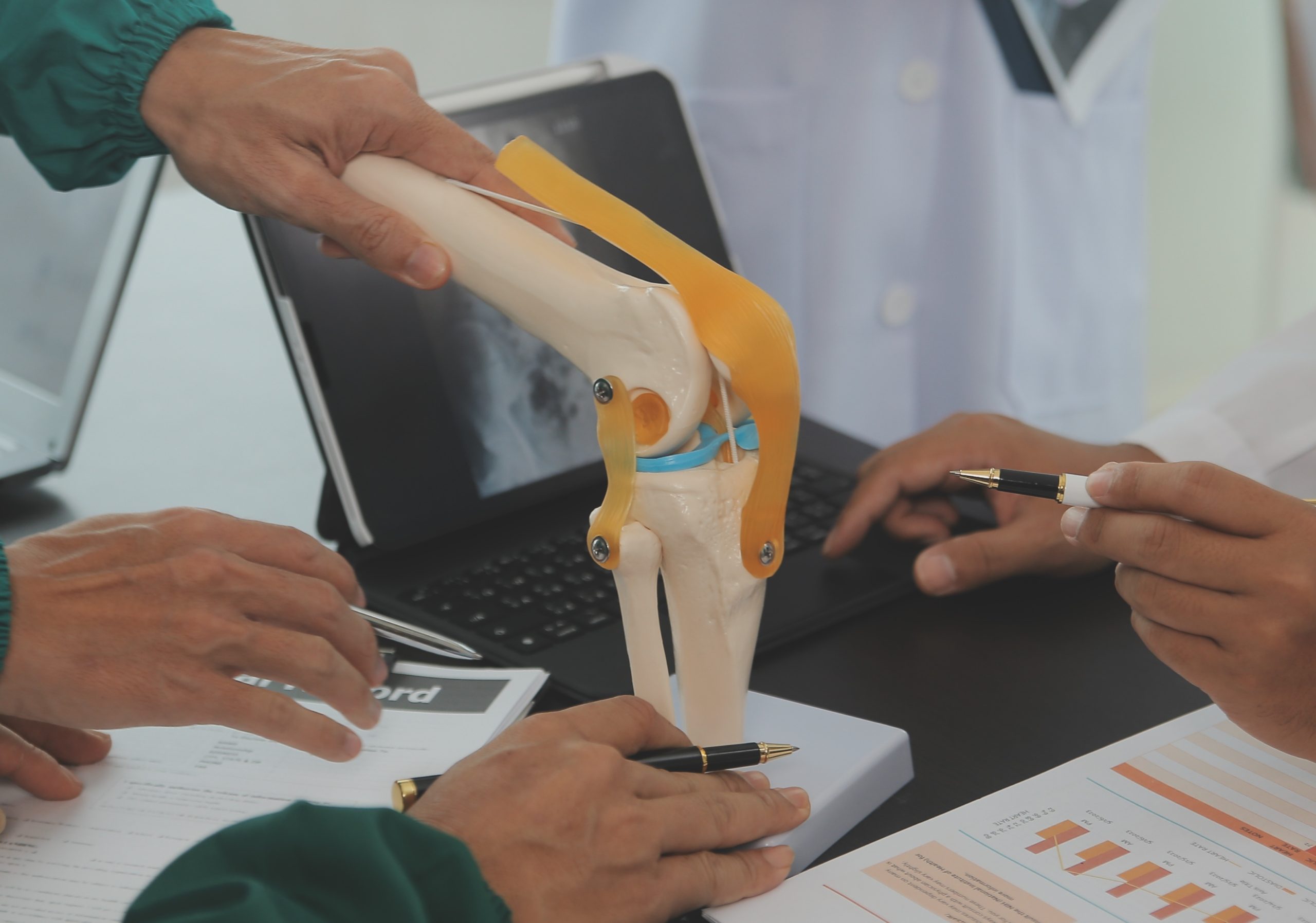
The knee is one of the largest and most complex joints in the body. It connects the thigh bone (femur) to the shin bone (tibia) and is supported by four main ligaments: 

  • Anterior Cruciate Ligament (ACL): Prevents the tibia from sliding forward. 
  • Posterior Cruciate Ligament (PCL): Prevents the tibia from sliding backward. 
  • Medial Collateral Ligament (MCL): Provides stability on the inner side of the knee. 
  • Lateral Collateral Ligament (LCL): Provides stability on the outer side of the knee. 

Together, these ligaments keep the knee stable during movement and absorb stress from activity. 

Different Types of Knee Ligament Injuries 

ACL Injury 

The ACL is the most commonly injured ligament. It’s often torn during sports that involve sudden stops, pivots, or awkward landings. 

PCL Injury 

The PCL is injured less frequently. It usually happens in car accidents or contact sports when the shin is forced backward. 

MCL Injury 

The MCL is commonly injured by a direct blow to the outside of the knee, pushing it inward. This is frequent in football, rugby, and skiing. 

LCL Injury 

The LCL is the least commonly injured ligament. Damage occurs when the inside of the knee is hit, forcing the joint outward. 

Causes of Knee Ligament Tears 

Knee ligament injuries are often caused by sudden, forceful movements that strain the knee beyond its normal range of motion. Some common causes include: 

  • Sudden changes in direction: Quick pivots, shifts, or sudden stops in sports can lead to a ligament tear, particularly in high-intensity activities like soccer, basketball, and skiing. 
  • Improper landings: Landing incorrectly after a jump can put excess stress on the ligaments, causing them to stretch or tear. 
  • Direct impacts: Contact sports such as football and rugby often involve direct blows to the knee, which can damage ligaments. 
  • Twisting or overstretching: Actions that cause the knee to twist awkwardly or stretch too far can result in tears, especially in the anterior cruciate ligament (ACL). 

Symptoms of Knee Ligament Tears 

The severity of symptoms can vary depending on the ligament affected and the extent of the injury. Some common symptoms include: 

  • Pain: This can range from mild to severe, often occurring immediately after the injury. Pain tends to be sharp and intense, especially with movement. 
  • Swelling: Swelling usually occurs within hours of the injury, and the knee may become noticeably larger and more tender. 
  • Instability: You may experience a feeling of the knee “giving way” or a loss of stability, making it difficult to walk or stand. 
  • Reduced range of motion: The knee may feel stiff, and bending or straightening it could be painful or difficult. 

Essential Recovery Steps and Treatments 

Treatment for knee ligament tears varies depending on the severity of the injury. Here’s an overview of the recovery steps and treatments available at Burjeel Royal Hospital Asharej: 

  1. Initial Treatment (RICE Protocol)
    For mild injuries or sprains, the first step in treatment is the RICE method: 
  • Rest: Avoid putting weight on the knee to prevent further damage. 
  • Ice: Apply ice packs to reduce swelling and manage pain. 
  • Compression: Use an elastic bandage or brace to support the knee and limit swelling. 
  • Elevation: Keep the knee elevated to help reduce swelling. 
  1. Physical Therapy
    For moderate injuries, physical therapy is essential to regain strength and mobility. A physical therapist will guide you through exercises that focus on strengthening the muscles around the knee, such as the hamstrings and quadriceps, to help stabilize the joint. 
  1. Bracing and Activity Modification
    In cases of moderate to severe tears, a knee brace may be used to provide support during movement. Activity modifications are also recommended to prevent further strain on the ligament while it heals. 
  1. Surgical Intervention
    For severe tears, particularly of the ACL, surgical intervention may be required. This is often followed by a lengthy rehabilitation period to restore the knee’s functionality. At Burjeel Royal Hospital Asharej, our orthopedic surgeons specialize in minimally invasive knee surgeries, such as ACL reconstruction, to ensure a quicker recovery with less risk of complications. 
  1. Rehabilitation
    Following surgery, rehabilitation is critical. A structured physical therapy program will help restore strength, flexibility, and stability to the knee. This phase of recovery may take several months, with most patients returning to normal activities within 6 to 12 months, depending on the severity of the tear. 

Treatments Offered at Burjeel Royal Hospital Asharej 

At Burjeel Royal Hospital Asharej, we offer advanced, personalized treatments for knee ligament injuries: 

  • Consultation with Expert Orthopedic Surgeons: Our team of orthopedic surgeons specializes in knee injuries and provides tailored treatment plans based on your unique condition. 
  • Advanced Imaging and Diagnostics: We utilize state-of-the-art imaging techniques, such as MRI and X-rays, to accurately diagnose the extent of the injury and create an effective treatment strategy. 
  • Minimally Invasive Surgery: We perform minimally invasive surgeries that reduce recovery time and minimize scarring, allowing for a faster return to activity. 
  • Comprehensive Rehabilitation Programs: Our physical therapists offer customized rehabilitation programs to ensure full recovery, improve range of motion, and strengthen the muscles around the knee. 

Prevention Strategies 

While not every injury can be avoided, you can lower your risk by: 

  • Strengthening muscles around the knee (especially hamstrings and quadriceps) 
  • Improving flexibility and balance 
  • Using proper sports techniques 
  • Wearing appropriate shoes and protective gear 
  • Avoiding sudden, uncontrolled movements 

Frequently Asked Questions 

1) What is the recovery time for a knee ligament injury? 

It depends on the ligament and severity. Mild sprains may heal in weeks, while ACL reconstruction can take 6–12 months. 

2) Can you walk with a knee ligament injury? 

Yes, in mild cases. But with severe tears, walking may be very difficult without support. 

3) What happens if a knee ligament injury is left untreated? 

Chronic instability, worsening pain, and long-term joint damage such as early arthritis. 

Conclusion 

Knee ligament tears can significantly impact your mobility, but with prompt treatment and proper rehabilitation, most people can fully recover and return to their normal activities. If you suspect you have a knee ligament injury, don’t wait. Contact Burjeel Royal Hospital Asharej to schedule a consultation with our specialized orthopedic doctors and begin your path to recovery today. 

Causes of Shoulder Pain: A Comprehensive Look at the Source of Your Discomfort 

Shoulder pain is a common issue that can disrupt work, hobbies, and even restful sleep. By understanding the causes of shoulder pain, you can take proactive steps to manage symptoms, prevent worsening, and find lasting relief. This guide explores the most frequent culprits—ranging from injuries and arthritis to shoulder bursitis. 

Table of Contents 

  1. Introduction to Shoulder Pain 
  1. Common Shoulder Injuries 
  1. Dislocations, Sprains, and Strains 
  1. Frozen Shoulder and Rotator Cuff Tears 
  1. Arthritis in the Shoulder 
  1. Understanding Shoulder Bursitis 
  1. FAQs 
  1. Conclusion 

Introduction to Shoulder Pain 

The shoulder is a complex joint with an exceptional range of motion. While this flexibility allows us to reach, lift, and rotate with ease, it also makes the shoulder more vulnerable to injuries and degenerative conditions. Identifying the root causes of shoulder pain is the first step toward proper treatment and long-term relief. 

Common Shoulder Injuries 

Because of its mobility, the shoulder is susceptible to both sudden trauma and chronic overuse injuries. 

Dislocations, Sprains, and Strains 

Falls, quick movements, or accidents often lead to shoulder dislocations, sprains, or strains. These injuries typically result in: 

  • Sudden, sharp pain 
  • Swelling and inflammation 
  • Limited mobility and difficulty lifting the arm 

Frozen Shoulder and Rotator Cuff Tears 

Some shoulder problems develop more gradually. Frozen shoulder (adhesive capsulitis) causes stiffness and reduced range of motion, often lasting months. Meanwhile, rotator cuff tears—either partial or complete—occur due to repetitive strain, aging, or acute injury. Both can significantly limit daily activities if left untreated. 

Arthritis in the Shoulder 

Arthritis in the shoulder is a common cause of chronic pain, especially in individuals over 50. However, it can also result from prior injuries or overuse earlier in life. Key symptoms include: 

  • Deep, aching shoulder pain 
  • Swelling around the joint 
  • Stiffness, particularly in the morning or after inactivity 

While arthritis cannot be reversed, treatments focus on managing symptoms and maintaining joint function. 

Understanding Shoulder Bursitis 

Shoulder bursitis occurs when the bursa—small, fluid-filled sacs that cushion the joint—becomes inflamed. This often develops from repetitive motion or direct trauma. Symptoms include: 

  • Tenderness at the top of the shoulder 
  • Swelling and warmth in the area 
  • Pain that worsens with overhead activity 

Proper rest, anti-inflammatory measures, and physical therapy can usually help resolve the condition. 

FAQs 

What are the most common causes of shoulder pain? 

The leading causes include acute injuries (dislocations, sprains, strains), chronic conditions such as frozen shoulder or rotator cuff tears, arthritis, and shoulder bursitis. 

How is shoulder pain diagnosed? 

Doctors use a combination of medical history, physical examination, and imaging tests like X-rays, ultrasound, or MRI to determine the underlying cause. 

How is shoulder pain treated? 

Treatment varies depending on the cause. Options range from rest, physiotherapy, and medications to injections or surgery in severe cases. 

Conclusion 

Knowing what’s behind your shoulder pain is the first step toward lasting relief. From sports injuries to joint conditions, there are effective treatment options to improve strength and flexibility. The orthopedic and physiotherapy experts at Burjeel Hospital Abu Dhabi combine advanced diagnostics with expert physiotherapy to ensure a faster, more complete recovery. Don’t let shoulder pain hold you back—get the right care today.

Book your consultation now and start your journey toward pain-free movement.

Understanding ACL Injury: Causes, Diagnosis, and Rehabilitation 

ACL injuries are among the most common sports-related injuries, affecting athletes of all levels—from recreational players to professionals. A torn ACL doesn’t just sideline you from your favorite activity; it also demands time, patience, and the right care to recover fully. This comprehensive guide explains everything you need to know about ACL injuries, from how they happen to how you can rehabilitate and return to your sport. 

What is an ACL Injury? 

The Anterior Cruciate Ligament (ACL) is one of four major ligaments in your knee. Its primary role is to stabilize the knee joint and control back-and-forth motion. 

An ACL injury, often called a torn ACL, occurs when the ligament is overstretched or torn due to sudden stress. These injuries are especially common in high-impact sports that require quick pivots, jumps, and direction changes. 

Causes of ACL Injury 

Most ACL injuries happen during sports or physical activities that involve: 

  • Sudden stops or rapid changes in direction 
  • Jumping and landing awkwardly 
  • Direct collisions with another player 
  • Twisting the knee beyond its normal range of motion 

Sports with higher ACL injury rates include soccer, basketball, football, and skiing

Risk Factors for ACL Injury 

While anyone can suffer an ACL tear, certain factors increase the risk: 

  • Gender differences: Female athletes are statistically more likely to experience ACL injuries due to anatomical and hormonal factors. 
  • Improper footwear: Shoes that don’t provide proper support increase instability. 
  • Playing surface conditions: Uneven or poorly maintained fields raise injury risk. 
  • Previous knee injuries: Past ligament or meniscus issues can make the ACL more vulnerable. 

Symptoms of ACL Injury 

Signs of an ACL tear are usually immediate and difficult to ignore. Common symptoms include: 

  • A loud “pop” at the moment of injury 
  • Sudden, severe knee pain 
  • Rapid swelling within hours 
  • Instability or the knee “giving way” 
  • Difficulty bearing weight on the injured leg 

Diagnosing an ACL Injury 

If an ACL tear is suspected, a healthcare professional will carry out: 

  • Physical examination: Tests like the Lachman test or pivot-shift test check for knee stability. 
  • Imaging tests: 
  • X-ray to rule out bone fractures. 
  • MRI to confirm ligament damage and evaluate associated injuries like meniscus tears. 

Accurate diagnosis is essential to creating the right treatment plan. 

Treatment Options for ACL Injury 

Treatment varies depending on the severity of the injury, the patient’s age, and activity level. 

Non-Surgical Options 

  • Rest, ice, compression, and elevation (RICE method
  • Physical therapy to restore strength and mobility 
  • Bracing to stabilize the knee during activity 

Surgical Options 

For athletes and highly active individuals, ACL reconstruction surgery is often recommended. This involves replacing the torn ligament with a graft from the patient’s own body (autograft) or a donor (allograft). 

ACL Rehabilitation 

Rehabilitation is the most critical part of ACL recovery—whether surgery is performed or not. The process typically involves: 

  • Phase 1: Reducing pain and swelling, regaining basic motion 
  • Phase 2: Strengthening exercises for quadriceps and hamstrings 
  • Phase 3: Balance and stability training 
  • Phase 4: Sport-specific drills and functional exercises 
  • Phase 5: Gradual return to play, usually 6–12 months after injury (post-surgery) 

Commitment to rehab is the single most important factor in regaining full knee function. 

FAQs about ACL Injury 

Q: How long does it take to recover from an ACL injury? 
A: Recovery time varies, but after surgery, most athletes return to sport within 6–12 months. Non-surgical cases may recover faster, depending on severity. 

Q: Is surgery always necessary for an ACL injury? 
A: Not always. Less active individuals may benefit from physiotherapy alone, but surgery is usually advised for athletes or those whose lifestyles demand high knee stability. 

Q: Can ACL injuries be prevented? 
A: While not all injuries can be prevented, you can reduce your risk by strengthening leg muscles, practicing proper landing techniques, using supportive footwear, and training on safe surfaces. 

Conclusion 

Understanding an ACL injury is the first step toward prevention and complete recovery. If you suspect a tear, seek prompt medical attention. At Burjeel Hospital, Abu Dhabi, our orthopedic and rehabilitation experts provide advanced diagnosis, treatment, and physiotherapy to help you regain full mobility and confidence.

Take care of your knees—they’re the foundation of every step, jump, and stride. 

Our Experts

Transforming Mobility – Liposuction for Grade 3 Lipedema

Introduction 

Living with Lipedema can be both painful and life-limiting. A 37-year-old patient came to Burjeel Hospital after years of struggling with massive swelling in both lower limbs, causing severe pain and restricting mobility. Despite seeking help from multiple physicians, her condition persisted until a specialized vascular surgery team diagnosed her with Grade 3 Lipedema

Understanding Lipedema 

Lipedema is a chronic medical condition marked by abnormal fat accumulation in the lower body, often misdiagnosed as obesity or lymphedema. In advanced stages, it severely impacts mobility and quality of life. 

Treatment Approach 

At Burjeel Hospital, the patient underwent suction-assisted liposuction—the only proven effective treatment for Lipedema. Non-surgical approaches such as compression therapy and diet changes offer limited relief but do not resolve the underlying issue. 

Surgical Success 

The first stage of her treatment involved a 4.5-hour procedure where 10 liters of pathological fat were removed from her thighs and calves. The surgery was performed with precision through multiple small incisions, ensuring minimal scarring and optimal outcomes. 

Recovery and Results 

Post-surgery, the patient enrolled in a comprehensive rehabilitation program, including manual lymphatic drainage and physiotherapy. Within weeks, she experienced significant pain relief and regained her mobility. Grateful for the holistic care, she shared her satisfaction with the multidisciplinary team at Burjeel Holdings. 

Conclusion 

This case demonstrates how advanced surgical care, rehabilitation, and patient-centered treatment can restore not only physical function but also confidence and quality of life for Lipedema patients. 

Our Doctors

Robotic Esophageal Cancer Surgery in High-Risk Elderly Patient: A Medical Breakthrough

When a 74-year-old man with multiple serious health conditions was diagnosed with aggressive esophageal cancer, many might have considered his case too risky for major surgery. However, the expert team at Burjeel Medical City proved that age and comorbidities don’t have to be barriers to life-saving treatment when advanced robotic surgery techniques are employed.

A Complex Cancer Diagnosis in a High-Risk Patient

The patient’s journey began with chest discomfort in his home country. What started as routine medical evaluation quickly revealed a serious diagnosis that would challenge even the most experienced oncological teams.

Initial Discovery

Routine endoscopy revealed concerning findings:

  • Mucosal lesion in the mid to lower esophagus
  • Hiatus hernia with Barrett’s esophagus
  • Histopathology results showing poorly differentiated signet ring cell adenocarcinoma

Signet ring cell adenocarcinoma is particularly aggressive, known for its poor prognosis and challenging treatment requirements, especially in elderly patients.

The Challenge of Multiple Comorbidities

What made this case extraordinarily complex was the patient’s extensive medical history:

  • Previous coronary artery bypass grafting (CABG)
  • Moderate aortic stenosis
  • Hypertension and Type 2 diabetes
  • Chronic kidney disease
  • Hyperlipidemia
  • Interstitial lung disease

This combination of conditions would typically make a patient unsuitable for major surgery, particularly the extensive procedure required for esophageal cancer treatment.

Advanced Staging and Treatment Planning

Upon arrival in the UAE for specialized care, comprehensive staging was performed:

Imaging Studies

  • CT scan revealed eccentric wall thickening of the mid-esophagus
  • PET scan showed lymph nodes in paratracheal, subcarinal, and paraesophageal regions
  • EBUS biopsy of suspicious lymph nodes was negative for malignancy

Multidisciplinary Team (MDT) Approach

The case was presented to Burjeel’s specialized oncology MDT, which includes:

  • Gastrointestinal surgeons
  • Medical oncologists
  • Radiation oncologists
  • Anesthesiologists
  • Pathologists

The team’s consensus was to proceed with neoadjuvant FLOT chemotherapy followed by surgical re-evaluation.

Neoadjuvant Chemotherapy: Preparing for Surgery

FLOT Chemotherapy Protocol

The patient received four courses of FLOT chemotherapy, a specialized regimen that includes:

  • 5-Fluorouracil
  • Leucovorin
  • Oxaliplatin
  • Docetaxel

This intensive treatment aims to:

  • Shrink the primary tumor
  • Eliminate microscopic disease
  • Improve surgical outcomes
  • Reduce risk of recurrence

Response Evaluation

After completing chemotherapy, comprehensive re-staging showed no disease progression, making the patient an excellent candidate for surgical intervention despite his high-risk profile.

Revolutionary Robotic Surgery: RAMIE Technique

Dr. Ali Iyoob Valiyaveettil, Consultant and Head of Gastrointestinal Surgery, led the team in performing Robot-Assisted Minimally Invasive Esophagectomy (RAMIE) – a cutting-edge technique that represents the future of esophageal cancer surgery.

Why Robotic Surgery for High-Risk Patients?

Traditional open esophagectomy is one of the most challenging surgeries in oncology, with significant risks including:

  • Major blood loss
  • Extended recovery times
  • High complication rates
  • Prolonged hospital stays

RAMIE offers revolutionary advantages:

  • Minimally invasive approach reduces surgical trauma
  • Enhanced precision with robotic arms
  • 3D high-definition visualization
  • Improved maneuverability in confined spaces
  • Reduced post-operative pain
  • Faster recovery times

The Complex Three-Stage Procedure

The surgery involved multiple phases, each requiring exceptional surgical skill:

Stage 1: Thoracoscopic Phase

  • Patient positioned prone for optimal access
  • Radical esophagectomy with complete tumor removal
  • Comprehensive lymph node dissection of mediastinal nodes
  • Preservation of vital structures

Stage 2: Abdominal Phase

  • Gastric conduit preparation using the stomach to replace the esophagus
  • ICG (Indocyanine Green) test to assess blood supply adequacy
  • Pyloroplasty to improve gastric emptying
  • Careful preservation of gastric blood vessels

Stage 3: Cervical Phase

  • Neck incision for anastomosis creation
  • Side-to-side connection between gastric conduit and remaining esophagus
  • Meticulous technique to prevent leakage
  • No feeding tubes required due to superior surgical technique

Advanced Intraoperative Technology

The procedure utilized state-of-the-art technology:

Da Vinci Xi Robotic System

  • Four robotic arms with enhanced dexterity
  • 3D high-definition visualization
  • Tremor elimination for precise movements
  • Scaled motion for fine tissue manipulation

ICG Fluorescence Imaging

  • Real-time assessment of tissue blood supply
  • Prevention of anastomotic complications
  • Immediate visualization of perfusion quality

Expert Surgical Team Excellence

The successful outcome was achieved through the collaboration of:

Remarkable Recovery and Outcomes

Despite the patient’s high-risk profile, the results exceeded expectations:

Immediate Post-Operative Period

  • ICU monitoring for 2 days as precaution
  • Nasogastric tube removal after 48 hours
  • Contrast swallow study showing normal passage with no leaks
  • Early mobilization reducing complications

Rapid Recovery Milestones

  • Oral feeding started within 48 hours
  • Progression to puree diet without problems
  • Chest tube removal after 72 hours
  • Abdominal drain removal after 72 hours
  • No voice changes or breathing difficulties
  • Clean wound healing

Hospital Discharge

The patient was discharged on the 7th post-operative day – remarkably fast for such extensive surgery, especially in an elderly patient with multiple comorbidities.

Pathology Results and Long-Term Prognosis

Histopathology Findings

  • Poorly differentiated adenocarcinoma (Grade 3)
  • ypT3N1 staging (1 out of 26 lymph nodes positive)
  • Good response to neoadjuvant chemotherapy
  • Clear surgical margins

Unexpected Finding

Multiple lymph nodes showed caseating granulomatous changes, indicating possible concurrent inflammatory conditions that were successfully managed.

The Advantages of Minimally Invasive Esophagectomy

This case demonstrates why RAMIE is becoming the gold standard for esophageal cancer surgery:

Reduced Morbidity

  • Lower complication rates compared to open surgery
  • Decreased blood loss during the procedure
  • Reduced post-operative pain
  • Faster return to normal activities

Superior Oncological Outcomes

  • Better lymph node clearance due to enhanced visualization
  • Reduced positive margin rates
  • Improved long-term survival in many studies

Enhanced Recovery

  • Shorter ICU stays reduce healthcare costs
  • Earlier hospital discharge improves patient satisfaction
  • Faster return to baseline function

Overcoming the Learning Curve Challenge

One of the traditional barriers to minimally invasive esophagectomy has been the steep learning curve for surgeons. However, robotic assistance helps overcome this challenge:

Robotic Advantages for Surgeons

  • Intuitive movements that mirror hand motions
  • Enhanced dexterity in confined spaces
  • Tremor elimination for precise dissection
  • 3D visualization improves depth perception

Training and Expertise

Burjeel Medical City’s commitment to surgical excellence includes:

  • Specialized training programs for robotic surgery
  • Continuous education in latest techniques
  • Volume requirements ensuring surgeon proficiency
  • Multidisciplinary collaboration

Why Choose Burjeel for Complex Cancer Surgery?

This successful case highlights several key advantages:

Advanced Technology

  • Latest Da Vinci Xi robotic system
  • Integrated OR technology for seamless procedures
  • Real-time imaging capabilities
  • Comprehensive monitoring systems

Specialized Expertise

  • High-volume esophageal surgery program
  • Experienced robotic surgeons
  • Multidisciplinary cancer care team
  • Specialized anesthesia for complex cases

Comprehensive Care

  • Neoadjuvant and adjuvant therapy coordination
  • Nutritional support services
  • Rehabilitation programs
  • Long-term follow-up care

The Future of Esophageal Cancer Treatment

This case represents the evolution toward personalized, minimally invasive cancer care:

Precision Medicine

  • Molecular profiling of tumors for targeted therapy
  • Personalized treatment protocols based on patient factors
  • Biomarker-guided therapy selection

Technological Advances

  • Enhanced robotic systems with improved capabilities
  • Artificial intelligence integration for surgical planning
  • Advanced imaging for better staging and monitoring

Patient Selection: Making High-Risk Surgery Safer

This case demonstrates that with proper evaluation and advanced techniques, many patients previously considered unsuitable for surgery can achieve excellent outcomes:

Comprehensive Pre-Operative Assessment

  • Cardiac evaluation and optimization
  • Pulmonary function testing
  • Nutritional assessment and support
  • Multidisciplinary consultation

Risk Mitigation Strategies

  • Enhanced recovery protocols
  • Specialized anesthesia techniques
  • Minimally invasive approaches
  • Expert post-operative care

Conclusion: Hope for Complex Cancer Patients

The successful treatment of this 74-year-old patient with multiple comorbidities demonstrates that:

  • Age alone should not exclude patients from curative surgery
  • Robotic technology can make complex procedures safer
  • Multidisciplinary care improves outcomes significantly
  • Expert surgical teams can achieve excellent results in challenging cases
  • Advanced techniques continue to expand treatment options

For patients and families facing similar diagnoses, this case provides hope that even complex situations can be successfully managed with the right expertise, technology, and comprehensive care approach.

The future of esophageal cancer treatment lies in this combination of advanced technology, surgical expertise, and personalized care – exactly what Burjeel Medical City provides to every patient.