Robotic Esophageal Cancer Surgery in High-Risk Elderly Patient: A Medical Breakthrough

When a 74-year-old man with multiple serious health conditions was diagnosed with aggressive esophageal cancer, many might have considered his case too risky for major surgery. However, the expert team at Burjeel Medical City proved that age and comorbidities don’t have to be barriers to life-saving treatment when advanced robotic surgery techniques are employed.

A Complex Cancer Diagnosis in a High-Risk Patient

The patient’s journey began with chest discomfort in his home country. What started as routine medical evaluation quickly revealed a serious diagnosis that would challenge even the most experienced oncological teams.

Initial Discovery

Routine endoscopy revealed concerning findings:

  • Mucosal lesion in the mid to lower esophagus
  • Hiatus hernia with Barrett’s esophagus
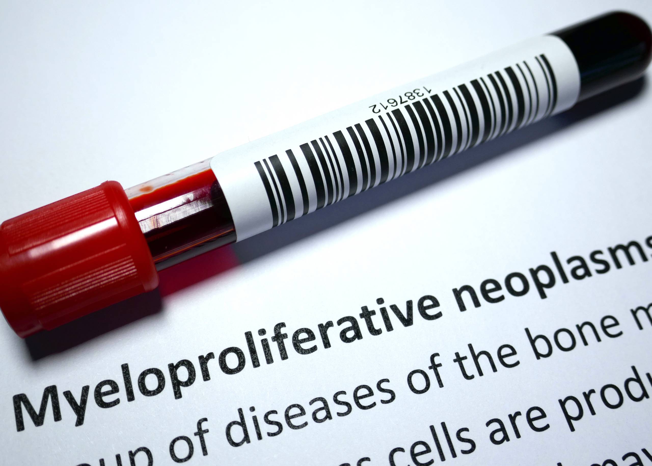
  • Histopathology results showing poorly differentiated signet ring cell adenocarcinoma

Signet ring cell adenocarcinoma is particularly aggressive, known for its poor prognosis and challenging treatment requirements, especially in elderly patients.

The Challenge of Multiple Comorbidities

What made this case extraordinarily complex was the patient’s extensive medical history:

  • Previous coronary artery bypass grafting (CABG)
  • Moderate aortic stenosis
  • Hypertension and Type 2 diabetes
  • Chronic kidney disease
  • Hyperlipidemia
  • Interstitial lung disease

This combination of conditions would typically make a patient unsuitable for major surgery, particularly the extensive procedure required for esophageal cancer treatment.

Advanced Staging and Treatment Planning

Upon arrival in the UAE for specialized care, comprehensive staging was performed:

Imaging Studies

  • CT scan revealed eccentric wall thickening of the mid-esophagus
  • PET scan showed lymph nodes in paratracheal, subcarinal, and paraesophageal regions
  • EBUS biopsy of suspicious lymph nodes was negative for malignancy

Multidisciplinary Team (MDT) Approach

The case was presented to Burjeel’s specialized oncology MDT, which includes:

  • Gastrointestinal surgeons
  • Medical oncologists
  • Radiation oncologists
  • Anesthesiologists
  • Pathologists

The team’s consensus was to proceed with neoadjuvant FLOT chemotherapy followed by surgical re-evaluation.

Neoadjuvant Chemotherapy: Preparing for Surgery

FLOT Chemotherapy Protocol

The patient received four courses of FLOT chemotherapy, a specialized regimen that includes:

  • 5-Fluorouracil
  • Leucovorin
  • Oxaliplatin
  • Docetaxel

This intensive treatment aims to:

  • Shrink the primary tumor
  • Eliminate microscopic disease
  • Improve surgical outcomes
  • Reduce risk of recurrence

Response Evaluation

After completing chemotherapy, comprehensive re-staging showed no disease progression, making the patient an excellent candidate for surgical intervention despite his high-risk profile.

Revolutionary Robotic Surgery: RAMIE Technique

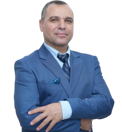
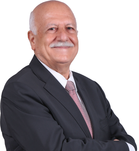
Dr. Ali Iyoob Valiyaveettil, Consultant and Head of Gastrointestinal Surgery, led the team in performing Robot-Assisted Minimally Invasive Esophagectomy (RAMIE) – a cutting-edge technique that represents the future of esophageal cancer surgery.

Why Robotic Surgery for High-Risk Patients?

Traditional open esophagectomy is one of the most challenging surgeries in oncology, with significant risks including:

  • Major blood loss
  • Extended recovery times
  • High complication rates
  • Prolonged hospital stays

RAMIE offers revolutionary advantages:

  • Minimally invasive approach reduces surgical trauma
  • Enhanced precision with robotic arms
  • 3D high-definition visualization
  • Improved maneuverability in confined spaces
  • Reduced post-operative pain
  • Faster recovery times

The Complex Three-Stage Procedure

The surgery involved multiple phases, each requiring exceptional surgical skill:

Stage 1: Thoracoscopic Phase

  • Patient positioned prone for optimal access
  • Radical esophagectomy with complete tumor removal
  • Comprehensive lymph node dissection of mediastinal nodes
  • Preservation of vital structures

Stage 2: Abdominal Phase

  • Gastric conduit preparation using the stomach to replace the esophagus
  • ICG (Indocyanine Green) test to assess blood supply adequacy
  • Pyloroplasty to improve gastric emptying
  • Careful preservation of gastric blood vessels

Stage 3: Cervical Phase

  • Neck incision for anastomosis creation
  • Side-to-side connection between gastric conduit and remaining esophagus
  • Meticulous technique to prevent leakage
  • No feeding tubes required due to superior surgical technique

Advanced Intraoperative Technology

The procedure utilized state-of-the-art technology:

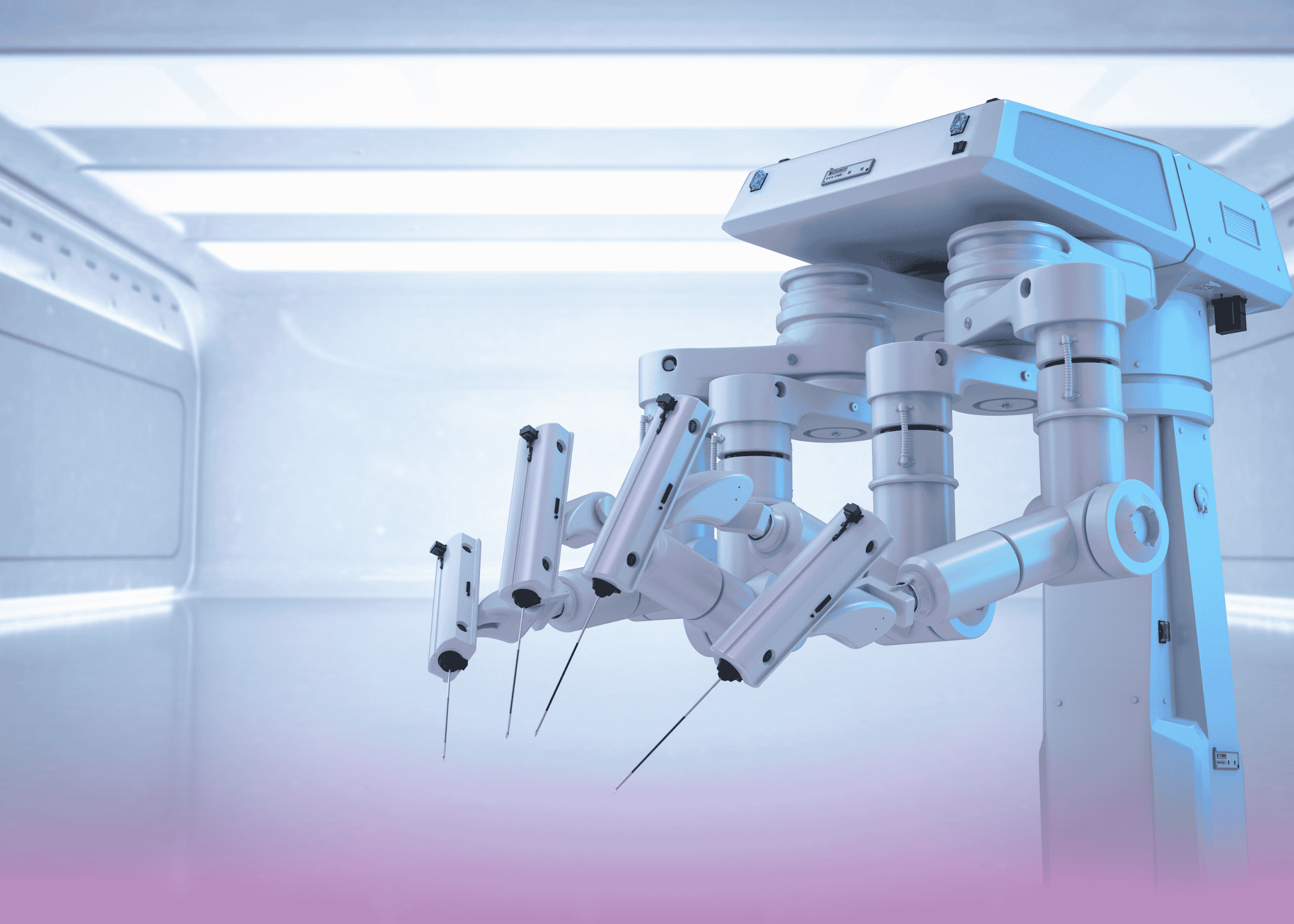
Da Vinci Xi Robotic System

  • Four robotic arms with enhanced dexterity
  • 3D high-definition visualization
  • Tremor elimination for precise movements
  • Scaled motion for fine tissue manipulation

ICG Fluorescence Imaging

  • Real-time assessment of tissue blood supply
  • Prevention of anastomotic complications
  • Immediate visualization of perfusion quality

Expert Surgical Team Excellence

The successful outcome was achieved through the collaboration of:

Remarkable Recovery and Outcomes

Despite the patient’s high-risk profile, the results exceeded expectations:

Immediate Post-Operative Period

  • ICU monitoring for 2 days as precaution
  • Nasogastric tube removal after 48 hours
  • Contrast swallow study showing normal passage with no leaks
  • Early mobilization reducing complications

Rapid Recovery Milestones

  • Oral feeding started within 48 hours
  • Progression to puree diet without problems
  • Chest tube removal after 72 hours
  • Abdominal drain removal after 72 hours
  • No voice changes or breathing difficulties
  • Clean wound healing

Hospital Discharge

The patient was discharged on the 7th post-operative day – remarkably fast for such extensive surgery, especially in an elderly patient with multiple comorbidities.

Pathology Results and Long-Term Prognosis

Histopathology Findings

  • Poorly differentiated adenocarcinoma (Grade 3)
  • ypT3N1 staging (1 out of 26 lymph nodes positive)
  • Good response to neoadjuvant chemotherapy
  • Clear surgical margins

Unexpected Finding

Multiple lymph nodes showed caseating granulomatous changes, indicating possible concurrent inflammatory conditions that were successfully managed.

The Advantages of Minimally Invasive Esophagectomy

This case demonstrates why RAMIE is becoming the gold standard for esophageal cancer surgery:

Reduced Morbidity

  • Lower complication rates compared to open surgery
  • Decreased blood loss during the procedure
  • Reduced post-operative pain
  • Faster return to normal activities

Superior Oncological Outcomes

  • Better lymph node clearance due to enhanced visualization
  • Reduced positive margin rates
  • Improved long-term survival in many studies

Enhanced Recovery

  • Shorter ICU stays reduce healthcare costs
  • Earlier hospital discharge improves patient satisfaction
  • Faster return to baseline function

Overcoming the Learning Curve Challenge

One of the traditional barriers to minimally invasive esophagectomy has been the steep learning curve for surgeons. However, robotic assistance helps overcome this challenge:

Robotic Advantages for Surgeons

  • Intuitive movements that mirror hand motions
  • Enhanced dexterity in confined spaces
  • Tremor elimination for precise dissection
  • 3D visualization improves depth perception

Training and Expertise

Burjeel Medical City’s commitment to surgical excellence includes:

  • Specialized training programs for robotic surgery
  • Continuous education in latest techniques
  • Volume requirements ensuring surgeon proficiency
  • Multidisciplinary collaboration

Why Choose Burjeel for Complex Cancer Surgery?

This successful case highlights several key advantages:

Advanced Technology

  • Latest Da Vinci Xi robotic system
  • Integrated OR technology for seamless procedures
  • Real-time imaging capabilities
  • Comprehensive monitoring systems

Specialized Expertise

  • High-volume esophageal surgery program
  • Experienced robotic surgeons
  • Multidisciplinary cancer care team
  • Specialized anesthesia for complex cases

Comprehensive Care

  • Neoadjuvant and adjuvant therapy coordination
  • Nutritional support services
  • Rehabilitation programs
  • Long-term follow-up care

The Future of Esophageal Cancer Treatment

This case represents the evolution toward personalized, minimally invasive cancer care:

Precision Medicine

  • Molecular profiling of tumors for targeted therapy
  • Personalized treatment protocols based on patient factors
  • Biomarker-guided therapy selection

Technological Advances

  • Enhanced robotic systems with improved capabilities
  • Artificial intelligence integration for surgical planning
  • Advanced imaging for better staging and monitoring

Patient Selection: Making High-Risk Surgery Safer

This case demonstrates that with proper evaluation and advanced techniques, many patients previously considered unsuitable for surgery can achieve excellent outcomes:

Comprehensive Pre-Operative Assessment

  • Cardiac evaluation and optimization
  • Pulmonary function testing
  • Nutritional assessment and support
  • Multidisciplinary consultation

Risk Mitigation Strategies

  • Enhanced recovery protocols
  • Specialized anesthesia techniques
  • Minimally invasive approaches
  • Expert post-operative care

Conclusion: Hope for Complex Cancer Patients

The successful treatment of this 74-year-old patient with multiple comorbidities demonstrates that:

  • Age alone should not exclude patients from curative surgery
  • Robotic technology can make complex procedures safer
  • Multidisciplinary care improves outcomes significantly
  • Expert surgical teams can achieve excellent results in challenging cases
  • Advanced techniques continue to expand treatment options

For patients and families facing similar diagnoses, this case provides hope that even complex situations can be successfully managed with the right expertise, technology, and comprehensive care approach.

The future of esophageal cancer treatment lies in this combination of advanced technology, surgical expertise, and personalized care – exactly what Burjeel Medical City provides to every patient.

Breast Cancer – Symptoms, Causes & Treatment

Breast cancer is a type of cancer that begins in the breast. It develops when cells in the breast begin to grow out of control. It can spread to other parts of the body, including lymph nodes and other organs. The most common types of breast cancer are ductal carcinoma and lobular carcinoma. These cancers usually start in the milk ducts or lobules. Other types include inflammatory breast cancer, Paget’s disease of the nipple, and atypical hyperplasia. On a global scale, breast cancer is women’s most common form of cancer. It affects more than 1.7 million people each year and accounts for 25% of all cancers diagnosed among women.

Triple Negative Breast Cancer

Triple Negative Breast Cancer Triple negative breast cancer is a rare form of cancer that does not have any of the three hormone receptors, estrogen, progesterone, and HER2 (human epidermal growth factor receptor 2). Triple negative breast cancers make up about 15% of all breast cancers. Triple-negative means that the cancer cells do not have receptors for three important hormones: estrogen, progesterone, or HER2.

This is different from other types of breast cancer that may be hormone-receptor positive or hormone-receptor negative. The absence of these receptors makes triple-negative breast cancers more difficult to treat than other types of breast cancer because they don’t respond to hormone therapy drugs that target these receptors. Treatment options for triple-negative breast cancer include chemotherapy and radiation therapy. But researchers are working on new treatments that target specific genetic mutations found in some patients with this type of breast cancer.

Symptoms of Breast Cancer

  • A lump or thickening in one or both breasts
  • Nipple discharge other than breast milk
  • Inverted nipple
  • Pain or tenderness in one or both breasts or underarm area
  • Skin irritation or dimpling of the breast skin
  • A change in the size or shape of one breast

Causes and Risk Factors

Breast cancer is the most common cancer among women. It occurs when cells in the breast start to grow uncontrollably. The most common type is ductal carcinoma, which forms in the breast milk ducts. Other types include lobular carcinoma, inflammatory carcinoma, medullary carcinoma, papillary carcinoma, and Paget’s disease. The exact cause isn’t known, but certain factors may increase your risk for developing it. These include:

  • Age – Most cases occur after age 40; however, breast cancer can develop in younger women. Women who have a family history of breast cancer or have had their ovaries removed before menopause have a higher risk of developing the disease than other women.
  • Family History – Having a close relative (mother, sister, or daughter) with breast cancer increases your risk of developing it yourself by up to 10 times that of women who don’t have this history in their family. Having two or more close relatives with breast cancer greatly increases your risk (by as much as 20 times).
  • Genetic disorders – Certain genetic disorders, such as BRCA1 and BRCA2, can increase your risk of developing the condition by up to 60%.
  • Lifestyle – Factors such as smoking, drinking alcohol, and being overweight can increase your risk for developing the condition.

Diagnosis

The diagnosis begins with a thorough physical examination. The doctor will look for lumps or abnormalities in your breasts and feel the lymph nodes in your armpits to see whether they have become swollen. If you have any suspicious lumps, the doctor may recommend a mammogram, ultrasound, breast MRI, or biopsy.

A mammogram is an X-ray of the breast used to detect tumors. An ultrasound uses sound waves to create images of your internal organs and tissues.

A breast MRI uses magnetic waves to create images of your internal organs and tissues.

A biopsy involves taking small samples of tissue from suspicious areas during an operation or from cells taken from a needle inserted into a lump or mass during a diagnostic test.

Treatment

Treatment depends on the stage at which it is detected. Treatment may include surgery, radiation therapy, chemotherapy, and hormone therapy. 

Surgery is one of the main treatments and can be performed either by lumpectomy or mastectomy. 

Lumpectomy involves removing only the tumor and surrounding tissue without removing any lymph nodes or other organs. 

Mastectomy involves removing all or part of the breast as well as some lymph nodes under the arm. Radiation therapy is a treatment option for all stages. It is used to shrink tumors and destroy cancer cells before surgery or when surgery is not possible due to tumor location or size. 

Chemotherapy is a treatment that uses drugs to destroy cancer cells in your body by stopping them from growing and dividing properly. 

Hormone therapy is a treatment that blocks estrogen production in women who have estrogen-receptor positive (ER+) breast cancer, which can help slow down or stop tumor growth in these patients by blocking estrogen receptors in breast tissue.

The Breast Care Clinic at Burjeel Medical City is a leading provider of comprehensive breast care services for women. Our team of expert physicians, surgeons, and nurses work together to provide the best possible care for every patient in a comfortable, supportive setting.

We offer a full range of services from diagnostic mammography through breast surgery, including lumpectomy, mastectomy, and breast reconstruction. Our facility is one of the most modern and advanced in the UAE, with state-of-the-art equipment that allows us to provide you with the best care.

Myeloproliferative Neoplasms (MPN) – Symptoms, Causes & Treatment

Myeloproliferative neoplasms (MPN) are a group of clonal hematopoietic disorders characterized by increased production of immature blood cells.

Types of Myeloproliferative Neoplasms

There are three types of MPN:

Polycythaemia Vera (PV) is a chronic blood disorder in which the body produces too many red blood cells, leading to an increase in the volume of blood and subsequent swelling. PV can also cause anemia, with insufficient red blood cells. Excessing red blood cells can lead to blockage in blood vessels, which may cause symptoms such as leg or abdominal pain or headaches.

Essential Thrombocythaemia (ET) is a rare genetic disorder that causes an increased number of platelets in the blood. This can lead to excessive bruising and bleeding, as well as clotting issues, if too many platelets are present in the bloodstream.

Myelofibrosis (MF) is a condition where bone marrow becomes abnormal and cannot produce normal amounts of blood cells. This condition can cause anemia due to abnormal red blood cell production and excessive bleeding due to low platelet counts.

Causes of Myeloproliferative Neoplasms

The causes of myeloproliferative neoplasms are not well understood. Myeloproliferative neoplasms are thought to occur when there is an imbalance between the body’s production and the destruction of blood cells.

Diagnosis

Myeloproliferative neoplasms (MPNs) can be diagnosed by a variety of approaches, including,

Symptoms: Patients with anemia or thrombocytopenia may present with fatigue, headaches, weakness, shortness of breath, or nosebleeds.

Physical Examination: The presence of splenomegaly (enlarged spleen) or hepatomegaly (enlarged liver) will be noted on physical exam. Additional findings may include enlarged lymph nodes or bone marrow hypercellularity.

Laboratory Tests: A complete blood count is a common laboratory test used to diagnose MPNs, as it can indicate anemia and thrombocytopenia. Other lab tests may include liver function tests, iron studies, and serum protein electrophoresis.

Bone Marrow Biopsy: A bone marrow biopsy is the gold standard for diagnosing MPNs; however, this test is invasive and can cause serious complications if not performed correctly by a trained physician who has experience performing these procedures in patients with compromised immune systems

Treatment

Treatment for MPNs depends on the disorder type and the symptoms’ severity. Treatment options may include medications, chemotherapy, radiation therapy, and stem cell transplantation.

Medications: Medications such as hydroxyurea, anagrelide, and interferon-alpha are commonly used to reduce the production of abnormal blood cells and reduce symptoms.

Chemotherapy: Chemotherapy destroys abnormal cells and reduces the risk of complications. It is often used in combination with other treatments.

Radiation Therapy: Radiation therapy is used to target and destroy abnormal cells. It is usually used in combination with other treatments.

Stem Cell Transplantation: Stem cell transplantation is used to replace abnormal cells with healthy ones. It is usually used in combination with other treatments.

Hematologists and Bone Marrow Transplantation experts at Burjeel Medical City, Abu Dhabi, treat Myeloproliferative Neoplasms with the latest medical advancements.

Bell’s Palsy – Symptoms, Causes & Treatment

Bell’s palsy is a medical condition in which one side of the face becomes paralyzed due to facial nerve damage. This condition was named after Sir Charles Bell, a Scottish surgeon who described it for the first time in 1821.

The facial nerve controls the muscles of the face, and when it is damaged, the muscles on that side of the face weaken or become paralyzed. This causes the affected side’s face, eyelids, and mouth to be immobile.

What Causes Bell’s Palsy?

The cause of Bell’s palsy is unknown, but it appears to be related to facial nerve inflammation. It is thought to be caused by a viral infection, such as herpes simplex virus or varicella-zoster virus, which can cause facial nerve inflammation and swelling.

Other potential triggers include facial trauma, diabetes, Lyme disease, and high blood pressure.

What are the Symptoms?

Bell’s palsy symptoms include facial weakness or paralysis on one side of the face, drooping eyelids or corners of the mouth, difficulty closing the eye on the affected side, an inability to make facial expressions, and eye or mouth dryness.

How is Bell’s Palsy Treated?

This condition can be extremely distressing for the person affected because it causes them to appear drastically different from their normal selves. Fortunately, it can be treated in a variety of ways.

Some treatment may be necessary, depending on the severity of the condition, while others may be extremely beneficial.

Corticosteroid medication is the most commonly used for treatment. This medication helps reduce the severity of the symptoms by reducing inflammation of the facial nerve. Other medications, such as antivirals, may be prescribed in addition to corticosteroids.

Physical therapy is another common Bell’s palsy treatment. Physical therapy helps to strengthen and improve the range of motion of the facial muscles. This can help to alleviate symptoms and improve the appearance of the affected side of the face.

Finally, the best Bell’s palsy treatment will be determined by the severity of the condition and the individual’s overall health. Working with a healthcare professional to determine the best treatment plan is essential. With the right treatment, you can lessen the severity of the symptoms and improve your quality of life.

Expert Neurologist in Abu Dhabi

Low Carbohydrate Diet for Diabetes – Here is the Truth

Low carbohydrate diet for diabetes typically limits total carbohydrate intake to between 130 and 150 grams per day, with at least half coming from vegetables and fruits. The remaining amount should come from whole grains, legumes and dairy products.

Did you know that restricting carbohydrates is not an excellent way to manage diabetes and may even cause hypoglycemia? 

All of the carbohydrates you consume are converted into glucose. The type and amount of sugar you consume can impact your blood glucose levels and diabetes management.

Carbohydrates come in various forms, including simple sugars (white sugar, white bread, white rice juice, and so on), complex carbohydrates (whole grain bread, brown rice, and so on), and fibers.

If you have diabetes and use insulin, you must keep track of your carbohydrate intake.

Learn which foods contain carbohydrates, how to estimate carbohydrate portions, and how to track the effect of carbohydrates on blood glucose levels.

Evidence suggests that the quality of carbohydrates, rather than the quantity, is more important for overall health and blood glucose control.

The glycemic index (GI), glycemic load, and fiber content were used to assess the quality of carbohydrates.

Foods with a lower glycemic index, in general, can help manage blood glucose levels by slowing the release of sugar into the blood.

More importantly, choosing foods high in fiber and whole grains over refined carbohydrates like white bread is better for our health and lowers our risk of type 2 diabetes mellitus.

It is also known that certain carbohydrate-containing foods, such as fruits and vegetables, are associated with good overall health.

Wholegrain bread and cereals are examples of low-glycemic-index foods.

Rather than fruit juices, eat whole fruits.

Eating an apple with the peel on, for example, contains more fiber and less sugar than drinking apple juice, which contains more sugar and fewer fibers.

As an alternative to white rice, try quinoa and bulgur wheat.

Low-carb sources of protein include legumes.

The carbohydrate serving size in your food is critical for blood glucose control.

Personal needs and body composition analysis are used to calculate the number of servings. This is thoroughly discussed with your dietitian and planned based on your preferences and needs.

Finally, by reducing carbohydrates or following a low carbohydrate diet for diabetes, we can reduce the calorie content of food; as a result, overweight or obese people with type 2 diabetes mellitus will benefit and lose weight, aiding blood glucose management.

Following a low carbohydrate diet for diabetes will result in good glycemic control in the long run.

Always remember to eat better, not less.

Experienced Diabetes Dietitian in Abu Dhabi

Ms. Dina Aldarweesh

Clinical Dietitian 


Years of Experience: 4

Nationality: Jordan

Languages Known: English, Arabic