Introduction
Sciatica—characterized by radiating pain from the lower back down the leg along the sciatic nerve distribution—affects millions of people worldwide and is a common cause of disability and decreased quality of life. The traditional understanding of sciatica has centered primarily around structural causes, particularly herniated discs and spinal stenosis mechanically compressing nerve roots. However, emerging evidence suggests that in some cases, sciatic pain may occur in the absence of visible structural compression on imaging studies, pointing to alternative mechanisms that are biochemical rather than mechanical in nature.
This case study explores the successful diagnosis and treatment of chemical radiculopathy, a fascinating yet underrecognized cause of sciatica, highlighting how advanced interventional pain management techniques can provide significant relief even when conventional imaging suggests no obvious mechanical compression.
Understanding Chemical Radiculopathy
Beyond Mechanical Compression
The conventional wisdom in spine medicine has long held that sciatic pain results primarily from physical compression of nerve roots, typically by herniated disc material or bony overgrowth. However, research has increasingly revealed that inflammation and biochemical irritation of nerve roots can produce identical symptoms even without visible compression on imaging studies.
Chemical radiculopathy occurs when inflammatory mediators from degenerated discs or surrounding structures leak through annular tears (fissures in the disc’s outer fibrous ring) and irritate nearby nerve roots. This represents an early stage of disc degeneration that precedes the development of frank herniation visible on imaging.
The Inflammatory Cascade
Several biochemical substances have been implicated in this inflammatory process:
- Phospholipase A2: An enzyme found in high concentrations in herniated disc material
- Cytokines: Particularly tumor necrosis factor-alpha (TNF-α) and interleukins
- Matrix metalloproteinases: Enzymes involved in disc degeneration
- Prostaglandins: Inflammatory mediators that sensitize nerve roots to pain
- Substance P: A neuropeptide involved in pain transmission
These inflammatory mediators can diffuse through annular tears and reach nerve roots without any significant structural displacement of disc material, explaining why some patients exhibit classic radicular symptoms despite minimal findings on imaging studies.
Case Presentation
Patient Profile
The patient was a female athlete in her 40s who was a long-distance runner. She presented with severe lower extremity pain that was more pronounced than her axial (back) pain, suggesting a predominantly radicular component to her symptoms.
Clinical Examination
The physical examination revealed several significant findings:
- Bilateral straight leg raise (SLR) was limited to 30 degrees due to pain
- Inability to bear weight on toes and heels because of pain
- No detectable neurological deficit (normal strength, sensation, and reflexes)
- Clinical pattern strongly suggestive of L5 and S1 nerve root involvement
These findings were consistent with bilateral radiculopathy affecting the L5 and S1 nerve roots, despite the absence of neurological deficits.
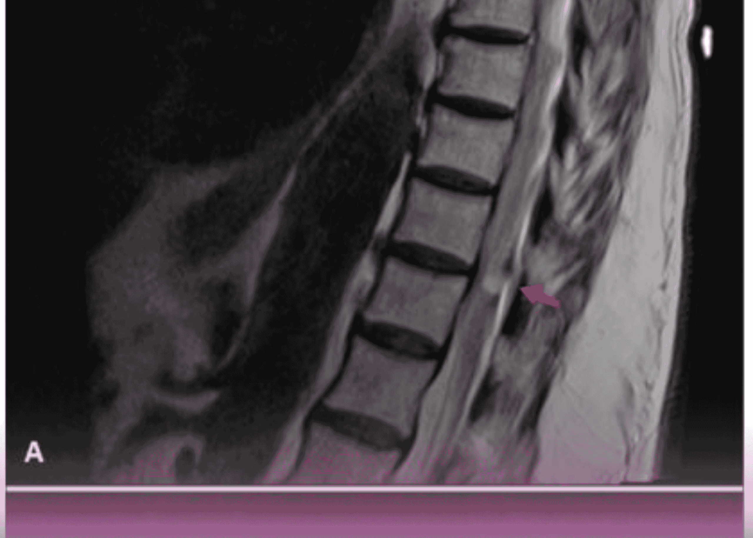
Diagnostic Imaging
Magnetic Resonance Imaging (MRI) of the lumbar spine revealed:
- Degenerated and desiccated L5-S1 disc (loss of normal hydration and height)
- Importantly, axial views showed the absence of mechanical compression
- Both exiting nerve roots appeared free from impingement
This created a clinical paradox: the patient had clear symptoms of nerve root irritation, but imaging showed no structural cause for these symptoms.
Interventional Management
Diagnostic-Therapeutic Procedure
Based on the clinical presentation and imaging findings, Dr. Shailendra Chauhan, Consultant in Anesthesia and Pain Management at Burjeel Day Surgery Center, performed a bilateral L5-S1 selective nerve root block under fluoroscopic guidance.
The procedure involved:
- Precise needle placement at the L5 and S1 nerve roots bilaterally
- Injection of non-ionic contrast to confirm appropriate spread and the absence of significant mechanical compression
- Administration of a therapeutic mixture containing lidocaine (a local anesthetic) and Depo-Medrol (a corticosteroid)
Immediate Outcome
The results were dramatic and immediate:
- Complete resolution of pain immediately following the procedure
- Bilateral straight leg raise improved from 30 degrees to 80 degrees
- Restored ability to bear weight on toes and heels
- Significant improvement in overall mobility
This dramatic response to selective nerve root block with anti-inflammatory medication strongly supported the diagnosis of chemical radiculopathy as the underlying cause of the patient’s sciatica.
Discussion
Diagnostic Challenges
Chemical radiculopathy presents several diagnostic challenges:
- Imaging-Clinical Mismatch: Patients exhibit classic radicular symptoms despite minimal or no compression on imaging.
- Overlooked Diagnosis: Many practitioners continue to focus exclusively on structural causes visible on imaging, potentially missing chemical causes.
- Early Intervention: Chemical radiculopathy may represent an earlier stage of disc pathology before structural herniation occurs, offering a window for preventive interventions.
Treatment Implications
Understanding chemical radiculopathy expands the treatment options available to patients with sciatica:
- Targeted Anti-inflammatory Therapy: Selective nerve root blocks deliver potent anti-inflammatory medications directly to the site of inflammation.
- Beyond Decompression: In cases where imaging shows no significant compression, pursuing surgical decompression may be unnecessary and unlikely to address the true cause of pain.
- Annular Repair Techniques: Emerging therapies aimed at sealing annular tears may help prevent leakage of inflammatory mediators.
- Systemic Anti-inflammatory Approaches: Oral medications, dietary modifications, and lifestyle interventions that reduce systemic inflammation may play a supportive role.
Future Management Plan
For this particular patient, a comprehensive follow-up plan was established:
- Recommendation to repeat the procedure after 3-4 months if pain recurs
- Consideration of discography (a diagnostic procedure to identify painful discs) in case of recurrence
- Possible annuloplasty (a procedure to repair annular tears) if indicated by discography
Clinical Implications
This case highlights several important clinical implications:
Expanded Diagnostic Framework
The recognition of chemical radiculopathy as a cause of sciatica encourages clinicians to move beyond the traditional structure-based paradigm when evaluating patients with radicular symptoms. A more comprehensive approach should include:
- Thorough clinical examination: Identifying patterns of pain and dysfunction that may suggest radicular involvement even in the absence of neurological deficits
- Critical interpretation of imaging: Recognizing that “normal” or “minimally abnormal” imaging does not rule out significant radicular pain
- Diagnostic injections: Utilizing selective nerve root blocks as diagnostic tools to identify pain generators and confirm chemical causes
Therapeutic Options
Understanding chemical radiculopathy expands the therapeutic options available to patients:
- Targeted injections: Delivering anti-inflammatory medications directly to the site of chemical irritation
- Pharmacological approaches: Medications specifically targeting neuroinflammation and neuropathic pain mechanisms
- Avoidance of unnecessary surgery: Preventing surgical interventions that may not address the underlying chemical causes
- Regenerative approaches: Emerging therapies aimed at improving disc health and repairing annular integrity
Evidence-Based Application
The case underscores the growing clinical evidence that anatomical abnormalities are not always required to cause radiculopathy, and that biochemical etiologies play a significant role in many cases. This paradigm shift is supported by:
- Basic science research demonstrating the presence of inflammatory mediators in disc material
- Clinical studies showing successful treatment of radicular pain with anti-inflammatory approaches
- Observations of patients with clear radicular symptoms despite minimal imaging findings
Conclusion
This case illustrates the importance of considering chemical radiculopathy in the differential diagnosis of patients with sciatic symptoms, particularly when imaging studies fail to demonstrate significant mechanical compression. The dramatic response to selective nerve root block with anti-inflammatory medication in this patient serves as a powerful reminder that pain mechanisms extend beyond simple mechanical models.
For clinicians, this case encourages a broader conceptual framework when evaluating patients with radicular pain, incorporating both structural and chemical etiologies. For patients suffering from sciatica without clear imaging findings, the recognition of chemical radiculopathy offers hope through targeted interventional approaches.
The successful management of this case at Burjeel Day Surgery Center demonstrates the value of a sophisticated, mechanism-based approach to pain diagnosis and treatment, ultimately leading to improved outcomes and quality of life for patients with complex pain conditions.

