A remarkable case of a forgotten double J stent retained for six years presented unique challenges for urologists at LLH Hospital, who successfully removed the heavily encrusted stent through a combination of innovative techniques.
Case Background
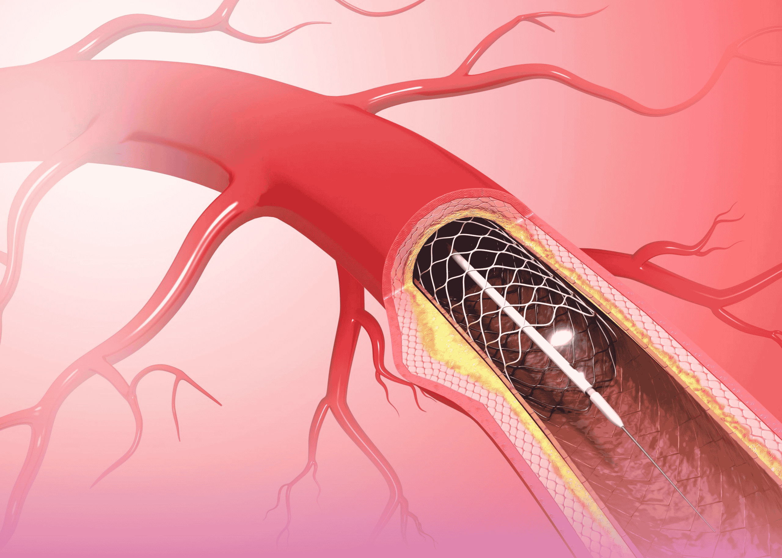
A 35-year-old female patient presented to LLH Hospital with right flank pain and burning during urination. Her medical history revealed that she had undergone ureteroscopy with double J stent placement six years prior while working outside the UAE. Due to various circumstances, the stent—which is typically removed after 4-6 weeks—had never been extracted.
“Double J stents are essential tools in urological practice, providing effective drainage from kidney to bladder in various conditions,” explains Dr. Jaipal Paryani, Specialist Urologist at LLH Hospital. “However, when forgotten or neglected, these temporary devices can create serious complications including stone formation, infection, and renal impairment.”
Diagnostic Findings
Initial ultrasound and X-ray KUB (kidney, ureter, bladder) examinations revealed a heavily encrusted double J stent with a large bladder stone measuring approximately 6 cm. CT scan confirmed the findings, showing calculus deposits along the entire length of the stent, with particular concentration around the upper coil in the kidney.
Laboratory tests revealed slightly elevated creatinine levels and a urinary tract infection with Staphylococcus aureus. This case represented one of the longest-retained stents with the highest stone burden documented in the UAE.
Treatment Approach
The management strategy developed by Dr. Paryani and Dr. Jana Kalyan Vijaya Kumar involved a multi-step approach:
- Initial Antibiotic Treatment: The patient received appropriate antibiotics to address the urinary infection before surgical intervention.
- Extracorporeal Shock Wave Lithotripsy: An initial attempt to break up the stones around the upper coil of the stent using ESWL proved unsuccessful in releasing it from the renal pelvis mucosa.
- Combined Endourological Procedure:
- Introduction of a 20 Fr nephroscope sheath to remove the large bladder stone using pneumatic lithotripsy
- Deployment of a 6 Fr ureteroscope alongside the encrusted stent within the ureter
- Careful internal lithotripsy to fragment stone formations throughout the ureter
- Delicate freeing of the upper coil from renal pelvis mucosa where it had become embedded
- Successful removal of the entire stent with all fragmented stones
Technical Excellence
The case presented extraordinary technical challenges. The stent had become an integrated part of the urinary tract after six years, with significant encrustations throughout its length. The bladder stone alone was substantial, while the embedded nature of the upper coil required exceptional care to avoid renal injury.
“Managing forgotten stents requires a careful, individualized approach,” notes Dr. Vijaya Kumar. “In this case, we utilized multiple techniques across several specialties, including endourology, lithotripsy, and minimally invasive approaches to achieve complete removal with minimal trauma.”
Preventive Strategies
This case underscores the importance of proper stent management systems in urological practice. Key preventive measures include:
- Comprehensive patient education about temporary nature of stents
- Clear follow-up protocols with reminder systems
- Maintenance of stent registries in hospitals
- Patient tracking mechanisms for those relocating between healthcare systems
The successful management of this complex case highlights the sophisticated urological capabilities available at LLH Hospital and serves as an important reminder about the potential complications of medical device oversight.

