Workplace eye injuries can happen in an instant, but the consequences can be life-altering. A recent case at Burjeel Hospital demonstrates how quick thinking, advanced surgical techniques, and specialized expertise can save both vision and prevent serious complications when dealing with penetrating orbital injuries.
A Workplace Accident with Serious Consequences
A 43-year-old construction worker was rushed to Burjeel Hospital’s emergency department after sustaining a severe eye injury while working with metal equipment. A metallic object had penetrated his left eye, causing immediate bleeding and excruciating pain.
The injury occurred during routine work activities, highlighting the importance of proper safety equipment and the unpredictable nature of workplace accidents involving the delicate structures around the eye.
Critical Assessment and Advanced Imaging
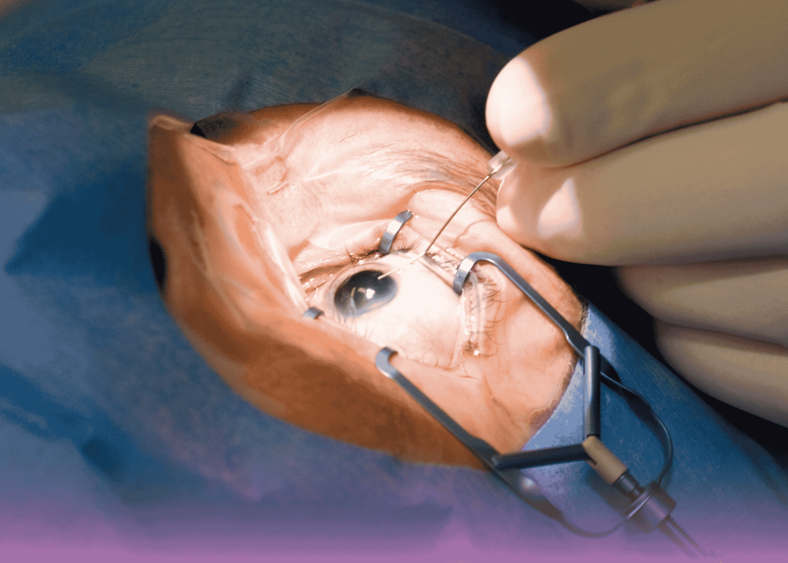
Upon arrival, Dr. Ann Divya Jacob, Specialist in Ophthalmology, conducted a thorough clinical examination that revealed a 7mm external wound on the medial aspect of the left eye. While concerning, the external wound didn’t reveal the full extent of the injury.
Advanced CT imaging was immediately ordered to assess the internal damage and locate the foreign object. The results were both surprising and alarming:
- A dense, metallic foreign body measuring 15 x 15 mm
- Located in the extraconal space near the insertion of the medial rectus muscle
- No immediate damage to the optic nerve or other critical structures
The Surgical Challenge: Precision and Safety
The diagnosis was clear: penetrating orbital injury with retained metallic foreign body. However, the treatment required exceptional surgical skill and careful planning.
Key Challenges Faced:
- Size of the foreign object – At 15x15mm, it was significantly larger than typical foreign bodies
- Location near critical structures – Close proximity to the medial rectus muscle
- Risk of posterior displacement – Any surgical error could push the object toward the optic nerve
- Preventing muscle damage – Maintaining extraocular muscle function was crucial
Innovative Surgical Technique Under Local Anesthesia
Dr. Pulak Puneet, Specialist Anesthetist, administered local anesthesia to keep the patient comfortable while maintaining their ability to cooperate during the procedure.
The surgical approach required innovative thinking when standard ophthalmic instruments proved inadequate:
The Solution:
- Standard ophthalmic instruments were too small and couldn’t provide secure grip
- Large curved artery forceps were selected for their superior grasping capability
- Single-piece extraction through the original entry wound
- Careful technique to prevent posterior displacement toward the optic nerve
Advanced Surgical Facilities and Equipment
This complex procedure was performed in Burjeel’s state-of-the-art operating theater equipped with:
- High-resolution surgical microscopes for precise visualization
- Advanced imaging integration for real-time guidance
- Specialized ophthalmic instruments and backup equipment
- Emergency protocols for potential complications
Successful Outcome and Vision Preservation
The foreign body was successfully extracted in one piece without causing additional damage to surrounding structures. Post-extraction testing included a forced duction test to ensure the extraocular muscles remained intact and functional.
Key Success Factors:
- Immediate medical attention prevented infection and further damage
- Advanced diagnostic imaging provided crucial pre-surgical planning
- Experienced surgical team with expertise in complex orbital procedures
- Innovative problem-solving when standard instruments proved inadequate
The Importance of Emergency Orbital Surgery
This case highlights several critical aspects of emergency eye care:
Timing is Critical
- Prompt medical attention prevents complications
- Delayed treatment can lead to infection, scarring, or vision loss
- Emergency surgery may be required to preserve sight
Specialized Expertise Required
- Orbital anatomy is complex and requires specialized knowledge
- Experience with foreign body removal is essential
- Understanding of both ophthalmology and emergency surgery
Advanced Imaging Essential
- CT scans provide detailed visualization of foreign objects
- Pre-surgical planning reduces complications
- Real-time imaging guidance improves outcomes
Prevention and Workplace Safety
While this case had a successful outcome, it serves as an important reminder about workplace eye safety:
- Always wear appropriate safety eyewear when working with metal tools
- Ensure proper safety protocols are followed in industrial settings
- Seek immediate medical attention for any eye injury, regardless of severity
- Regular safety training can prevent many workplace injuries
Why Choose Burjeel for Emergency Eye Care?
Burjeel Hospital’s ophthalmology department offers:
- 24/7 emergency eye care services
- Advanced diagnostic imaging including high-resolution CT
- Experienced orbital surgeons trained in complex procedures
- State-of-the-art surgical facilities with latest technology
- Comprehensive follow-up care for optimal recovery
Our multidisciplinary approach ensures that patients receive the highest level of care for even the most challenging eye emergencies.
Recovery and Follow-up Care
The patient’s recovery was smooth and uncomplicated. Follow-up examinations confirmed:
- Complete healing of the entry wound
- Normal extraocular muscle function
- No signs of infection or other complications
- Preserved vision in the affected eye
Regular follow-up appointments ensure continued monitoring and early detection of any potential late complications.

