Patient Presentation
A 62-year-old female presented with irregular left flank pain persisting over several months. The pain pattern was intermittent and occasionally relieved with analgesics. The patient also reported occasional dysuria but no visible hematuria. Despite these concerning symptoms, she had not sought earlier medical attention.
Diagnostic Findings
Initial ultrasound examination revealed a large 10 cm diameter tumor in the left kidney. Subsequent CT scanning confirmed a 10 x 10 cm mass with features suspicious for malignancy, accompanied by multiple prominent collateral vessels in the perinephric region.
Preoperative assessment identified the patient as having high surgical risk, supported by abnormal laboratory parameters including:
- Significantly elevated APTT
- Increased INR and prothrombin levels
- Elevated creatinine indicating impaired renal function
Surgical Approach and Outcome
After thorough discussion with the patient and her family regarding available options, Dr. Humam Qaraschouli, Consultant Urologist, proceeded with an open radical tumor nephrectomy. The kidney removal surgery was completed without complications, and the patient experienced an uneventful postoperative course, allowing for discharge as scheduled.
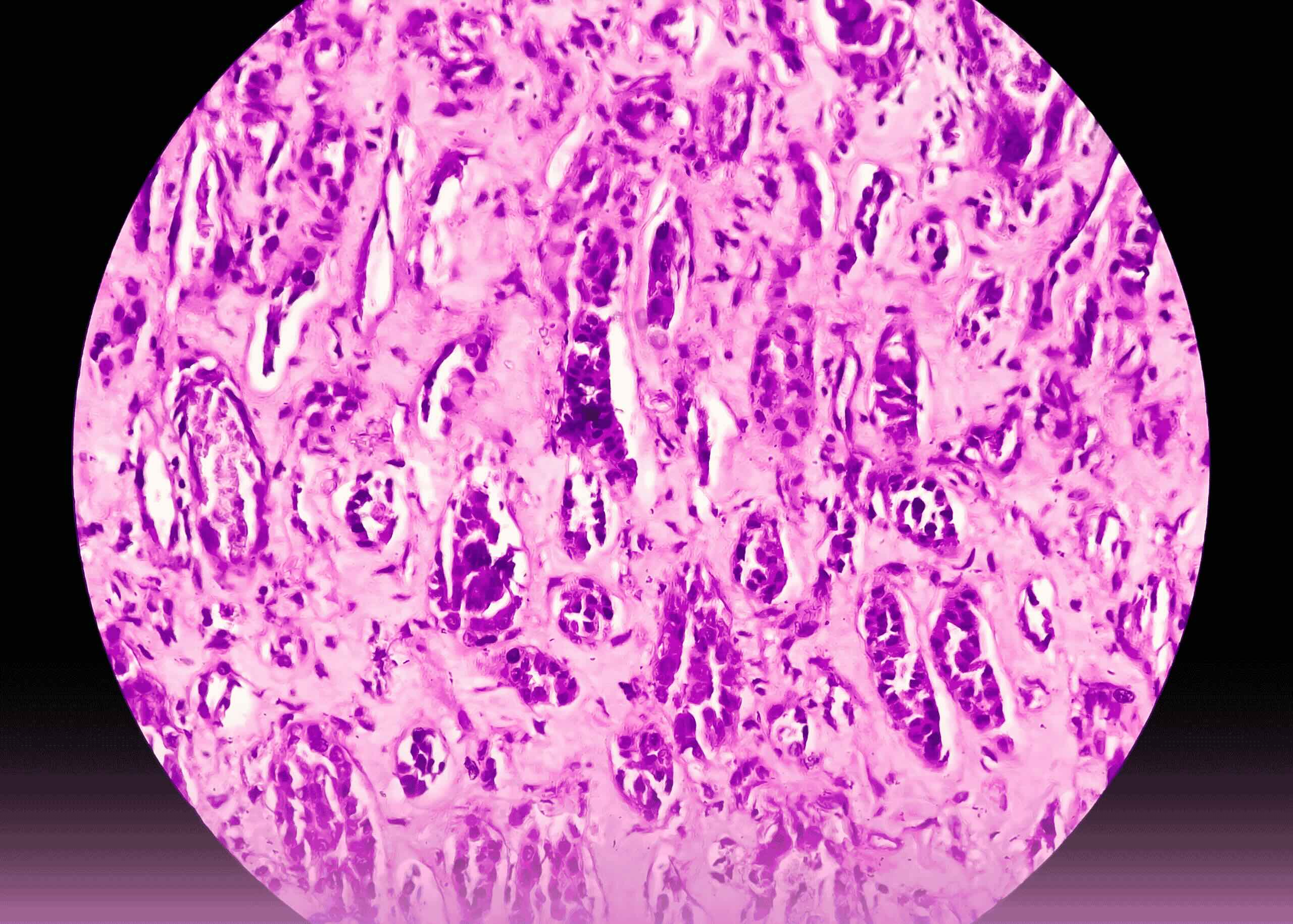
Histopathological examination confirmed renal cell carcinoma (pT2a, size 9.6 cm, G4, R0). Postoperative CT scanning of the thorax and abdomen showed no evidence of metastatic disease and demonstrated a satisfactory appearance of the left renal fossa. The oncology team of Burjeel Royal Asharej subsequently established a regular follow-up protocol.

